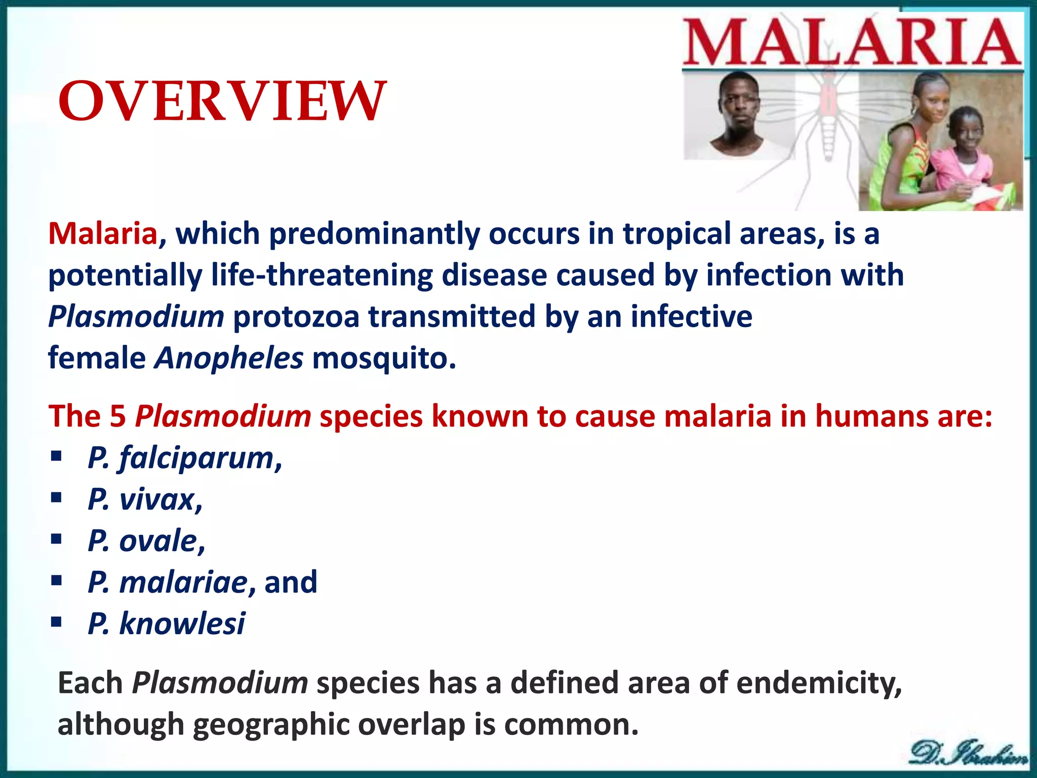
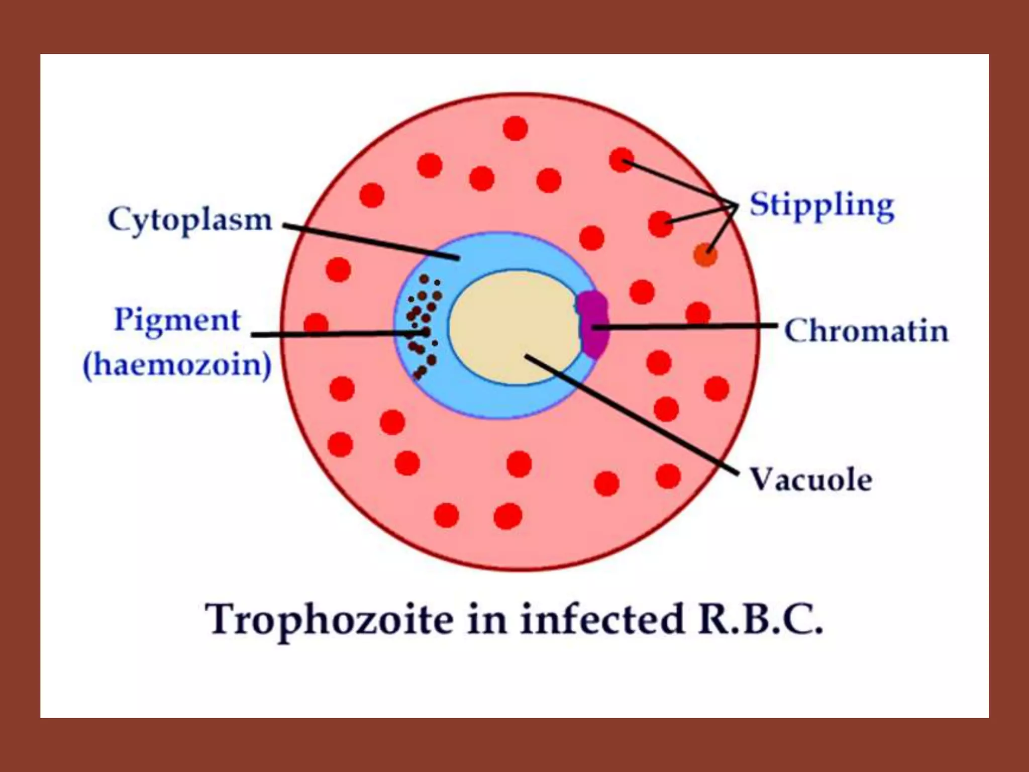
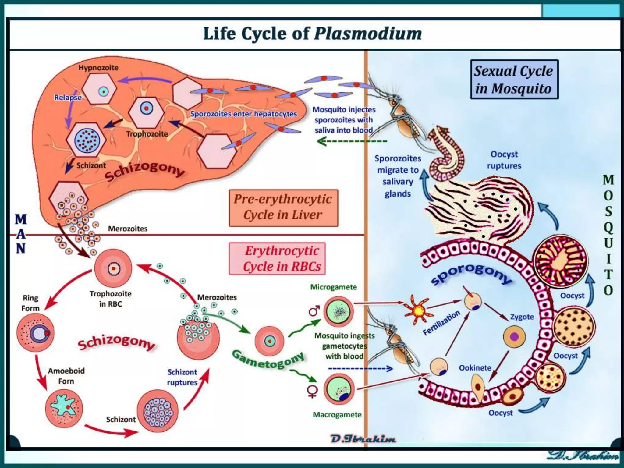
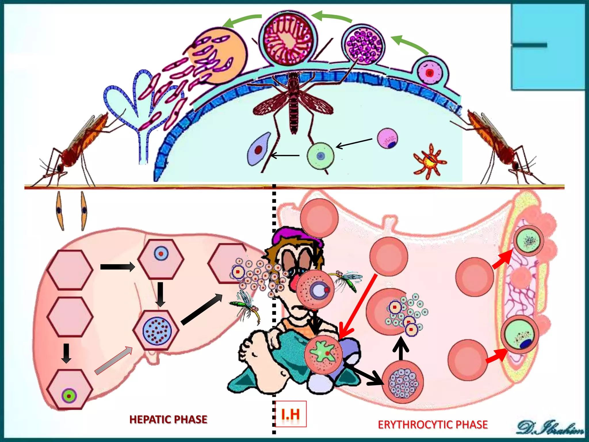
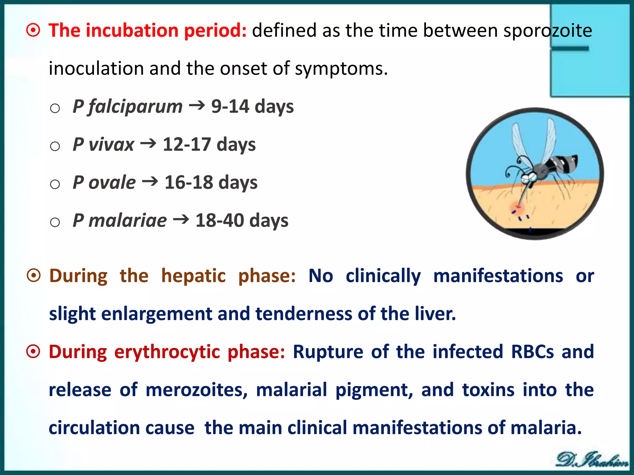
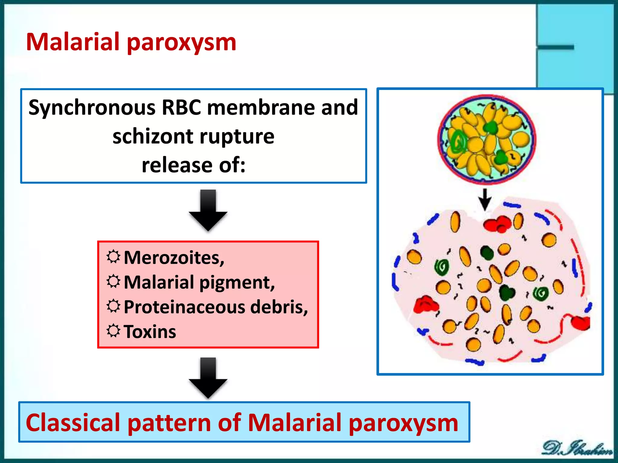
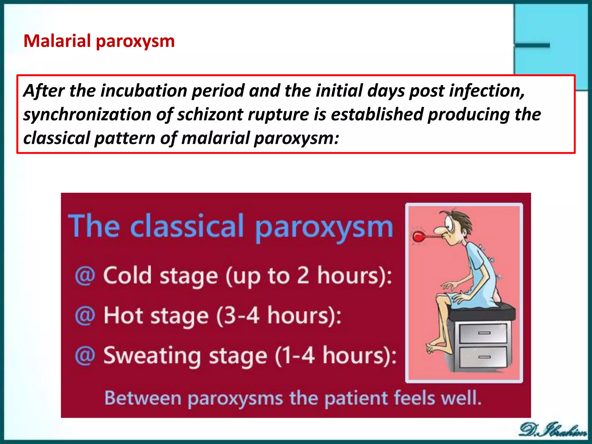
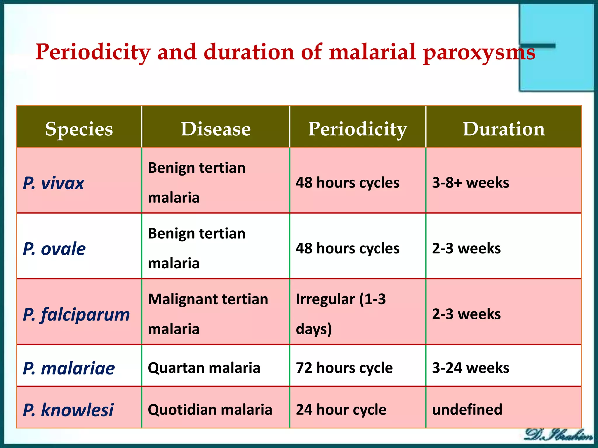
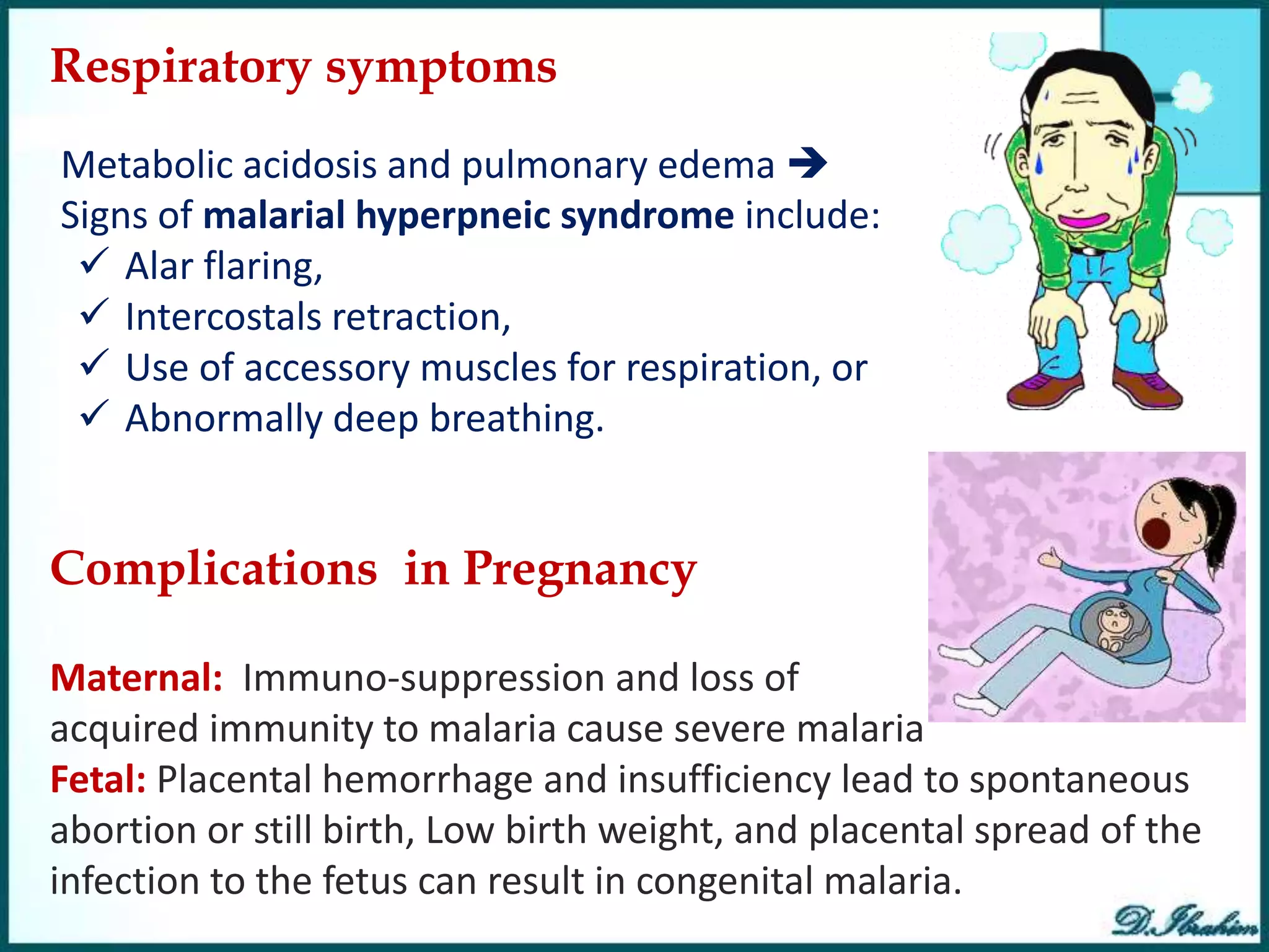
Malaria is caused by infection with Plasmodium parasites transmitted through the bites of infected Anopheles mosquitoes. There are five Plasmodium species that cause malaria in humans, with P. falciparum being the most deadly. The life cycle involves an initial hepatic phase followed by an erythrocytic phase where the parasites multiply within red blood cells. This causes cyclical fevers, chills, and other symptoms. P. falciparum malaria can progress to severe complications due to adhesion of infected cells and blockage of small blood vessels. Untreated malaria remains a major global health problem, especially among young children in sub-Saharan Africa.
Malaria is a mosquito-borne parasitic disease caused by Plasmodium parasites. Physiotherapists play a role in educating patients about malaria, its types and symptoms. They should refer patients to physicians if they suspect malaria based on symptoms and history. Malaria symptoms include fever, chills, headaches and fatigue. It can cause severe complications like cerebral malaria if caused by P. falciparum. Diagnosis involves examining blood smears under a microscope for malaria parasites. Treatment depends on the Plasmodium species and may involve medications like chloroquine, primaquine, artemisinin or quinine.
Cestodes, or tapeworms, are flat segmented parasitic worms that infect the intestines of humans and other animals. They range in size from a few millimeters to several meters in length. The body consists of a head (scolex) and chain of segments (proglottids) that contain reproductive organs. Two major orders that infect humans are Pseudophyllidea and Cyclophyllidea. Pseudophyllidea have slit-like grooves instead of suckers, while Cyclophyllidea have cup-like suckers. Common tapeworms infecting humans include Diphyllobothrium latum (fish tapeworm), Taenia saginata (beef tapeworm), Taenia sol
The document summarizes information about malaria from its causative parasite Plasmodium to its complex life cycle involving both human and mosquito hosts. It describes the four main Plasmodium species that infect humans and cause malaria, and outlines their distinct life cycles including exoerythrocytic schizogony in the liver, asexual erythrocytic schizogony in red blood cells, and sexual reproduction through gametocytes in mosquitoes leading to sporogony and infective sporozoites. It also reviews the morphological appearance of different parasite stages in blood for each Plasmodium species.
Dracunculus medinensis, also known as guinea worm, is a parasitic nematode transmitted through contaminated water. It causes the disease dracunculiasis. The parasite has a two-host lifecycle, infecting humans and water fleas. People in rural areas who depend on open water sources for drinking are most at risk. Symptoms appear about a year after infection as a blister forms on the skin and the female worm emerges to release larvae into water, causing intense pain. Diagnosis involves detecting the emerging worm or larvae microscopically. Treatment focuses on slowly removing the worm and preventing secondary infections. Contaminated water prevention through filtration and use of boiled water is key to control.
This document discusses Giardia lamblia, a unicellular parasite that causes giardiasis. Some key points:
- G. lamblia was first observed by Van Leeuwenhoek in 1681 and causes traveler's diarrhea or beaver fever. It inhabits the small intestine as a trophozoite attached to the mucosa.
- The infective stage is the cyst, which is ingested through contaminated food or water or from contact with infected flies or food handlers. The cyst contains 4 nuclei and can survive outside the host.
- Symptoms of giardiasis include diarrhea, flatulence, and greasy stool. The infection is common in children. The
The document summarizes Trichinella spiralis, a parasitic roundworm that can infect humans and cause trichinosis. Key points:
- T. spiralis has a complex life cycle involving an intestinal phase in hosts and an encapsulated muscle phase that can persist for decades.
- Common symptoms in humans include nausea, vomiting, fever and muscle pain as the larvae migrate and cysts form in muscles.
- Transmission occurs through ingesting undercooked meat from infected pigs and other reservoir hosts like bears that harbor the parasite.
This document provides information on Toxoplasma gondii, a protozoan parasite that can infect humans and cause the disease toxoplasmosis. It discusses the introduction, taxonomy, structure/morphology, life cycle, epidemiology/transmission, clinical significance/pathogenesis, diagnosis, and treatment of T. gondii. Key points include that T. gondii has three stages in its life cycle (tachyzoite, bradyzoite, sporozoite), cats are the definitive host, and infection is often asymptomatic but can cause severe disease in immunocompromised or congenitally infected individuals. Diagnosis involves serologic tests or direct detection of the parasite, and treatment focuses on drugs that target
The document discusses Plasmodium, the parasite that causes malaria. It describes the different Plasmodium species that infect humans, including P. falciparum which causes the most severe form of malaria. The life cycle is explained, involving an asexual replication phase in humans and a sexual phase in mosquitos. Symptoms include fever, anemia, and enlarged spleen. Diagnosis involves examining blood smears under a microscope to identify the Plasmodium species and recommend treatment, usually chloroquine for sensitive strains.
Balantidium coli is the largest protozoan parasite that inhabits the large intestines of humans and other primates like pigs and monkeys. It exists in two stages - the trophozoite, which is the invasive stage, and the cyst, which is the infective transmission stage. Balantidium coli is found worldwide and can be transmitted through ingestion of contaminated food, water, or undercooked meat containing the cysts. In the intestines, the cysts excyst into trophozoites that colonize and cause damage to the large intestine mucosa through enzyme production, potentially resulting in ulcers, diarrhea, dysentery, and other gastrointestinal symptoms. Diagnosis involves stool examination and treatment focuses on hy
The document discusses malaria, caused by parasites of the Plasmodium genus transmitted via mosquito bites. It affects over 100 countries and kills approximately 2,000 people per day. The most common species causing malaria in India are P. vivax, P. falciparum, P. ovale, and P. knowlesi, with P. falciparum being the most lethal. Malaria symptoms include fever, fatigue, nausea, and in severe cases can include cerebral malaria, acidosis, anemia, renal failure, pulmonary edema, hypoglycemia, and death. Diagnosis involves examining blood smears under a microscope for parasites. Treatment depends on the Plasmodium species and may include chloroquine,
The document describes Dracunculus medinensis, commonly known as the Guinea worm. It provides details on the morphology of the female and male worms, noting females are elongated and cylindrical with a recurved caudal end, while males are smaller and coiled posteriorly. The life cycle involves a cyclops intermediate host, with larvae released from females when submerged in water and infecting humans who ingest water contaminated with the larvae. Infection causes the painful disease dracunculiasis, presenting as a cutaneous blister that emerges from the skin, often followed by secondary bacterial infections like tetanus.
The document describes and classifies various medically important trematode parasites. It discusses their general characteristics, classification, life cycles, and morphology of adults and eggs. Key points include: trematodes are flatworms with oral and ventral suckers, require two intermediate hosts, and eggs are operculated; major groups discussed are blood flukes (Schistosoma), liver flukes (Fasciola, Clonorchis, Opisthorchis), intestinal flukes (Fasciolopsis, Heterophyids, Echinostoma), and lung flukes (Paragonimus). Adult and egg morphology and sizes are provided for identification purposes.
Plasmodium is a protozoan parasite that causes malaria in humans. It has a complex life cycle involving two hosts. In humans, it infects liver cells and red blood cells in an asexual reproductive cycle. In mosquitoes, it undergoes sexual reproduction resulting in sporozoites that can infect other humans and continue the cycle. The four main Plasmodium species that infect humans are P. vivax, P. falciparum, P. malariae, and P. ovale, with P. falciparum being the most lethal.
This document describes the key characteristics of four species of Plasmodium that infect humans and cause malaria: P. falciparum, P. vivax, P. malariae, and P. ovale. It covers their morphology, life cycles, symptoms caused, and distinguishing features. P. falciparum is the most dangerous species and causes malignant tertian malaria. P. vivax can cause relapse of infection through dormant liver stages called hypnozoites. The life cycles involve alternating asexual replication phases in the human host and sexual phases in the Anopheles mosquito vector.
This document discusses Balantidium coli, a ciliated protozoan parasite that causes the disease balantidiasis in humans. It has two life stages, a motile trophozoite stage that inhabits the large intestine and reproduces, and an infective cyst stage that is transmitted through fecal contamination. Symptoms include diarrhea, dysentery, abdominal pain and ulceration of the intestinal wall. Diagnosis is made by examining stool samples under a microscope. Treatment involves oral antibiotics such as tetracycline or metronidazole.
Fasciola hepatica, also known as the common liver fluke, is a parasitic flatworm that infects the livers of sheep and cattle. Its complex life cycle involves freshwater snails acting as intermediate hosts. Humans can become accidentally infected by consuming raw freshwater plants containing the fluke larvae. The flukes mature and reproduce in the bile ducts of the liver, causing a disease called fascioliasis. Symptoms range from asymptomatic to abdominal pain and liver damage. Diagnosis involves examining stool samples for fluke eggs or conducting imaging tests and antibody tests. Treatment primarily involves administering deworming medications like triclabendazole or bithionol.
Giardia lamblia is a protozoan parasite that infects the small intestine. It has two stages - the trophozoite stage, which actively multiplies in the small intestine, and the cyst stage, which is passed in feces and can survive outside the body. Infection occurs when cysts are ingested and excyst in the small intestine, releasing trophozoites. Trophozoites attach to the intestine and cause symptoms like diarrhea and abdominal cramps. Cysts form when trophozoites reach the colon and are passed in stool, allowing transmission to new hosts. Diagnosis involves examining stool samples microscopically for cysts or trophozoites. Treatment involves antibiotics like metronidaz
Malaria is a vector-borne infectious disease caused by protozoan parasites that is transmitted through the bites of infected Anopheles mosquitoes. It is widespread in tropical and subtropical regions and affects around 40% of the world's population. The most common and deadly type is Plasmodium falciparum malaria, which can lead to complications like cerebral malaria. Signs and symptoms include periodic fevers, headaches, chills, and anemia. Diagnosis involves examining blood smears under a microscope to look for the parasites. Treatment involves antimalarial drugs like chloroquine, primaquine, and artemisinin combination therapies.
Malaria is a mosquito-borne infectious disease caused by Plasmodium parasites. It is transmitted via the bite of an infected female Anopheles mosquito and symptoms typically include fever and headache that can progress to coma and death in severe cases. While not directly transmissible between humans, malaria can be transmitted through blood transfusions or shared needles. Resistance and susceptibility to malaria is partly determined by certain red blood cell polymorphisms. Control methods include removing mosquito breeding grounds, insecticide spraying, and use of bed nets.
This document summarizes key information about the filarial nematode Wuchereria bancrofti, which causes lymphatic filariasis or elephantiasis. It describes the parasite's life cycle within the human host and mosquito vector, the pathology it causes, its geographic distribution mainly in tropical areas, and its diagnosis and treatment. Prevention focuses on mosquito control and mass drug administration to eliminate microfilariae from infected individuals.
This document provides an overview of schistosomiasis (snail fever) including its history, epidemiology, life cycle, pathogenesis, clinical features, diagnosis, treatment and complications. It notes that schistosomiasis is commonly found in places with poor sanitation and affects school-aged children. The main types that infect humans are S. mansoni, S. haematobium, and S. japonicum, which are found throughout Africa and parts of Asia. Acute schistosomiasis presents with fever and myalgia while chronic infection can lead to organ damage like hepatic fibrosis, bladder cancer, and pulmonary hypertension. Diagnosis involves finding parasite eggs microscopically in stool or
1. Ascaris lumbricoides is the largest roundworm that commonly infects humans, inhabiting the small intestine. It is highly prevalent in areas with poor sanitation.
2. The adult worms can cause intestinal obstruction, while migrating larvae can cause aspiration pneumonia. Symptoms range from none to severe abdominal pain.
3. Diagnosis involves finding the eggs in stool samples. Treatment involves anthelmintic drugs like albendazole or mebendazole. Maintaining good sanitation is important for prevention.
local names, definition, etiology,epidemiology lifecycle, pathogenesis, clinical findings, necropsy finding, diagnosis,treatment, control and prevention
Ascaris lumbricoides, commonly known as the large roundworm, is the most prevalent intestinal nematode parasite of humans. It inhabits the small intestine and can cause complications like intestinal obstruction. The adult female worm is 20-35cm long and lays hundreds of thousands of eggs per day that are passed in feces. When ingested, the eggs hatch in the intestines releasing larvae that penetrate the intestinal wall, travel to the lungs, and are then swallowed making their way back to the small intestine where they mature into adult worms.
Fasciola hepatica, commonly known as the common liver fluke or sheep liver fluke, is a parasitic flatworm that infects the livers of various mammals. It has a complex life cycle involving an intermediate snail host and transmission through metacercariae encysted on aquatic plants. In humans, F. hepatica infection can cause acute, chronic, or obstructive phases of disease depending on the fluke's life stage and location. Diagnosis is typically made by identifying eggs in stool or bile samples, though serological tests can detect antibodies earlier. Treatment involves anthelmintic drugs while prevention focuses on limiting the parasite's transmission between hosts.
E. granulosus and E. multilocularis are tapeworm parasites that cause hydatid disease. The main differences between their larvae stages are that E. granulosus forms large, spherical cysts while E. multilocularis grows invasively, forming many small cysts that spread. The definitive hosts are dogs, wolves, and coyotes for E. granulosus and mostly foxes for E. multilocularis.
Schistosomiasis is caused by parasitic worms of the genus Schistosoma. There are several species that cause distinct forms of the disease depending on the organs affected. People become infected through contact with freshwater contaminated with larvae from infected snails. Symptoms vary depending on the species and stage of infection but commonly include abdominal pain, diarrhea, blood in urine or stool. Praziquantel is the treatment used to cure all forms of the disease. Prevention relies on avoiding contact with contaminated freshwater sources.
Malaria is caused by protozoan parasites of the genus Plasmodium which belong to the phylum Apicomplexa. It remains a major global health problem affecting over 40% of the world's population, with estimates of 350-500 million cases annually. The malaria parasite has a complex life cycle involving sexual reproduction in mosquitos and asexual replication in human hosts. Symptoms in humans include fever, chills, sweats and headaches. Complications can be severe especially with P. falciparum infection, potentially causing cerebral malaria, respiratory distress or kidney failure. Diagnosis involves identification of the parasite in blood smears and treatment depends on the infecting species, such as chloroquine for P. viv
This document discusses malaria, caused by Plasmodium parasites transmitted through mosquito bites. It covers the epidemiology, risk factors, pathophysiology, clinical presentation, complications, diagnosis, treatment and prognosis of malaria. Malaria affects hundreds of millions annually and causes over 400,000 deaths per year. The most dangerous species is P. falciparum, which can cause severe complications like cerebral malaria, acute respiratory distress syndrome, kidney failure and death if not promptly treated. Physical examination may reveal fever, pallor, jaundice, enlarged liver or spleen. Diagnosis involves blood smear examination and rapid diagnostic tests. Treatment depends on the species but involves antimalarial drugs.
Malaria is caused by Plasmodium parasites and transmitted via mosquito bites. It is endemic in over 100 countries, infecting around 2 billion people annually and killing 0.5-3 million people per year, mostly children in Africa. The four main species that infect humans are P. falciparum, P. vivax, P. ovale, and P. malariae. P. falciparum is the most deadly and can cause severe complications such as cerebral malaria, pulmonary edema, and renal failure. Malaria symptoms include periodic fevers, chills, sweats, and flu-like symptoms that occur on a 48-72 hour cycle depending on the Plasmodium species.
The document discusses the phylum Apicomplexa, which includes the protozoan parasites that cause malaria. It describes the key structures in their apical complex, including polar rings and organelles like rhoptries, micronemes, and micropores. It notes these structures likely aid in motility and altering the host cell membrane. The document then provides information on the four Plasmodium species that cause human malaria, their geographic distribution, life cycles, clinical manifestations, methods of diagnosis, and relapsing/recrudescent infections.
This document summarizes malaria, caused by protozoan parasites of the genus Plasmodium. Four species can infect humans: P. vivax, P. malariae, P. falciparum, and P. ovale. P. falciparum causes the most severe and potentially fatal form of malaria. The parasite has a complex life cycle involving both human and mosquito hosts. Symptoms in humans include febrile paroxysms characterized by chills, fever, and sweating. Complications can include severe anemia, cerebral malaria, and blackwater fever. Laboratory diagnosis is made by identifying the parasite in blood smears. Treatment involves antimalarial drugs while prevention relies on vector control and chemopro
This document summarizes information about malaria in Bangladesh, including:
- The main Plasmodium species that cause malaria in different regions of Bangladesh. P. vivax causes benign tertian malaria in flat lands, while P. falciparum causes malignant tertian malaria in hilly areas.
- The life cycle of Plasmodium, which involves both an intermediate human host and definitive mosquito host.
- The pathogenesis of malaria, including the infective forms, hosts, vectors, reservoirs, and modes of transmission.
- The clinical features of malaria, including periodic fevers, anemia, splenomegaly, and potential complications like cerebral malaria and blackwater fever.
- Methods for laboratory
Plasmodium parasites are transmitted via the bite of an infected Anopheles mosquito and undergo development stages in the human liver and red blood cells. There are four main species that cause malaria in humans: P. falciparum, P. vivax, P. malariae, P. ovale. Symptoms include fever, chills and sweating and vary depending on the species. Malaria is diagnosed via examination of Giemsa stained thin and thick blood films under a microscope to identify the parasite species, stage of development and density.
This document provides an overview of malaria, including its definition, causative organisms, life cycle, signs and symptoms, risk factors, complications, diagnosis, and treatment. Malaria is caused by protozoan parasites of the genus Plasmodium and transmitted via mosquito bites. It presents with fever, chills, and flu-like symptoms. Risk factors include living in endemic areas and pregnancy. Complications can include severe anemia, renal failure, liver dysfunction, and death if falciparum malaria is not treated. Diagnosis involves examining blood smears under a microscope. Treatment depends on the parasitic species but generally involves antimalarial medications like chloroquine or artemesinin combination therapies.
Malaria is a protozoan disease transmitted by mosquitoes that infects over 350 million people annually, killing over 1 million mostly young children in sub-Saharan Africa. It is caused by Plasmodium parasites including P. falciparum which is responsible for the majority of deaths. The parasite undergoes asexual reproduction in the liver and blood cells of humans. Infected mosquitoes can then transmit the parasite to other humans, perpetuating the cycle. Symptoms include fevers, anemia, and enlarged spleen. Without treatment, P. falciparum malaria can lead to severe complications and death.
Malaria is a protozoan disease transmitted by mosquitoes. It is caused by Plasmodium parasites and affects over 3 billion people worldwide, causing approximately 1,200 deaths per day. The most severe form is caused by P. falciparum. Malaria symptoms include fever, chills, vomiting and headaches. Severe malaria can involve cerebral malaria, hypoglycemia, acidosis, pulmonary edema, renal failure and other complications affecting multiple organ systems. Malaria in pregnancy poses risks of low birth weight, stillbirth and maternal mortality.
The document summarizes information about malaria parasites, including Plasmodium falciparum, Plasmodium vivax, Plasmodium ovale, and Plasmodium malariae. It describes the morphology, life cycle, pathology, diagnosis, epidemiology, prevention, treatment, and control of malaria. Malaria is caused by protozoan parasites of the genus Plasmodium which are transmitted via mosquito bites. It causes intermittent fevers and remains a major public health problem in many developing countries, with children being most at risk of death from the disease.
1. Malaria is transmitted through the bite of an infected female Anopheles mosquito which injects malaria parasites into the bloodstream.
2. The parasites multiply in the liver and infect red blood cells, causing symptoms like fever, chills, and sweating in cyclical patterns.
3. Complications from severe malaria can include cerebral malaria, respiratory distress, kidney and liver failure, and death if not promptly treated.
This presentation includes definition, epidemiology, etiology, pathophysiology (life cycle), diagnosis, clinical features of uncomplicated & severe malaria and treatment of malaria.
1. Typhoid is caused by Salmonella typhi bacteria which is ingested through contaminated food or water. It initially incubates for 2 weeks before invading the intestines and bloodstream, causing symptoms.
2. Malaria is caused by Plasmodium parasites transmitted via mosquito bites. The parasites infect the liver then red blood cells, multiplying and causing cycles of fever as they burst out of cells. P. falciparum can cause severe complications by clogging blood vessels.
3. Both diseases are diagnosed via blood tests detecting the pathogens, and can cause organ damage if severe.
Malaria is a life-threatening disease caused by Plasmodium parasites transmitted via the bites of infected Anopheles mosquitoes. It is a major public health issue in many developing countries, especially in sub-Saharan Africa. The most severe and deadly form is caused by P. falciparum. Symptoms include fevers, chills, and flu-like illness. Without prompt treatment, P. falciparum malaria can progress to severe complications and death. Diagnosis is by microscopy of blood films to detect the parasites. Treatment depends on the parasite species, but may include chloroquine, quinine, artemesinin or antifolate combinations. Prevention focuses on mosquito avoidance, control,
Malaria is caused by a parasite called Plasmodium, which is transmitted via the bites of infected mosquitoes. The parasite has a complex life cycle, involving stages in both the human and mosquito hosts. In humans, the parasites multiply in the liver and then infect red blood cells, causing symptoms like fever, chills, and flu-like illness. Malaria remains a major global health problem and is widespread in tropical and subtropical regions. Microscopic examination of blood smears remains the gold standard for diagnosis, and treatment involves antimalarial drugs.
This document provides information about malaria, including what it is, the species that cause it in humans, its prevalence and severity, symptoms, life cycle, transmission, immunity, diagnosis and treatment. Malaria is a mosquito-borne infectious disease caused by Plasmodium parasites. Four species infect humans: P. vivax, P. malariae, P. falciparum, and P. ovale. It is very common in developing countries, with 300-500 million cases and over 1 million deaths per year. Symptoms include periodic fevers, chills, sweating and flu-like illness. The parasite has stages in both humans and mosquitoes during its life cycle. Diagnosis is by blood smear microscopy
Malaria remains a major global health problem, infecting hundreds of millions annually. It is caused by Plasmodium parasites transmitted via Anopheles mosquito bites. The malaria lifecycle involves both sexual reproduction in mosquitoes and asexual replication in human hosts. In humans, parasites initially develop in the liver before infecting and destroying red blood cells. Common symptoms include fever, chills, and sweats. Without treatment, P. falciparum malaria can cause severe complications like cerebral malaria. Diagnosis involves examining blood smears microscopically or using rapid diagnostic tests. Prevention focuses on reducing mosquito bites through clothing, bednets, and insecticides.
RADIOLOGY and US Imaging for Protozoal Diseases.pptxIbrahimAboAlasaad
To understand the basic principles of imaging techniques used in medical parasitology, including X-ray imaging, CT scanning, MRI, ultrasound, endoscopy, and radionuclide imaging.
To identify the common imaging findings in protozoal diseases, such as malaria, leishmaniasis, amoebiasis, trypanosomiasis, toxoplasmosis, cryptosporidiosis, giardiasis, pneumocystis pneumonia, and babesiosis.
Imaging RADIOKLOGY and US for Protozoal Diseases.pptxIbrahimAboAlasaad
To identify the common imaging findings in protozoal diseases, such as malaria, leishmaniasis, amoebiasis, trypanosomiasis, toxoplasmosis, cryptosporidiosis, giardiasis, pneumocystis pneumonia, and babesiosis.
To discuss the strengths and limitations of each imaging technique in the diagnosis and management of protozoal diseases.
Ultrasound has several applications in diagnosing and assessing parasitic diseases:
1) It can identify characteristic ultrasound features of parasites that help in diagnosis of diseases like ascariasis and hydatid cysts.
2) It can detect characteristic pathology caused by parasites, as seen in schistosomiasis and amoebic liver abscess.
3) It guides needle biopsies and aspirates of organs infected by parasites like the liver.
4) It assesses the severity of conditions like schistosomiasis-induced liver fibrosis and monitors changes with treatment.
5) Portable ultrasound enables large-scale epidemiological studies in parasite-endemic areas.
1. The document discusses various parasitic diseases that can infect the central nervous system (CNS), including their clinical manifestations and imaging features.
2. Common parasitic infections that can affect the CNS discussed include neurocysticercosis, toxoplasmosis, strongyloidiasis, baylisascariasis, angiostrongyliasis, and gnathostomiasis.
3. Imaging modalities like CT and MRI play an important role in the diagnosis of parasitic CNS infections by revealing characteristic lesion patterns and anatomical involvement that can help differentiate between infections.
2) chest soft tisue Radiological for helminth infections.pptxIbrahimAboAlasaad
helminthic infections with thoracic involvement, including
Pulmonary pathology and pathogenesis in cases of parasitic infections can be attributed to:
Soft Tissue and Bone Parasites
radiography
1) Abdomen & Pelvis Radiography for helminthic diseases.pptxIbrahimAboAlasaad
To spotlight on the techniques used for imaging as a diagnostic tool for parasitic diseases.
To outline that imaging techniques can play an important role in diagnosis and management of Helminthic Infections.
To illustrate the characteristic radiological findings of some Helminthic Infections.
the mechanisms of parasite evolution,
the factors that influence the rate and direction of evolution,
the implications of evolution for the control and management of parasitic diseases, and finally
the dynamic of Host-Parasite Coevolution.
This document discusses various types of host-parasite relationships including commensalism, mutualism, parasitism, predation, competition, and amensalism. It provides examples of each type of relationship between humans and microorganisms. The document also covers classifications of parasites based on their relationship to the host such as obligate vs facultative parasites, and classifications based on host specificity such as generalist vs specialist parasites. Understanding host-parasite relationships is important for fields like ecology, evolution, medicine, and controlling parasitic diseases.
Overview of arthropod allergies
Routes of exposure for arthropod allergens and target organ of arthropod allergic diseases
Immune response to arthropod allergens
Pathogenesis of arthropod allergies
Arthropod species involved in allergic diseases
Immuno-diagnosis, skin prick tests. Allergen-specific immunotherapy, desensitization
Prevention and prophylaxis
Impacts of arthropod allergy on individual and community health
This document discusses the diagnosis of parasitic diseases through clinical, laboratory, and imaging techniques. It covers the importance of clinical evaluation as the initial step, noting that many parasitic infections present with nonspecific symptoms. Laboratory tests play a pivotal role through microscopic examination of samples and newer PCR assays. Imaging such as X-rays, ultrasound, CT and MRI can detect parasitic infections in difficult to access organs. The document also discusses specific clinical manifestations of parasitic diseases, including fever, jaundice, abdominal signs, and genitourinary symptoms, as well as different parasites that can cause similar manifestations.
This document discusses Toxoplasma gondii and toxoplasmosis. It covers the parasite's complex life cycle between definitive and intermediate hosts, the molecular mechanisms it uses to invade host cells and evade the immune system, and how host-parasite interactions influence disease pathogenesis. It also examines the genetic diversity of T. gondii populations and emerging research topics, like the potential links between toxoplasmosis and neuropsychiatric disorders. The goal is to provide an in-depth examination of T. gondii and toxoplasmosis for PhD students and researchers.
Recomendações da OMS sobre cuidados maternos e neonatais para uma experiência pós-natal positiva.
Em consonância com os ODS – Objetivos do Desenvolvimento Sustentável e a Estratégia Global para a Saúde das Mulheres, Crianças e Adolescentes, e aplicando uma abordagem baseada nos direitos humanos, os esforços de cuidados pós-natais devem expandir-se para além da cobertura e da simples sobrevivência, de modo a incluir cuidados de qualidade.
Estas diretrizes visam melhorar a qualidade dos cuidados pós-natais essenciais e de rotina prestados às mulheres e aos recém-nascidos, com o objetivo final de melhorar a saúde e o bem-estar materno e neonatal.
Uma “experiência pós-natal positiva” é um resultado importante para todas as mulheres que dão à luz e para os seus recém-nascidos, estabelecendo as bases para a melhoria da saúde e do bem-estar a curto e longo prazo. Uma experiência pós-natal positiva é definida como aquela em que as mulheres, pessoas que gestam, os recém-nascidos, os casais, os pais, os cuidadores e as famílias recebem informação consistente, garantia e apoio de profissionais de saúde motivados; e onde um sistema de saúde flexível e com recursos reconheça as necessidades das mulheres e dos bebês e respeite o seu contexto cultural.
Estas diretrizes consolidadas apresentam algumas recomendações novas e já bem fundamentadas sobre cuidados pós-natais de rotina para mulheres e neonatos que recebem cuidados no pós-parto em unidades de saúde ou na comunidade, independentemente dos recursos disponíveis.
É fornecido um conjunto abrangente de recomendações para cuidados durante o período puerperal, com ênfase nos cuidados essenciais que todas as mulheres e recém-nascidos devem receber, e com a devida atenção à qualidade dos cuidados; isto é, a entrega e a experiência do cuidado recebido. Estas diretrizes atualizam e ampliam as recomendações da OMS de 2014 sobre cuidados pós-natais da mãe e do recém-nascido e complementam as atuais diretrizes da OMS sobre a gestão de complicações pós-natais.
O estabelecimento da amamentação e o manejo das principais intercorrências é contemplada.
Recomendamos muito.
Vamos discutir essas recomendações no nosso curso de pós-graduação em Aleitamento no Instituto Ciclos.
Esta publicação só está disponível em inglês até o momento.
Prof. Marcus Renato de Carvalho
www.agostodourado.com
Osteoporosis - Definition , Evaluation and Management .pdfJim Jacob Roy
Osteoporosis is an increasing cause of morbidity among the elderly.
In this document , a brief outline of osteoporosis is given , including the risk factors of osteoporosis fractures , the indications for testing bone mineral density and the management of osteoporosis
Basavarajeeyam is an important text for ayurvedic physician belonging to andhra pradehs. It is a popular compendium in various parts of our country as well as in andhra pradesh. The content of the text was presented in sanskrit and telugu language (Bilingual). One of the most famous book in ayurvedic pharmaceutics and therapeutics. This book contains 25 chapters called as prakaranas. Many rasaoushadis were explained, pioneer of dhatu druti, nadi pareeksha, mutra pareeksha etc. Belongs to the period of 15-16 century. New diseases like upadamsha, phiranga rogas are explained.
NVBDCP.pptx Nation vector borne disease control programSapna Thakur
NVBDCP was launched in 2003-2004 . Vector-Borne Disease: Disease that results from an infection transmitted to humans and other animals by blood-feeding arthropods, such as mosquitoes, ticks, and fleas. Examples of vector-borne diseases include Dengue fever, West Nile Virus, Lyme disease, and malaria.
ABDOMINAL TRAUMA in pediatrics part one.drhasanrajab
Abdominal trauma in pediatrics refers to injuries or damage to the abdominal organs in children. It can occur due to various causes such as falls, motor vehicle accidents, sports-related injuries, and physical abuse. Children are more vulnerable to abdominal trauma due to their unique anatomical and physiological characteristics. Signs and symptoms include abdominal pain, tenderness, distension, vomiting, and signs of shock. Diagnosis involves physical examination, imaging studies, and laboratory tests. Management depends on the severity and may involve conservative treatment or surgical intervention. Prevention is crucial in reducing the incidence of abdominal trauma in children.
Integrating Ayurveda into Parkinson’s Management: A Holistic ApproachAyurveda ForAll
Explore the benefits of combining Ayurveda with conventional Parkinson's treatments. Learn how a holistic approach can manage symptoms, enhance well-being, and balance body energies. Discover the steps to safely integrate Ayurvedic practices into your Parkinson’s care plan, including expert guidance on diet, herbal remedies, and lifestyle modifications.
These lecture slides, by Dr Sidra Arshad, offer a quick overview of the physiological basis of a normal electrocardiogram.
Learning objectives:
1. Define an electrocardiogram (ECG) and electrocardiography
2. Describe how dipoles generated by the heart produce the waveforms of the ECG
3. Describe the components of a normal electrocardiogram of a typical bipolar lead (limb II)
4. Differentiate between intervals and segments
5. Enlist some common indications for obtaining an ECG
6. Describe the flow of current around the heart during the cardiac cycle
7. Discuss the placement and polarity of the leads of electrocardiograph
8. Describe the normal electrocardiograms recorded from the limb leads and explain the physiological basis of the different records that are obtained
9. Define mean electrical vector (axis) of the heart and give the normal range
10. Define the mean QRS vector
11. Describe the axes of leads (hexagonal reference system)
12. Comprehend the vectorial analysis of the normal ECG
13. Determine the mean electrical axis of the ventricular QRS and appreciate the mean axis deviation
14. Explain the concepts of current of injury, J point, and their significance
Study Resources:
1. Chapter 11, Guyton and Hall Textbook of Medical Physiology, 14th edition
2. Chapter 9, Human Physiology - From Cells to Systems, Lauralee Sherwood, 9th edition
3. Chapter 29, Ganong’s Review of Medical Physiology, 26th edition
4. Electrocardiogram, StatPearls - https://www.ncbi.nlm.nih.gov/books/NBK549803/
5. ECG in Medical Practice by ABM Abdullah, 4th edition
6. Chapter 3, Cardiology Explained, https://www.ncbi.nlm.nih.gov/books/NBK2214/
7. ECG Basics, http://www.nataliescasebook.com/tag/e-c-g-basics
2. OVERVIEW
Malaria, which predominantly occurs in tropical areas, is a
potentially life-threatening disease caused by infection with
Plasmodium protozoa transmitted by an infective
female Anopheles mosquito.
The 5 Plasmodium species known to cause malaria in humans are:
P. falciparum,
P. vivax,
P. ovale,
P. malariae, and
P. knowlesi
Each Plasmodium species has a defined area of endemicity,
although geographic overlap is common.
3. P. Vivax and P. falciparum are the most common.
P. Falciparum cause the most severe disease
P. knowlesi: The natural primate host of P. knowlesi is
the macaque. Naturally acquired human infections were thought to
be extremely rare until a large focus of human infections was
reported in Malaysian
P. malariae is a relatively mild form of malaria. Deaths associated with P.
malariae are not from acute infection but rather caused by end-stage renal
disease (nephrotic syndrome caused by an immune complex),
P. ovale causes a relatively mild form of malaria that is very rarely severe
or fatal. Although P. ovale has been reported from all continents, it is
edemic only in tropical Africa
OVERVIEW
4. Cosmopolitan especially in temperate, subtropical and tropical
zones. In Egypt, P. vivax malaria cases were identified in village of
the Aswan and Faiyum Governorates
Worldwide, an estimated 300-500 million cases occurring annually.
Malaria is responsible for approximately 1-3 million deaths per year,
typically in children in sub-Saharan Africa. P. falciparum is the
primary species responsible for increased morbidity and mortality.
Epidemiology
Young children aged 6 months to 3 years who live in endemic areas
are at an increased risk of death due to malaria. Travelers without
immunity are at an increased mortality risk, regardless of age.
Geographical distribution:
5. P. vivax P. ovale P. malariae P. falciparum
Infected R.B.C:
size
shape
+
distorted
+
Oval
-
Round
-
Round
Trophozoite:
Ring
Ameboid
Large
vacuoled
Large
compact
Large
Band like
Small, 2 or
more. Compact
Schizont
(merozoites No.) 12-24 4-12 6-12
rare
12-30
Gametocytes Large, round Large, round Compact, round Crescent shape
Stippling Schuffner’s
dots
Schuffner’s
dots
Ziemann’s dots Maurer’s dots
Hemozoin Fine granules
Light brown
Scatered
Light brown
Coarse granules
Dark brown
One or 2 solid
masses. Dark
Morphology
6.
7.
8. In the cytoplasm of the parasite
Malarial pigment (hemozoin):
Hemozoin is formed in the developing intra-erythrocytic parasites, as toxic heme
remaining after digestion of hemoglobin. Hemozoin released after the lysis of
infected RBC is phagocytosed by the RES, where it is readily observed within
macrophages of the bone marrow and spleen.
In the cytoplasm of the erythrocyte
Stippling (Punctate granulations):
Granules appear in the cytoplasm of the infected RBCs demonstrated in stained
thin blood film, due to alterations in/on the erythrocyte (as a consequence of
being parasitized by the plasmodia) and are not in the trophozoites. It is named
Schuffner’s dots (in vivax & ovale infections), Ziemann’s dots (in malariae
infection) and Maurer’s dots (in falciparum infection)
9. Habitat R.B.Cs. and liver cells
Hosts: D.H.: (invertebrate host): female Anopheles
mosquito; where sexual cycle takes place
(sporogony).
I.H.: man; where the asexual cycle occurs (asexual
multiplication by schizogony).
R.H.: Chimpanzee may be a reservoir host for P.
malariae in some parts of Africa.
Infective
stage:
Sporozoite transmitted by female Anopheles
Erythrocytic stages (except gametocytes) in induced
malaria
10. Modes of
infection:
• Biological transmission by female Anopheles mosquito.
• Blood transfusion from an infected donor (No hepatic phase).
• The use of contaminated syringes as in addicts (No hepatic phase).
• Rarely congenital transmission through the placenta (No hepatic
phase).
• Organ transplantation from a donor infected with P. falciparum or
with P. vivax and P. oval (hypnozoites in the liver).
14. The incubation period: defined as the time between sporozoite
inoculation and the onset of symptoms.
o P falciparum 9-14 days
o P vivax 12-17 days
o P ovale 16-18 days
o P malariae 18-40 days
During the hepatic phase: No clinically manifestations or
slight enlargement and tenderness of the liver.
During erythrocytic phase: Rupture of the infected RBCs and
release of merozoites, malarial pigment, and toxins into the
circulation cause the main clinical manifestations of malaria.
15. The erythrocytic phase: Pathology associated with all malarial
species is related to the rupture of infected erythrocytes and
the release of parasite material and metabolites, malaria
pigment and cellular debris clinical manifestations of
malaria:
Pathogenesis and clinical picture
After the incubation period (8-30 days according to the species),
the following clinical aspects occur in all types of malaria:
The pre-erythrocytic phase: Affects only a very few
hepatocytes. This phase passes off as a ‘silent’ phase
without any symptoms or very slight enlargement and
tenderness of the liver.
16. Clinical Picture
During the initial days of following the incubation period:
The classical clinical manifestations of malaria may not be seen
in many patients, as schizogonic cycles are not synchronized
and parasite populations are heterogeneous, resulting in
irregular or continuous fever, or no fever with prodromal
symptoms.
18. Malarial paroxysm
After the incubation period and the initial days post infection,
synchronization of schizont rupture is established producing the
classical pattern of malarial paroxysm:
19. Cold stage: feeling of extreme cold, rigors and
chattering of teeth.
Hot stage: fever (39-41°C), flushed face,
restlessness, rapid pulse, intense frontal headache,
nausea and vomiting, disorientation or even
delirium and convulsions especially in children.
Sweating stage: profuse sweating, decline of fever
and relief of symptoms.
Following the sweating stage, the temperature becomes normal, the
patient is exhausted and falls asleep.
Between paroxysms the patient feels well, whereas, the trophozoites
complete their cycle in erythrocytes till its rupture.
Malarial paroxysm
20. Periodicity and duration of malarial paroxysms
Species Disease Periodicity Duration
P. vivax
Benign tertian
malaria
48 hours cycles 3-8+ weeks
P. ovale
Benign tertian
malaria
48 hours cycles 2-3 weeks
P. falciparum
Malignant tertian
malaria
Irregular (1-3
days)
2-3 weeks
P. malariae Quartan malaria 72 hours cycle 3-24 weeks
P. knowlesi Quotidian malaria 24 hour cycle undefined
21. 1) Hemolytic anaemia and jaundice due to destruction of
a large number of red blood cells.
2) Hepatosplenomegaly due to hyperplasia of the
reticuloendothelial cells as a result of engulfment of
merozoites, malarial pigment, proteinaceous and toxins
3) Grey or black pigmentation of visceral organs due to
deposition of malarial pigment.
The consequences of the repeated malaria paroxysms:
In areas with endemic malaria, older children and adults are
immune to clinical illness and hence may not have fever despite
parasitemia. However, loss of immunity due to pregnancy or
immunosuppression can result in severe disease.
22. o Severe malaria primarily involves P falciparum infection.
o P. falciparum infection causes malignant malaria due to:
1) Short hepatic cycle
2) Asynchronized maturation and rupture of the schizonts.
3) Adhesion phenomenon
4) Pernicious malaria
Malignant Malaria
Short hepatic cycle (6 days) with large numbers of merozoites in
liver schizont (40,000). These merozoites invade RBCs of all ages
resulting in high parasitaemia, more severe anaemia and jaundice.
Erythrocytic schizogony is completed in 36-48 hours, so, schizogonic
cycles are not synchronized leading to irregular paroxysm pattern
and difficult diagnosis.
23. Adhesion phenomenon:
Infected erythrocytes tend to adhere to each other
and to the capillary endothelium in internal organs
forming thrombi and obstruction of the small blood
vessels. Ischemia and anoxia of these organs in
addition to rupture of the blocked capillaries and
haemorrhage cause destruction of the surrounding
tissue
Pernicious malaria:
are the result of capillary blockage arises from
agglutination of parasitized erythrocytes in the
internal organs. So, it is a consequence of adhesion
phenomenon. The clinical manifestations are
variable according to the affected organ and the
degree of ischemia and tissue damage
24. Complications of malaria:
• Severe anemia
• Pernicious syndrome
• Respiratory symptoms
• Complications in Pregnancy
• Black water fever
• Nephrotic syndrome
• Metabolic complications
• Hyperreactive malarial
splenomegaly
• Recrudescence and Relapse
25. The anemia associated with malaria is multifactorial due to:
1) Rupture of infected RBCs may occur with all Plasmodium
species especially P. falciparum infection.
2) Autoimmune hemolysis of uninfected RBCs due to
hypersplenism
3) Impaired hemopoiesis due to toxic bone marrow suppression.
Severe anemia
Pernicious syndrome
In P. falciparum Infection:
The adhesion phenomenon (vascular obstruction +
ischemia + hemorrhage) Pernicious syndrome
In Pernicious syndrome the clinical manifestations are
variable according to the affected organ and the degree of
tissue damage as follows:
26. Cerebral malaria: severe headache, hyperpyrexia, delirium,
paralysis, convulsions, coma, and death.
Gastrointestinal manifestation: thrombosis in the capillary bed of
the intestinal wall, ischemia and bleeding leading to:
Hematemesis
Cholera-like diarrhoea
Dysentery
Algid malaria: shock, collapse and peripheral circulatory failure due
to generalized vascular thrombosis, haemorrhage in gastrointestinal
tract and adrenal glands necrosis .
Renal failure: Infected erythrocytes adhere to the
microvasculature in the renal cortex, often resulting in acute
tubular necrosis and renal failure.
Loss of vision: due to malarial retinopathy or retinal haemorrhage
27. Maternal: Immuno-suppression and loss of
acquired immunity to malaria cause severe malaria
Fetal: Placental hemorrhage and insufficiency lead to spontaneous
abortion or still birth, Low birth weight, and placental spread of the
infection to the fetus can result in congenital malaria.
Complications in Pregnancy
Metabolic acidosis and pulmonary edema
Signs of malarial hyperpneic syndrome include:
Alar flaring,
Intercostals retraction,
Use of accessory muscles for respiration, or
Abnormally deep breathing.
Respiratory symptoms
28. Black water fever:
Acute massive intravascular haemolysis as
an autoimmune reaction occurs with
o inadequate quinine treatment or
o repeated infection with P. falciparum in
partially immune (previously infected)
patients.
It usually occurs during the malarial
paroxysm and is characterized by fever,
severe hemolytic anemia, jaundice,
haemoglobinaemia and haemoglobinurea
with dark red or black urine.
Blocking of the kidney tubules may lead to
anurea, renal failure and death.
29. Immune complex deposition in the renal glomeruli may occur in
chronic Plasmodium malariae infection especially in children.
It is characterized by oedema and ascites, hypertension and
proteinuria.
Nephrotic syndrome
Hypoglycemia; Hypoglycemia often occurs in young children and
pregnant women
Lactic acidosis; This occurs when the microvasculature becomes
clogged with P falciparum
Metabolic complications
30. Also known as Tropical splenomegaly syndrome
Etiology: immunological over-stimulation to repeated attacks
of malarial infection over a long period of time.
Condition is usually seen in malaria-endemic areas
like Africa and India.
Hyperreactive malarial splenomegaly
It is characterized by:
Massive splenomegaly, hepatomegaly,
marked elevations in levels of serum anti-
malarial antibodies.
Peripheral smear for malarial parasite is
usually negative.
Condition may show features
of hypersplenism
Splenic rupture may occur spontaneously or
after a minor trauma.
31. Recrudescence and Relapse
Relapse: In P. vivax and P. ovale, some of the sporozoites remain
dormant in the liver cells (as hypnozoites) to be reactivated later
resulting in relapses usually 3~6 mon after “cured”.
Recrudescence: In all species of plasmodium, persistence of drug
resistant parasite leads to a low-grade parasitaemia that can not
initiate a paroxysm and may persist for a long period (even up to
20 years in P. malariae) leading to or reappearance of paroxysms if
the patient becomes debilitated or immunosuppressed for any
reason.
32. Diagnosis
• Clinical Diagnosis:
o History of visiting or living in endemic
areas
o Characteristic clinical manifestations.
Clinical findings should always be confirmed
by a laboratory test for malaria.
• Direct Laboratory diagnosis:
o Microscopic examination of blood films,
o The quantitative buffy coat technique.
o Ascoli’s test
o Bone marrow puncture
• Indirect Laboratory diagnosis (immunologic
tests):
o Antibodies Detection
o Antigen Detection (Rapid diagnostic
test)
• Molecular Diagnosis:
o PCR is useful for diagnosis and
identification of species of Plasmodium.
• Imaging studies:
33. Microscopic examination of blood films
Identification of the parasites on a thin or thick blood smear
(stained with Giemsa or leishman stains) reveal all erythrocytic
stages except in P. falciparum (only ring & gametocyte stages).
Thick smears are 20 times more sensitive than thin smears,
but speciation may be more difficult. The parasitemia can be
calculated based on the number of infected RBCs, so, it is a
quantitative test.
Thin smears are less sensitive than thick smears, but they
allow identification of the different species. This should be
considered a qualitative test.
37. Alternative direct diagnostic tests:
1) The quantitative buffy coat technique.
2) Ascoli’s test.
3) Bone marrow puncture.
The quantitative buffy coat (QBC): Detection of acridine
orange-stained erythrocytic stages that fluoresce when
viewed by a fluorescent microscope.
It is a technique that is as sensitive as thick smears.
Ascoli’s test: Helpful in case of light chronic infections
0.5 ml of 1/1000 adrenaline injected subcutaneously will lead
to contraction of the spleen and passage of the parasites to
the circulation.
38. Bone marrow puncture:
Bone marrow aspiration studies are of vital importance in
diagnosing malarial infection in endemic areas as being one of
the cause of pancytopenia or thrombocytopenia.
Plasmodium can be detected in the bone marrow aspirate
Bone marrow aspirate demonstrates hemozoin pigment.
Bone marrow aspirate can be used for PCR analysis
39. Indirect Laboratory diagnosis (immunologic tests)
Antibodies Detection:
Serology detects antibodies against malaria parasites, using
either indirect immunofluorescence (IFA) or enzyme-linked
immunosorbent assay (ELISA).
Disadvantage: Serology does not detect current infection but
rather measures past exposure.
Antigen Detection (Rapid Diagnostic Test)
(RDT) is an alternate way of quickly establishing the diagnosis of
malaria infection by detecting specific malaria antigens in a
person’s blood.
Disadvantage: The RDT may not be able to detect some infections
with lower numbers of malaria parasites circulating in the patient’s
bloodstream.
40.
41. Molecular Diagnosis: Polymerase chain reaction assay;
PCR assay testing is a very specific and sensitive means of
diagnosis.
It is very effective at detecting Plasmodium species in patients
with low parasitemia.
However, PCR assay tests are not available in most clinical
situations.
42. Imaging studies
Chest radiography may be helpful if respiratory symptoms are
present.
Computed tomography of the head, if central nervous system
symptoms are present, to evaluate evidence of cerebral
edema or hemorrhage.
43. oOnce the diagnosis of malaria has been made,
appropriate antimalarial treatment must be
initiated immediately.
oTreatment should be guided by the following four
main factors:
Treatment of Malaria
1) Infecting Plasmodium species;
2) Clinical status of the patient;
3) Expected drug susceptibility of the infecting parasite as
determined by the geographic area where the infection
was acquired; and
4) Previous use of antimalarials, including those taken for
malaria chemoprophylaxis. In this case, the treatment
regimen should not involve the drug or drug combination
used for prophylaxis.
44. Determination of the infecting Plasmodium species for treatment purposes is
important for three main reasons:
1) P. falciparum infections can cause rapidly progressive severe illness or death,
while the other species, P. vivax, P. ovale, and P. malariae, are less likely to
cause severe disease.
2) P. vivax and P. ovale infections also require treatment for the hypnozoites,
which remain dormant in the liver and can cause relapsing episodes.
3) P. falciparum and P. vivax species have different drug resistance patterns in
different geographic regions of the world.
Clinical status of the patient: Patients diagnosed with malaria are generally
categorized as having either uncomplicated or severe malaria:
Patients diagnosed with uncomplicated malaria can be effectively treated
with oral antimalarials.
patients who have one or more of complications, are considered to have
manifestations of severe disease and should be treated aggressively with
intravenous antimalarial therapy
45. Antimalarial Therapy
Objective Explanation Drugs
Causal prophylaxis Tissue schizonticides:
eliminate developing or
dormant liver forms.
Primaquine, proguanil and
pyrimethamine
Clinical cure Blood schizonticides
act on erythrocytic parasite
Chloroquine, quinine,
tetracyclines, mefloquine,
pyrimethamine-sulfadoxine
and artemisinin
Radical cure Eliminate both hepatic &
erythrocytic stages
Primaquine
Tafenoquine
Prevent
transmission
Gametocytocidal: prevent
transmission of infection to
the mosquito
Primaquine, proguanil
pyrimethamine and quinine
Chemoprophylaxis Protection of non-infected
people traveling to an
endemic area
Chloroquine, doxycycline,
mefloquine and primaquine
46. chloroquine-sensitive malaria: Chloroquine phosphate; Dose: orally total dose 1.5
gm (10 tablets)
4 tablets [600 mg] initial dose.
2 tablets [300 mg] after 6 hours
2 tablets [300 mg] daily x 2 days
For prevention of relapses in vivax and ovale malaria, using primaquine 15mg /
day x 14 days to destroy hypnozoites in the liver.
Therapy of Uncomplicated malaria (P.
falciparum or species not identified):
chloroquine-resistant malaria:
A. Mefloquine: 15mg/kg in a single oral dose.
B. Combined chemotherapy is the best:
1) Quinine sulfate 650 mg tds x 3-7 days orally + one of the followings:
Doxycycline, Tetracycline or Clindamycin
2) Artemisinin-based combination therapy: Companion drugs with
Artemisinin in one tablet include lumefantrine, mefloquine, amodiaquine, or
sulfadoxine/pyrimethamine,
3) Atovaquone(250 mg )/ proguanil (100 mg) (Malarone®) 4 tabs qd x 3 days
47. Therapy of Uncomplicated P. malariae
Chloroquine as above
Therapy of Uncomplicated P. vivax or P. ovale
Chloroquine-sensitive:
Chloroquine plus primaquine: Chloroquine treatment as above, and
primaquine: 30 mg base qd x 14 days.
Chloroquine-resistant:
A. Quinine plus either doxycycline or tetracycline. For prevention
of relapses in vivax and ovale malaria. For radical cure
primaquine is using (15mg / day x 14 days) to destroy
hypnozoites in the liver.
B. Mefloquine plus primaquine for radical cure
48. Therapy of Complicated P. falciparum
(A) Quinine: 16.7 mg/kg loading diluted in 10 ml/ kg isotonic fluid by IV infusion
over 4 hrs, then 8.3 mg base/ kg by IV infusion over 4 hrs, repeated 8
hourly for up to 72 hrs or until can swallow, then quinine tablets to
complete 3-7 days of treatment
Or, Quinidine: 15 mg/kg loading diluted in 10 ml/ kg isotonic fluid by IV
infusion over 4 hrs, then 7.5 mg/kg by IV infusion over 4 hrs,
repeated 8 hourly for up to 72 hrs or until can swallow, then
Quinidine tablets to complete 3-7 days of treatment
Plus, Doxycycline, Tetracycline, or Clindamycin, (either concurrently with
quinine/ quinidine or immediately after)
(B) Alternative: Artimesinin or one of its derivatives as artemether (in a dose of 3.2
mg/ kg IM followed by 1.6mg/kg daily for one week) are used for
multi-resistant malaria or when quinine is contraindicated as in
black water fever.
49. Is essential for non-immune travelers to an endemic area.
Starts 2 weeks before exposure, during stay, and 1-4 weeks after
leaving the endemic area.
Chemoprophylaxis
Drug indication contraindication
Chloroquine, 300
mg/week
Chloroquine sensitive Chloroquine resistant
Mefloquine, 250 mg
once weekly
Can be used in
pregnancy
Mefloquine resistant
Doxycycline, 100 mg
daily
For last minute travelers Child and pregnant
Primaquine, 15 mg/ day Terminal prophylaxis: for
vivax and ovale malaria
on leaving
G-6-PD (Severe
Hemolysis)
50. Prevention and
control:
o Mass treatment of cases +
eradication of gametocytes by
giving a single dose of 4 tablets of
primaquine to prevent transmission
of infection to mosquito.
o Mosquito control.
o Personal protection by:
• Using repellants and
insecticides.
• Screening of doors and
windows.
• Chemoprophylaxis for non-
infected persons traveling to
an endemic area.
o Vaccination: still under trial.