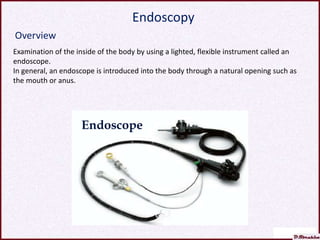
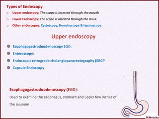
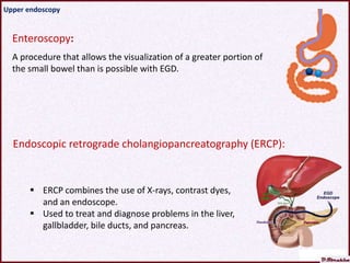
Prof. Dr. Ibrahim Aboul Asaad
Lecture Doctorate
degree Diagnosis of Parasitic Diseases
Approach to Diagnosis of Parasite Diseases
Internal Approach
External Approach
I) Clinical diagnosis:
1) History of exposure to infection
2) Clinical Aspect
II) Laboratory investigations:
1) Direct laboratory exam.
2) Indirect laboratory exam.
3) Advanced laboratory exam.
a) Molecular techniques
b) Cultivations
III) Imaging techniques
1) Radiography
a) X-rays.
b) Computed tomography (CT).
2) Magnetic resonance imaging (MRI).
3) Ultrasound imaging (US).
Endoscopy For:
1) Obtaining diagnostic specimens
2) Imaging of sites of infections.
Duodenal content can be obtained by:
1) Duodenal intubation
2) Duodenal capsule (Entero-test)
3) Upper endoscopy and sampling.
Colonic content can be obtained by:
1) Rectal swap or snip
2) Lower endoscopy
Bladder content can be obtained by:
1) Bladder snip or biopsy
2) Cystoscopy
Bronchial content can be obtained by:
1) Broncho-alveolar lavage
2) Bronchoscopy
Examination of the inside of the body by using a lighted, flexible instrument called an
endoscope.
In general, an endoscope is introduced into the body through a natural opening such as
the mouth or anus.
Endoscope
Overview
Endoscopy
Disadvantages
Endoscopy is an invasive procedure with possible complications include:
1) Perforation of an organ
2) Excessive bleeding (hemorrhage)
3) Infection
4) Allergic reaction to the anesthesia.
General Indications
Diagnostic procedure:
a) Obtaining diagnostic specimens
b) Visualization and Imaging the organisms or the visual evidence of infections , such as
ulceration, inflammation,
Therapeutic procedure: For complications of some parasitic infections such as
a) Remove of polyps.
b) Therapy of Esophageal varices .
c) Remove of the parasite from ectopic sites
 Endoscopy is an invasive procedure, but it may be of great value in management of parasitic
diseases.
 Its value as a direct diagnostic procedure.
 Its value in assessment of pathology.
 Its value in assessment of complications.
 Its value in management of complications.
Types of Endoscopy
o Upper endoscopy: The scope is inserted through the mouth
o Lower Endoscopy: The scope is inserted through the anus.
o Other endoscopes: Cystoscopy, Bronchoscope & laparoscope.
 Esophagogastroduodenoscopy EGD:
 Enteroscopy:
 Endoscopic retrograde cholangiopancreatography (ERCP
 Capsule Endoscopy
Upper endoscopy
Esophagogastroduodenoscopy (EGD):
Used to examine the esophagus, stomach and upper few inches of
the jejunum
Enteroscopy:
A procedure that allows the visualization of a greater portion of
the small bowel than is possible with EGD.
Upper endoscopy
Endoscopic retrograde cholangiopancreatography (ERCP):
 ERCP combines the use of X-rays, contrast dyes,
and an endoscope.
 Used to treat and diagnose problems in the liver,
gallbladder, bile ducts, and pancreas.
o Patient swallows a pill-sized camera. The capsule camera travels along the
digestive tract for 8 to 12 hrs.
o By this capsule, images and video can be captured, but samples cannot be
delivered.
Capsule Endoscopy
Wireless endoscopy system
• Proctoscopy
Used to examine rectum and sigmoid colon
• Sigmoidoscopy
Used to examine sigmoid colon
• Colonoscopy
Used to examine the entire length of the colon
Lower Endoscopy:
The scope is inserted through the anus.
o Cystoscopy is used to examine the bladder. The scope is inserted through the
urethra,
o Bronchoscopy is used to examine the lungs. The scope is inserted into the nose
or mouth.
o Laparoscopy is used to examine the abdominal or pelvic area. The scope is
inserted through a small incision near the area that’s being examined
Other Endoscopies:
Upper Endoscopy
Platyhelminthes Nematodes Protozoa
Fasciola
Clonorchis sinensis
Taenia
Diphyllobothrium latum
Ascaris lumbricoides
Hookworms
Strongyloides stercoralis
Capillaria philipinensis
Anisakis simplex
Giardia trophozoites.
Cryptosporidia
oocysts.
Isospora bell
Can help diagnosis of the following parasitic infections:
Ancylostoma
duodenale
Hookworm in the duodenum
Under magnifying endoscopy, a worm is seen hooking its head into the
intestinal mucosa. The parasite was removed and identified as Ancylostoma
ceylanicum, a nematode that is endemic to Southeast Asia.
Ancylostoma ceylanicum
Ascaris lumbricoides
in intestine
Ascaris worm in the intestine
Ascaris worm moving inside human stomach
Ascaris Worm in Common Bile Duct (CBD),
and endoscopic removal.
• Upper gastrointestinal endoscopy showed multiple white granular
lesions resembling xanthoma in the antrum (a, b) and widespread
erosion surrounded by granular lesions in the gastric body (c). Direct
microscopic examination of gastric fluid revealed crawling filariform
larvae of Strongyloides stercoralis
Strongyloides stercoralis
Enteroscopy
• Fasciola
• Endoscopic appearance of Fasciola
hepatica showing a leaf-like trematode
extracted by ERCP using a balloon
catheter.
ERCP-Removal of live Fasciola hepatica from Common bile duct
Clonorchis
sinensis
• A flattened (dorsoventrally) and leaf-shaped fluke was
observed repeatedly in the small intestinal lumen by capsule
endoscopy (A & B)
• It is a rare case that C sinensis was in the bowel lumen, being
found by capsule endoscopy.
• Percutaneous transhepatic cholangioscopy findings. (A) Dark
brown, soft obstructive mass is found at stricture site of left
intrahepatic duct. (B) Flat, leaf-shaped worm indentified
as Clonorchis sinensis is observed after extraction of bile sludge
Clonorchis
sinensis
Endoscopic retrograde cholangiogram in an adult Korean with clonorchiasis
shows marked dilatation of the intrahepatic bile ducts. These are numerous
oval, elliptical or crescentic filling defects representing adult C. sinensis in
small and medium-sized bile ducts.
Clonorchis sinensis
Clonorchis
sinensis
• Endoscopic retrograde
cholangiogram in patient showing
multiple small 1- to 2-cm crescentic
or oval filling defects from C.
sinensis flukes (arrows) in large
hepatic ducts and branches.
Clonorchis
sinensis
• Cholangiocarcinoma in the mid common duct, with
clonorchiasis, in a 64-year-old man. MR cholangiography shows
a diffuse dilatation of the intrahepatic bile ducts and common
duct. Note an intraluminal tumor (arrows) at the level of the mid
common duct.
Taenia
Saginata
Taenia Saginata in Third-Generation Capsule Endoscopy
Taenia saginata in the Small Intestine
Taenia
solium
• Capsule endoscopy demonstrated a long, thin,
segmented, flat structure consistent with a pork tapeworm
(Taenia solium) at least 1 meter in length.
Taenia solium in the small intestine
Diphyllobothrium
latum
Enteroscopy
A case of diphyllobothriasis is detected by video capsule endoscopy.
Diphyllobothrium latum
Capillaria
philipinensis
• Endoscopic finding of the stomach showing an exudative flat erosive
change on antral mucosa.
• Sectioned worms (C. philipinensis) in the superficial portion of the gastric
mucosa. Cross and tangential sections of eggs observed in the uterus. In
egg cross-sections, eggshell striations were arranged radially. In tangential-
sectioned eggs, striations formed a network resembling a net with irregular
meshes (inset: ×400) (H&E stain, ×200).
Anisakis
simplex
• (a) Endoscopic view of an Anisakis larva
sticking in the wall of the stomach.
• (b) Endoscopic treatment - visible
worms removed with biopsy forceps.
Endoscopic treatment: visible worms removed with biopsy forceps.
Anisakis in stomach
Lower Endoscopy
The following parasites can be detected:
1) S. mansoni
2) T. trichiura
3) Migrating Ascaris
4) Migrating Taenia
5) E. histolytica.
Intestinal
schistosomiasis
• A) Endoscopic manifestation of acute intestinal schistosomiasis:
Mucosal hyperemia and edema, mucus exudation, vague
vascular striation and scattered small ulcers and yellow nodules.
• B) Endoscopic manifestation of chronic intestinal
schistosomiasis, mucosal thickening and cicatrize with polypoid
protrusion.
A) Vascular net like map of sigmoid colon in chronic Schistosomiasis
B) Giant flat, lobulated polypus in rectum in chronic Schistosomiasis
Intestinal Schistosomiasis
Colonoscopy showing T. trichiura worm in the cecum.
Trichuris trichiura
Colonoscopy showing Ascaris worm migrating in a woman's colon.
Ascaris lumbricoides
colonoscopy showing Taenia Solium in cecum
Taenia Solium
Cystoscopy
A cystoscope is a thin tube with a camera and light on the end. During a
cystoscopy, this tube is inserted through the urethra and into the bladder so
the doctor can visualize the inside of the bladder.
Doctor can pass tiny surgical tools through the scope to take a sample
of bladder tissue.
(A) Bilharzial pseudotubercles and adjacent ulcer;
(B) Bilharzial sessile mass covered by psudotubercles;
Cystoscopic appearances of bilharzial lesions in the urinary bladder
Urinary Schistosomiasis
(C) Sandy patches
(D) Cystitis cystica: a series of mucosal cysts form in the bladder wall
Urinary Schistosomiasis
(E) Malignant ulcer (squamous cell carcinoma) with adjacent phosphate
encrustations and sandy patches;
(F) Fungating malignant mass (squamous cell carcinoma).
Urinary Schistosomiasis
Endoscopy helminth.pptx

Endoscopy helminth.pptx

  • 1.
    Prof. Dr. IbrahimAboul Asaad Lecture Doctorate degree Diagnosis of Parasitic Diseases
  • 2.
    Approach to Diagnosisof Parasite Diseases Internal Approach External Approach I) Clinical diagnosis: 1) History of exposure to infection 2) Clinical Aspect II) Laboratory investigations: 1) Direct laboratory exam. 2) Indirect laboratory exam. 3) Advanced laboratory exam. a) Molecular techniques b) Cultivations III) Imaging techniques 1) Radiography a) X-rays. b) Computed tomography (CT). 2) Magnetic resonance imaging (MRI). 3) Ultrasound imaging (US). Endoscopy For: 1) Obtaining diagnostic specimens 2) Imaging of sites of infections. Duodenal content can be obtained by: 1) Duodenal intubation 2) Duodenal capsule (Entero-test) 3) Upper endoscopy and sampling. Colonic content can be obtained by: 1) Rectal swap or snip 2) Lower endoscopy Bladder content can be obtained by: 1) Bladder snip or biopsy 2) Cystoscopy Bronchial content can be obtained by: 1) Broncho-alveolar lavage 2) Bronchoscopy
  • 3.
    Examination of theinside of the body by using a lighted, flexible instrument called an endoscope. In general, an endoscope is introduced into the body through a natural opening such as the mouth or anus. Endoscope Overview Endoscopy
  • 4.
    Disadvantages Endoscopy is aninvasive procedure with possible complications include: 1) Perforation of an organ 2) Excessive bleeding (hemorrhage) 3) Infection 4) Allergic reaction to the anesthesia. General Indications Diagnostic procedure: a) Obtaining diagnostic specimens b) Visualization and Imaging the organisms or the visual evidence of infections , such as ulceration, inflammation, Therapeutic procedure: For complications of some parasitic infections such as a) Remove of polyps. b) Therapy of Esophageal varices . c) Remove of the parasite from ectopic sites  Endoscopy is an invasive procedure, but it may be of great value in management of parasitic diseases.  Its value as a direct diagnostic procedure.  Its value in assessment of pathology.  Its value in assessment of complications.  Its value in management of complications.
  • 5.
    Types of Endoscopy oUpper endoscopy: The scope is inserted through the mouth o Lower Endoscopy: The scope is inserted through the anus. o Other endoscopes: Cystoscopy, Bronchoscope & laparoscope.  Esophagogastroduodenoscopy EGD:  Enteroscopy:  Endoscopic retrograde cholangiopancreatography (ERCP  Capsule Endoscopy Upper endoscopy Esophagogastroduodenoscopy (EGD): Used to examine the esophagus, stomach and upper few inches of the jejunum
  • 6.
    Enteroscopy: A procedure thatallows the visualization of a greater portion of the small bowel than is possible with EGD. Upper endoscopy Endoscopic retrograde cholangiopancreatography (ERCP):  ERCP combines the use of X-rays, contrast dyes, and an endoscope.  Used to treat and diagnose problems in the liver, gallbladder, bile ducts, and pancreas.
  • 7.
    o Patient swallowsa pill-sized camera. The capsule camera travels along the digestive tract for 8 to 12 hrs. o By this capsule, images and video can be captured, but samples cannot be delivered. Capsule Endoscopy Wireless endoscopy system
  • 8.
    • Proctoscopy Used toexamine rectum and sigmoid colon • Sigmoidoscopy Used to examine sigmoid colon • Colonoscopy Used to examine the entire length of the colon Lower Endoscopy: The scope is inserted through the anus. o Cystoscopy is used to examine the bladder. The scope is inserted through the urethra, o Bronchoscopy is used to examine the lungs. The scope is inserted into the nose or mouth. o Laparoscopy is used to examine the abdominal or pelvic area. The scope is inserted through a small incision near the area that’s being examined Other Endoscopies:
  • 9.
    Upper Endoscopy Platyhelminthes NematodesProtozoa Fasciola Clonorchis sinensis Taenia Diphyllobothrium latum Ascaris lumbricoides Hookworms Strongyloides stercoralis Capillaria philipinensis Anisakis simplex Giardia trophozoites. Cryptosporidia oocysts. Isospora bell Can help diagnosis of the following parasitic infections:
  • 10.
  • 11.
  • 12.
    Under magnifying endoscopy,a worm is seen hooking its head into the intestinal mucosa. The parasite was removed and identified as Ancylostoma ceylanicum, a nematode that is endemic to Southeast Asia. Ancylostoma ceylanicum
  • 13.
  • 14.
    Ascaris worm inthe intestine
  • 15.
    Ascaris worm movinginside human stomach
  • 16.
    Ascaris Worm inCommon Bile Duct (CBD), and endoscopic removal.
  • 17.
    • Upper gastrointestinalendoscopy showed multiple white granular lesions resembling xanthoma in the antrum (a, b) and widespread erosion surrounded by granular lesions in the gastric body (c). Direct microscopic examination of gastric fluid revealed crawling filariform larvae of Strongyloides stercoralis Strongyloides stercoralis Enteroscopy
  • 18.
    • Fasciola • Endoscopicappearance of Fasciola hepatica showing a leaf-like trematode extracted by ERCP using a balloon catheter.
  • 19.
    ERCP-Removal of liveFasciola hepatica from Common bile duct
  • 20.
    Clonorchis sinensis • A flattened(dorsoventrally) and leaf-shaped fluke was observed repeatedly in the small intestinal lumen by capsule endoscopy (A & B) • It is a rare case that C sinensis was in the bowel lumen, being found by capsule endoscopy.
  • 21.
    • Percutaneous transhepaticcholangioscopy findings. (A) Dark brown, soft obstructive mass is found at stricture site of left intrahepatic duct. (B) Flat, leaf-shaped worm indentified as Clonorchis sinensis is observed after extraction of bile sludge Clonorchis sinensis
  • 22.
    Endoscopic retrograde cholangiogramin an adult Korean with clonorchiasis shows marked dilatation of the intrahepatic bile ducts. These are numerous oval, elliptical or crescentic filling defects representing adult C. sinensis in small and medium-sized bile ducts. Clonorchis sinensis
  • 23.
    Clonorchis sinensis • Endoscopic retrograde cholangiogramin patient showing multiple small 1- to 2-cm crescentic or oval filling defects from C. sinensis flukes (arrows) in large hepatic ducts and branches.
  • 24.
    Clonorchis sinensis • Cholangiocarcinoma inthe mid common duct, with clonorchiasis, in a 64-year-old man. MR cholangiography shows a diffuse dilatation of the intrahepatic bile ducts and common duct. Note an intraluminal tumor (arrows) at the level of the mid common duct.
  • 25.
    Taenia Saginata Taenia Saginata inThird-Generation Capsule Endoscopy
  • 26.
    Taenia saginata inthe Small Intestine
  • 27.
    Taenia solium • Capsule endoscopydemonstrated a long, thin, segmented, flat structure consistent with a pork tapeworm (Taenia solium) at least 1 meter in length.
  • 28.
    Taenia solium inthe small intestine
  • 29.
  • 30.
    A case ofdiphyllobothriasis is detected by video capsule endoscopy. Diphyllobothrium latum
  • 31.
    Capillaria philipinensis • Endoscopic findingof the stomach showing an exudative flat erosive change on antral mucosa. • Sectioned worms (C. philipinensis) in the superficial portion of the gastric mucosa. Cross and tangential sections of eggs observed in the uterus. In egg cross-sections, eggshell striations were arranged radially. In tangential- sectioned eggs, striations formed a network resembling a net with irregular meshes (inset: ×400) (H&E stain, ×200).
  • 32.
    Anisakis simplex • (a) Endoscopicview of an Anisakis larva sticking in the wall of the stomach. • (b) Endoscopic treatment - visible worms removed with biopsy forceps.
  • 33.
    Endoscopic treatment: visibleworms removed with biopsy forceps. Anisakis in stomach
  • 34.
    Lower Endoscopy The followingparasites can be detected: 1) S. mansoni 2) T. trichiura 3) Migrating Ascaris 4) Migrating Taenia 5) E. histolytica.
  • 35.
    Intestinal schistosomiasis • A) Endoscopicmanifestation of acute intestinal schistosomiasis: Mucosal hyperemia and edema, mucus exudation, vague vascular striation and scattered small ulcers and yellow nodules. • B) Endoscopic manifestation of chronic intestinal schistosomiasis, mucosal thickening and cicatrize with polypoid protrusion.
  • 36.
    A) Vascular netlike map of sigmoid colon in chronic Schistosomiasis B) Giant flat, lobulated polypus in rectum in chronic Schistosomiasis Intestinal Schistosomiasis
  • 37.
    Colonoscopy showing T.trichiura worm in the cecum. Trichuris trichiura
  • 38.
    Colonoscopy showing Ascarisworm migrating in a woman's colon. Ascaris lumbricoides
  • 39.
    colonoscopy showing TaeniaSolium in cecum Taenia Solium
  • 40.
    Cystoscopy A cystoscope isa thin tube with a camera and light on the end. During a cystoscopy, this tube is inserted through the urethra and into the bladder so the doctor can visualize the inside of the bladder. Doctor can pass tiny surgical tools through the scope to take a sample of bladder tissue.
  • 41.
    (A) Bilharzial pseudotuberclesand adjacent ulcer; (B) Bilharzial sessile mass covered by psudotubercles; Cystoscopic appearances of bilharzial lesions in the urinary bladder Urinary Schistosomiasis
  • 42.
    (C) Sandy patches (D)Cystitis cystica: a series of mucosal cysts form in the bladder wall Urinary Schistosomiasis
  • 43.
    (E) Malignant ulcer(squamous cell carcinoma) with adjacent phosphate encrustations and sandy patches; (F) Fungating malignant mass (squamous cell carcinoma). Urinary Schistosomiasis