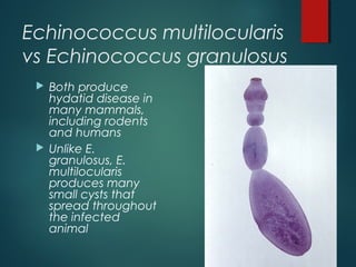
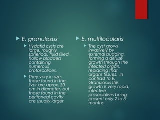
Echinococcus granulosus
(and multilocularis)
SUBMITTED BY
SAKIR
GR 21 SEM 7TH
Three Species
 Echinococcus granulosus: causing hydatid
disease
 Echinococcus multilocularis: causing alveolar
hydatid disease
 Echinococcus vogeli: causing hydatid disease
Echinococcus multilocularis
vs Echinococcus granulosus
 Both produce
hydatid disease in
many mammals,
including rodents
and humans
 Unlike E.
granulosus, E.
multilocularis
produces many
small cysts that
spread throughout
the infected
animal
Geographic Distribution
 Worldwide
 Higher prevalence in South America (Argentina,
Uruguay), Europe (Mediterranean bassin),
Northern Africa, Middle East, South Central and
East Asia
Larvae Stage
 E. granulosus
 Hydatid cysts are
large, roughly
spherical, fluid filled
hollow bladders
containing
numerous
protoscolices.
 They vary in size;
those found in the
liver are aprox. 20
cm in diameter, but
those found in the
peritoneal cavity
are usually larger
 E. multilocularis
 The cyst grows
invasively by
external budding,
forming a diffuse
growth through the
infected organ,
replacing that
organs tissues. In
contrast to E.
Granulosus this
growth is very rapid,
infective
prosocialises being
present only 2 to 3
months.
Adult Stage
 E. multilocaris
 Slightly smaller
than E.
granulosus (max.
length of aprox 4
mm and
consisting of 4-5
proglottids).
 E. granulosus
 The adult
parasites in the
dog represent
one of the
smallest of the
tapeworms (3-9
mm in length,
usually with 3
proglottids).
 Scolex is globular
in shape and has
a prominent
rostellum
Definitive Hosts
E. granulosus
 Dogs
 Coyotes
 Wolves
E. multilocularis
 Mostly foxes
Intermediate Hosts
E. granulosus
 Sheep
 Horses
 Camels
 Pigs
 Humans
E. multilocularis
 Small rodents
Life Cycle: E. granulosus
 The adult is in the small bowel of the definitive host
 Gravid proglottids release eggs that are passed in the
feces
 In the intermediate host the egg hatches in the small
bowel and releases an oncosphere
 The oncosphere penetrates the intestinal wall and
moves through the circulatory system to various organs
 In the organs they develop into cysts and enlarge
gradually
 The cysts produce protoscolices and daughter cysts
 Definitive host eats the infected organs and becomes
infected
 After ingestion, the protoscolices evaginate, attach to
the intestinal mucosa and develop into adult stages
 In 32-80 days, the cycle starts over
Life Cycle: E. multilocularis
 The life cycle is
basically the same E.
granulosus
Except
 There are different
definitive and
intermediate hosts
 Larval growth in the
liver remain indefinitely
in the proliferative
stage, which causes
invasion of the
surrounding tissues
(sometimes this can
take over the whole
organ)
Life Cycle (cont’d)
Diagnosis
 Diagnosis in the definitive host is
difficult by ordinary microscopy
because it will look a lot like Taenia
and Echinococcus eggs
 Detection of antigens in feces by
ELISA is currently the best available
technique
 Newer techniques like polymerase
chain reaction (PCR) is also used to
identify the parasite from DNA isolated
from eggs or feces
Treatment
 Surgery: with the goal of leaving the cyst intact so
new cysts do not form
 Mebendazole can be taken over a long period of
time at low dosages
 Praziquantel
Prevention
 The best way to keep
dogs from being
infected is to prevent
them from eating
infected feces, or
contaminated meat
 The best way to avoid
human infection is to
avoid ingesting food
or other substances
contaminated with
dog feces
Prevention (cont’d)
 The best method is to disrupt the lifecycle
 Basic hygiene practices
 Avoid feeding raw offal (internal organs of
butchered animals) to dogs
 By doing this hydatids have been virtually eliminated in
New Zealand (a once common place for this parasite)
 1. What are the main differences between the
larvae stage of E. granulosus and E. multilocularis?
 E. granulosus
 Hydatid cysts are
large, roughly
spherical, fluid filled
hollow bladders
containing
numerous
protoscolices.
 They vary in size;
those found in the
liver are aprox. 20
cm in diameter, but
those found in the
peritoneal cavity
are usually larger
 E. multilocularis
 The cyst grows
invasively by
external budding,
forming a diffuse
growth through the
infected organ,
replacing that
organs tissues. In
contrast to E.
Granulosus this
growth is very rapid,
infective
prosocialises being
present only 2 to 3
months.
2. What are the definitive hosts for each parasite?
E. granulosus
 Dogs
 Coyotes
 Wolves
E. multilocularis
 Mostly foxes
Echinoccocus
  is caused by infection with the larval stage of Echinococcus
granulosus. CE is found in Africa, Europe, Asia, the Middle East,
Central and South America, and in rare cases, North America. The
parasite is transmitted to dogs when they ingest the organs of
other animals that contain hydatid cysts. The cysts develop into
adult tapeworms in the dog. Infected dogs shed tapeworm eggs
in their feces which contaminate the ground. Sheep, cattle, goats,
and pigs ingest tapeworm eggs in the contaminated ground;
once ingested, the eggs hatch and develop into cysts in the
internal organs. The most common mode of transmission to
humans is by the accidental consumption of soil, water, or food
that has been contaminated by the fecal matter of an infected
dog. Echinococcus eggs that have been deposited in soil can
stay viable for up to a year. The disease is most commonly found in
people involved in raising sheep, as a result of the sheep's role as
an intermediate host of the parasite and the presence of working
dogs that are allowed to eat the offal of infected sheep.
Alveoccocus
 is caused by infection with the larval
stage of Echinococcus multilocularis. AE is
found across the globe and is especially
prevalent in the northern latitudes of
Europe, Asia, and North America. The
adult tapeworm is normally found in
foxes, coyotes, and dogs. Infection with
the larval stages is transmitted to people
through ingestion of food or water
contaminated with tapeworm eggs.

echinococcus

  • 1.
  • 2.
    Three Species  Echinococcusgranulosus: causing hydatid disease  Echinococcus multilocularis: causing alveolar hydatid disease  Echinococcus vogeli: causing hydatid disease
  • 3.
    Echinococcus multilocularis vs Echinococcusgranulosus  Both produce hydatid disease in many mammals, including rodents and humans  Unlike E. granulosus, E. multilocularis produces many small cysts that spread throughout the infected animal
  • 4.
    Geographic Distribution  Worldwide Higher prevalence in South America (Argentina, Uruguay), Europe (Mediterranean bassin), Northern Africa, Middle East, South Central and East Asia
  • 5.
    Larvae Stage  E.granulosus  Hydatid cysts are large, roughly spherical, fluid filled hollow bladders containing numerous protoscolices.  They vary in size; those found in the liver are aprox. 20 cm in diameter, but those found in the peritoneal cavity are usually larger  E. multilocularis  The cyst grows invasively by external budding, forming a diffuse growth through the infected organ, replacing that organs tissues. In contrast to E. Granulosus this growth is very rapid, infective prosocialises being present only 2 to 3 months.
  • 6.
    Adult Stage  E.multilocaris  Slightly smaller than E. granulosus (max. length of aprox 4 mm and consisting of 4-5 proglottids).  E. granulosus  The adult parasites in the dog represent one of the smallest of the tapeworms (3-9 mm in length, usually with 3 proglottids).  Scolex is globular in shape and has a prominent rostellum
  • 7.
    Definitive Hosts E. granulosus Dogs  Coyotes  Wolves E. multilocularis  Mostly foxes
  • 8.
    Intermediate Hosts E. granulosus Sheep  Horses  Camels  Pigs  Humans E. multilocularis  Small rodents
  • 9.
    Life Cycle: E.granulosus  The adult is in the small bowel of the definitive host  Gravid proglottids release eggs that are passed in the feces  In the intermediate host the egg hatches in the small bowel and releases an oncosphere  The oncosphere penetrates the intestinal wall and moves through the circulatory system to various organs  In the organs they develop into cysts and enlarge gradually  The cysts produce protoscolices and daughter cysts  Definitive host eats the infected organs and becomes infected  After ingestion, the protoscolices evaginate, attach to the intestinal mucosa and develop into adult stages  In 32-80 days, the cycle starts over
  • 10.
    Life Cycle: E.multilocularis  The life cycle is basically the same E. granulosus Except  There are different definitive and intermediate hosts  Larval growth in the liver remain indefinitely in the proliferative stage, which causes invasion of the surrounding tissues (sometimes this can take over the whole organ)
  • 11.
  • 12.
    Diagnosis  Diagnosis inthe definitive host is difficult by ordinary microscopy because it will look a lot like Taenia and Echinococcus eggs  Detection of antigens in feces by ELISA is currently the best available technique  Newer techniques like polymerase chain reaction (PCR) is also used to identify the parasite from DNA isolated from eggs or feces
  • 13.
    Treatment  Surgery: withthe goal of leaving the cyst intact so new cysts do not form  Mebendazole can be taken over a long period of time at low dosages  Praziquantel
  • 14.
    Prevention  The bestway to keep dogs from being infected is to prevent them from eating infected feces, or contaminated meat  The best way to avoid human infection is to avoid ingesting food or other substances contaminated with dog feces
  • 15.
    Prevention (cont’d)  Thebest method is to disrupt the lifecycle  Basic hygiene practices  Avoid feeding raw offal (internal organs of butchered animals) to dogs  By doing this hydatids have been virtually eliminated in New Zealand (a once common place for this parasite)
  • 16.
     1. Whatare the main differences between the larvae stage of E. granulosus and E. multilocularis?
  • 17.
     E. granulosus Hydatid cysts are large, roughly spherical, fluid filled hollow bladders containing numerous protoscolices.  They vary in size; those found in the liver are aprox. 20 cm in diameter, but those found in the peritoneal cavity are usually larger  E. multilocularis  The cyst grows invasively by external budding, forming a diffuse growth through the infected organ, replacing that organs tissues. In contrast to E. Granulosus this growth is very rapid, infective prosocialises being present only 2 to 3 months.
  • 18.
    2. What arethe definitive hosts for each parasite?
  • 19.
    E. granulosus  Dogs Coyotes  Wolves E. multilocularis  Mostly foxes
  • 20.
    Echinoccocus   is causedby infection with the larval stage of Echinococcus granulosus. CE is found in Africa, Europe, Asia, the Middle East, Central and South America, and in rare cases, North America. The parasite is transmitted to dogs when they ingest the organs of other animals that contain hydatid cysts. The cysts develop into adult tapeworms in the dog. Infected dogs shed tapeworm eggs in their feces which contaminate the ground. Sheep, cattle, goats, and pigs ingest tapeworm eggs in the contaminated ground; once ingested, the eggs hatch and develop into cysts in the internal organs. The most common mode of transmission to humans is by the accidental consumption of soil, water, or food that has been contaminated by the fecal matter of an infected dog. Echinococcus eggs that have been deposited in soil can stay viable for up to a year. The disease is most commonly found in people involved in raising sheep, as a result of the sheep's role as an intermediate host of the parasite and the presence of working dogs that are allowed to eat the offal of infected sheep.
  • 21.
    Alveoccocus  is causedby infection with the larval stage of Echinococcus multilocularis. AE is found across the globe and is especially prevalent in the northern latitudes of Europe, Asia, and North America. The adult tapeworm is normally found in foxes, coyotes, and dogs. Infection with the larval stages is transmitted to people through ingestion of food or water contaminated with tapeworm eggs.