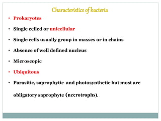
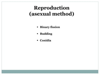
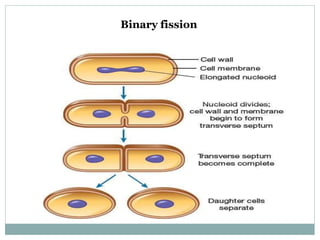
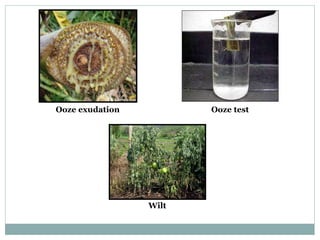
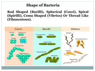
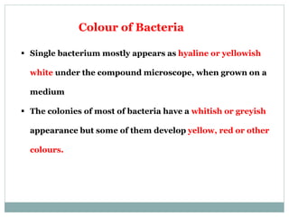
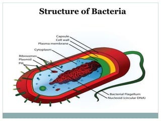
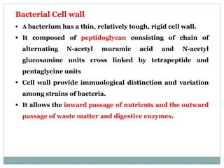
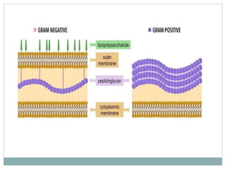
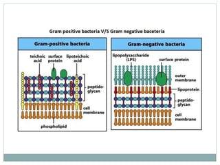
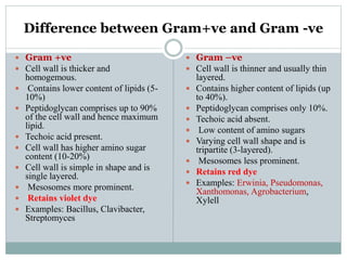
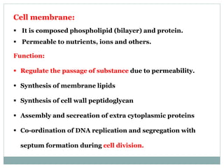
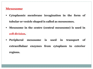
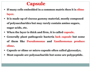
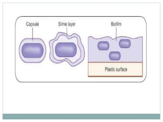
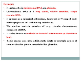
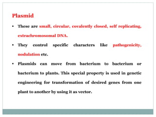
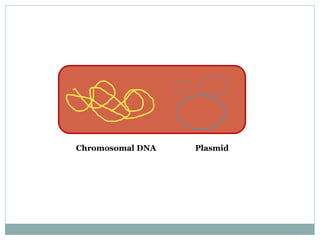
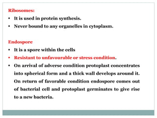
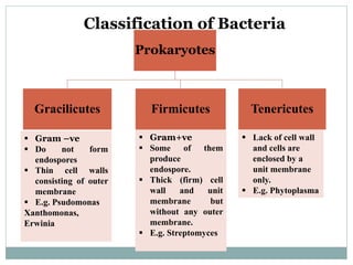
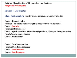
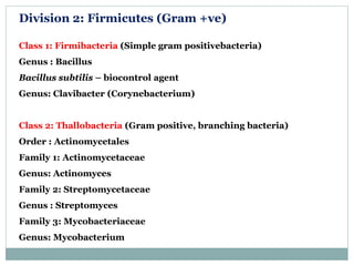
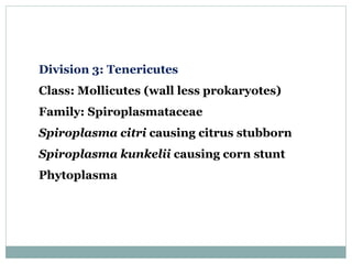
The document provides a comprehensive overview of plant pathogenic bacteria, detailing their characteristics, classifications, and the mechanisms by which they cause disease in plants. It describes structural components, such as the cell wall, flagella, and pili, alongside their reproductive methods and survival strategies. Additionally, it outlines management strategies for bacterial diseases in plants, emphasizing prevention and control measures.


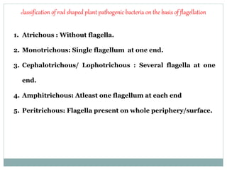


![06_Kingdom_Prokaryotae769[1].pptx](https://cdn.slidesharecdn.com/ss_thumbnails/06kingdomprokaryotae7691-240127144445-3a0d06ed-thumbnail.jpg?width=640&height=640&fit=bounds)


![Polymer [ बहुलक ] Chemistry Notes PDF - Irfanullah Mehar - JJ Sir Chemistry.pdf](https://cdn.slidesharecdn.com/ss_thumbnails/polymerchemistrynotespdf-irfanullahmehar-jjsirchemistry-260210172118-3f9b37f7-thumbnail.jpg?width=640&height=640&fit=bounds)


