The document discusses the physiology of pacemaker and contractile cells in the heart. It begins by describing the characteristics of pacemaker cells, including their automaticity due to unstable membrane potentials. It then discusses how sympathetic and parasympathetic activity can alter the activity of pacemaker cells to increase or decrease heart rate. The document next describes the characteristics of contractile myocardial cells and their action potentials. It compares skeletal and cardiac action potentials. Finally, it discusses excitation-contraction coupling in contractile cells and the roles of calcium in both contraction and relaxation.
Regulation of arterial blood pressure (The Guyton and Hall Physiology)Maryam Fida
BLOOD PRESSURE
The pressure exerted by the blood on vessel wall is known as blood pressure.
SYSTOLIC BLOOD PRESSURE
The maximum pressure exerted in the arteries during systole of heart.
Normal systolic pressure: 120 mm Hg.
DIASTOLIC BLOOD PRESSURE
The minimum pressure exerted in the arteries during diastole of heart.
Normal diastolic pressure: 80 mm Hg.
PULSE PRESSURE
The difference between the systolic pressure and diastolic pressure.
Normal pulse pressure: 40 mm Hg (120 – 80 = 40).
MEAN ARTERIAL BLOOD PRESSURE
The average pressure existing in the arteries.
Mean Arterial Blood Pressure = Diastolic Pressure + 1/3 Pulse Pressure
Pulse Pressure = (Systolic – Diastolic)
Mean Arterial Blood Pressure =Diastolic Pressure+1/3(Systolic – Diastolic)
Cardiac output (The Guyton and Hall Physiology)Maryam Fida
The volume of blood pumped by each ventricle per minute is called cardiac output
Cardiac output = Stroke Volume X Heart Rate
Normal value = 5 Liters /Minute
Cardiac output = Stroke Volume X Heart Rate
The factors which regulate stroke volume and Heart rate are basically regulating Cardiac output
Volume of blood ejected by each ventricle in single systole; Normal Value = 70 ml/beat
Stroke Volume = End diastolic Volume – End Systolic Volume
So stroke volume is mainly controlled by
EDV
ESV
VENOUS RETURN: What ever blood volume returns to the heart, same is pumped forward through the Frank’s Starlings Law. According to this law 13- 15 liters of blood volume can be pumped out without cardiac stimulation.
DURATION OF DIASTOLE OR FILLING TIME: ventricular filling occurs during diastole, so there must be adequate ventricular filling time.
DISTENSIBILITY OF THE VENTRICLES: Normally ventricles are distensible to accommodate adequate blood volume. Infarction decreases the distensibility which decreases the EDV.
ATRIAL CONTRACTION: There must be adequate atrial contraction to have adequate EDV. If atrial function is not adequate then EDV will decrease.
E.S.V is basically CONTROLLED BY MYOCARDIAL CONTRACTION
FORCE OF MYOCARDIAL CONTRACTION: It depends upon the initial length of muscle fibers according to frank’s starlings law.
PRELOAD: The effect of EDV on initial length is called preload. So EDV also effects the ESV.
AFTER LOAD: Force of contraction is also dependant upon the resistance against which the ventricles have to pump
CONDITION OF THE MYOCARDIUM : It also effects the force of contraction.
AUTONOMIC NERVES : Sympathetic stimulation increases and parasympathetic stimulation decreases force of contraction
HORMONES: Catecholamines, thyroxine, glucagon, digitalis, calcium, increased temp, caffeine, theophyline increase the force.
Force decreases by hypoxia, acidosis, barniturates, procainamide and quinidine decrease the force of contraction.
“Cardiac output refers to the volume of blood pumped out per ventricle per minute.”
Cardiac output is the function of heart rate and stroke volume.
STROKE VOLUME:
The amount of blood pumped by the left ventricle in one compression is called the stroke volume.
Heart Rate
The cardiac output increases with the increase in heart rate.
Cardiac muscle (The Guyton and Hall Physiology)Maryam Fida
In the heart there is Atrial muscle and Ventricular muscle which are separated from each other by the fibrous AV Rings containing Valves.
ATRIAL MUSCLE: thin walled. There are two sheets, superficial and deep sheet. Superficial sheet is common over both atria. Deep sheet is separate for each atrium. Muscle fibers in the deep sheet are at right angle to the muscle fibers in the superficial sheet.
FUNCTIONS OF THE ATRIUM:
1. Receive venous blood from large veins. So atria act as reservoir.
2. Conduct the blood into the ventricles.
3. Atrial contraction is responsible for last 25 % of ventricular filling.
4. In the right atrium there is SA Node(Pace maker) and AV node.
5. In the wall of the atria, there are low pressure stretch receptors and these are involved in various reflexes like brain bridge reflex and left atrial reflex.
6. Atria also produce a hormone i.e. Atrial Natriuretic Hormone. Whenever NaCl increases in ECF, it causes release of ANH which causes natriuresis.
VENTRICULAR MUSCLE:
Much thicker than atrial muscle. Thickness of right ventricle wall is 3-4 mm and thickness of left ventricle is 8 – 12 mm.
1.Involuntary
2.Has cross striations
3.Each cardiac muscle fiber consists of a number of cardiac cells, united at ends in series. Where as in skeletal muscle each muscle fiber is individual cell.
4.Cardiac muscle cells are branching and interdigitate.
5.Single central nucleus in each cell.
6. Atrial muscle and ventricular muscle act as separate functional syncytium and impulses from atria are conducted to ventricles through the AV Node and AV Bundle.
7. Sarcoplasmic system is present. In skeletal muscle triad is at the junction of A and I bands. In cardiac muscle T Tubules are much large and thus in cardiac muscle if we take a section it may form a diad or a triad. And these diads and triads are present at the level of Z Disks.
8.Between adjacent cardiac cells there are side to side and end to end connections and these are the intercellular junctions. These junctions are Gap Junctions. Or intercalated discs
9.When one part of myocardium is excited the whole muscle is excited.
10.Whole myocardium obeys all or none law as a whole.
11.No spike potential but action potential with plateau.
12.Has got long refractory period.
Absolute refractory period in ventricular muscle is 250 – 300 milli sec.
In atrial muscle Absolute refractory period is 150 milli sec
Because of long refractory period cardiac muscle cannot be tetanized.
Properties of cm, plateau potential & pacemaker by Pandian M this PPT for I ...Pandian M
Describe the properties of cardiac muscle including its morphology, electrical, mechanical and metabolic functionsSLOs: After attending lecture & studying the assigned materials, the student will: 1.Describe the general features of cardiac muscle.2.Discuss the light and electron microscopic appearance of cardiac muscle, characteristic features of sarcotubular system.3.Enlist the electrical properties of heart muscle.4.Explain the phases of cardiac muscle action potential5.Explain the nodal action potential.6.Differentiate between cardiac muscle A.P. and nodal A.P., effect of nervous innervation and ions on AP.7.Enumerate and explain the mechanical properties of heart muscle, metabolic functions, characteristic features.
Anatomy and physiology of the cardiac system
The electrocardiogram a, curves and interpretation of the first and second heart sounds. Generation of action potential within the myocardium ,the gap junctions and how they propagate electrical pilese from sinoatrial mode and ectopoic heartbeat.
Cardiac muscle has three types of membrane ion channels that play important roles in causing the voltage changes of the action potential. They are (1) fast sodium channels, (2) slow sodium-calcium channels, and (3) potassium channels
Depolarization: First, the action potential of cardiac muscle is caused almost entirely by sudden opening of large numbers of so-called fast sodium channels that allow tremendous numbers of sodium ions to enter the cardiac muscle fiber from the extracellular fluid. These channels are called “fast” channels because they remain open for only a few thousandths of a second and then abruptly close. After depolarization, there's a brief repolarization that takes place with the efflux of potassium through fast acting potassium channels.
Plateau: Secondly, another entirely different population of slow calcium channels, which are also called calcium-sodium channels. This second population of channels differs from the fast sodium channels in that they are slower to open and, even more important, remain open for several tenths of a second. During this time, a large quantity of both calcium and sodium ions flows through these channels to the interior of the cardiac muscle fiber, and this maintains a prolonged period of depolarization, causing the plateau in the action potential.
Repolarization: When the slow calcium-sodium channels do close at the end of 0.2 to 0.3 second and the influx of calcium and sodium ions ceases, the membrane permeability for potassium ions also increases rapidly; this rapid loss of potassium from the fiber immediately returns the membrane potential to its resting level, thus ending the action potential.
Report Back from SGO 2024: What’s the Latest in Cervical Cancer?bkling
Are you curious about what’s new in cervical cancer research or unsure what the findings mean? Join Dr. Emily Ko, a gynecologic oncologist at Penn Medicine, to learn about the latest updates from the Society of Gynecologic Oncology (SGO) 2024 Annual Meeting on Women’s Cancer. Dr. Ko will discuss what the research presented at the conference means for you and answer your questions about the new developments.
Title: Sense of Smell
Presenter: Dr. Faiza, Assistant Professor of Physiology
Qualifications:
MBBS (Best Graduate, AIMC Lahore)
FCPS Physiology
ICMT, CHPE, DHPE (STMU)
MPH (GC University, Faisalabad)
MBA (Virtual University of Pakistan)
Learning Objectives:
Describe the primary categories of smells and the concept of odor blindness.
Explain the structure and location of the olfactory membrane and mucosa, including the types and roles of cells involved in olfaction.
Describe the pathway and mechanisms of olfactory signal transmission from the olfactory receptors to the brain.
Illustrate the biochemical cascade triggered by odorant binding to olfactory receptors, including the role of G-proteins and second messengers in generating an action potential.
Identify different types of olfactory disorders such as anosmia, hyposmia, hyperosmia, and dysosmia, including their potential causes.
Key Topics:
Olfactory Genes:
3% of the human genome accounts for olfactory genes.
400 genes for odorant receptors.
Olfactory Membrane:
Located in the superior part of the nasal cavity.
Medially: Folds downward along the superior septum.
Laterally: Folds over the superior turbinate and upper surface of the middle turbinate.
Total surface area: 5-10 square centimeters.
Olfactory Mucosa:
Olfactory Cells: Bipolar nerve cells derived from the CNS (100 million), with 4-25 olfactory cilia per cell.
Sustentacular Cells: Produce mucus and maintain ionic and molecular environment.
Basal Cells: Replace worn-out olfactory cells with an average lifespan of 1-2 months.
Bowman’s Gland: Secretes mucus.
Stimulation of Olfactory Cells:
Odorant dissolves in mucus and attaches to receptors on olfactory cilia.
Involves a cascade effect through G-proteins and second messengers, leading to depolarization and action potential generation in the olfactory nerve.
Quality of a Good Odorant:
Small (3-20 Carbon atoms), volatile, water-soluble, and lipid-soluble.
Facilitated by odorant-binding proteins in mucus.
Membrane Potential and Action Potential:
Resting membrane potential: -55mV.
Action potential frequency in the olfactory nerve increases with odorant strength.
Adaptation Towards the Sense of Smell:
Rapid adaptation within the first second, with further slow adaptation.
Psychological adaptation greater than receptor adaptation, involving feedback inhibition from the central nervous system.
Primary Sensations of Smell:
Camphoraceous, Musky, Floral, Pepperminty, Ethereal, Pungent, Putrid.
Odor Detection Threshold:
Examples: Hydrogen sulfide (0.0005 ppm), Methyl-mercaptan (0.002 ppm).
Some toxic substances are odorless at lethal concentrations.
Characteristics of Smell:
Odor blindness for single substances due to lack of appropriate receptor protein.
Behavioral and emotional influences of smell.
Transmission of Olfactory Signals:
From olfactory cells to glomeruli in the olfactory bulb, involving lateral inhibition.
Primitive, less old, and new olfactory systems with different path
Tom Selleck Health: A Comprehensive Look at the Iconic Actor’s Wellness Journeygreendigital
Tom Selleck, an enduring figure in Hollywood. has captivated audiences for decades with his rugged charm, iconic moustache. and memorable roles in television and film. From his breakout role as Thomas Magnum in Magnum P.I. to his current portrayal of Frank Reagan in Blue Bloods. Selleck's career has spanned over 50 years. But beyond his professional achievements. fans have often been curious about Tom Selleck Health. especially as he has aged in the public eye.
Follow us on: Pinterest
Introduction
Many have been interested in Tom Selleck health. not only because of his enduring presence on screen but also because of the challenges. and lifestyle choices he has faced and made over the years. This article delves into the various aspects of Tom Selleck health. exploring his fitness regimen, diet, mental health. and the challenges he has encountered as he ages. We'll look at how he maintains his well-being. the health issues he has faced, and his approach to ageing .
Early Life and Career
Childhood and Athletic Beginnings
Tom Selleck was born on January 29, 1945, in Detroit, Michigan, and grew up in Sherman Oaks, California. From an early age, he was involved in sports, particularly basketball. which played a significant role in his physical development. His athletic pursuits continued into college. where he attended the University of Southern California (USC) on a basketball scholarship. This early involvement in sports laid a strong foundation for his physical health and disciplined lifestyle.
Transition to Acting
Selleck's transition from an athlete to an actor came with its physical demands. His first significant role in "Magnum P.I." required him to perform various stunts and maintain a fit appearance. This role, which he played from 1980 to 1988. necessitated a rigorous fitness routine to meet the show's demands. setting the stage for his long-term commitment to health and wellness.
Fitness Regimen
Workout Routine
Tom Selleck health and fitness regimen has evolved. adapting to his changing roles and age. During his "Magnum, P.I." days. Selleck's workouts were intense and focused on building and maintaining muscle mass. His routine included weightlifting, cardiovascular exercises. and specific training for the stunts he performed on the show.
Selleck adjusted his fitness routine as he aged to suit his body's needs. Today, his workouts focus on maintaining flexibility, strength, and cardiovascular health. He incorporates low-impact exercises such as swimming, walking, and light weightlifting. This balanced approach helps him stay fit without putting undue strain on his joints and muscles.
Importance of Flexibility and Mobility
In recent years, Selleck has emphasized the importance of flexibility and mobility in his fitness regimen. Understanding the natural decline in muscle mass and joint flexibility with age. he includes stretching and yoga in his routine. These practices help prevent injuries, improve posture, and maintain mobilit
New Drug Discovery and Development .....NEHA GUPTA
The "New Drug Discovery and Development" process involves the identification, design, testing, and manufacturing of novel pharmaceutical compounds with the aim of introducing new and improved treatments for various medical conditions. This comprehensive endeavor encompasses various stages, including target identification, preclinical studies, clinical trials, regulatory approval, and post-market surveillance. It involves multidisciplinary collaboration among scientists, researchers, clinicians, regulatory experts, and pharmaceutical companies to bring innovative therapies to market and address unmet medical needs.
Prix Galien International 2024 Forum ProgramLevi Shapiro
June 20, 2024, Prix Galien International and Jerusalem Ethics Forum in ROME. Detailed agenda including panels:
- ADVANCES IN CARDIOLOGY: A NEW PARADIGM IS COMING
- WOMEN’S HEALTH: FERTILITY PRESERVATION
- WHAT’S NEW IN THE TREATMENT OF INFECTIOUS,
ONCOLOGICAL AND INFLAMMATORY SKIN DISEASES?
- ARTIFICIAL INTELLIGENCE AND ETHICS
- GENE THERAPY
- BEYOND BORDERS: GLOBAL INITIATIVES FOR DEMOCRATIZING LIFE SCIENCE TECHNOLOGIES AND PROMOTING ACCESS TO HEALTHCARE
- ETHICAL CHALLENGES IN LIFE SCIENCES
- Prix Galien International Awards Ceremony
Ozempic: Preoperative Management of Patients on GLP-1 Receptor Agonists Saeid Safari
Preoperative Management of Patients on GLP-1 Receptor Agonists like Ozempic and Semiglutide
ASA GUIDELINE
NYSORA Guideline
2 Case Reports of Gastric Ultrasound
Ethanol (CH3CH2OH), or beverage alcohol, is a two-carbon alcohol
that is rapidly distributed in the body and brain. Ethanol alters many
neurochemical systems and has rewarding and addictive properties. It
is the oldest recreational drug and likely contributes to more morbidity,
mortality, and public health costs than all illicit drugs combined. The
5th edition of the Diagnostic and Statistical Manual of Mental Disorders
(DSM-5) integrates alcohol abuse and alcohol dependence into a single
disorder called alcohol use disorder (AUD), with mild, moderate,
and severe subclassifications (American Psychiatric Association, 2013).
In the DSM-5, all types of substance abuse and dependence have been
combined into a single substance use disorder (SUD) on a continuum
from mild to severe. A diagnosis of AUD requires that at least two of
the 11 DSM-5 behaviors be present within a 12-month period (mild
AUD: 2–3 criteria; moderate AUD: 4–5 criteria; severe AUD: 6–11 criteria).
The four main behavioral effects of AUD are impaired control over
drinking, negative social consequences, risky use, and altered physiological
effects (tolerance, withdrawal). This chapter presents an overview
of the prevalence and harmful consequences of AUD in the U.S.,
the systemic nature of the disease, neurocircuitry and stages of AUD,
comorbidities, fetal alcohol spectrum disorders, genetic risk factors, and
pharmacotherapies for AUD.
HOT NEW PRODUCT! BIG SALES FAST SHIPPING NOW FROM CHINA!! EU KU DB BK substit...GL Anaacs
Contact us if you are interested:
Email / Skype : kefaya1771@gmail.com
Threema: PXHY5PDH
New BATCH Ku !!! MUCH IN DEMAND FAST SALE EVERY BATCH HAPPY GOOD EFFECT BIG BATCH !
Contact me on Threema or skype to start big business!!
Hot-sale products:
NEW HOT EUTYLONE WHITE CRYSTAL!!
5cl-adba precursor (semi finished )
5cl-adba raw materials
ADBB precursor (semi finished )
ADBB raw materials
APVP powder
5fadb/4f-adb
Jwh018 / Jwh210
Eutylone crystal
Protonitazene (hydrochloride) CAS: 119276-01-6
Flubrotizolam CAS: 57801-95-3
Metonitazene CAS: 14680-51-4
Payment terms: Western Union,MoneyGram,Bitcoin or USDT.
Deliver Time: Usually 7-15days
Shipping method: FedEx, TNT, DHL,UPS etc.Our deliveries are 100% safe, fast, reliable and discreet.
Samples will be sent for your evaluation!If you are interested in, please contact me, let's talk details.
We specializes in exporting high quality Research chemical, medical intermediate, Pharmaceutical chemicals and so on. Products are exported to USA, Canada, France, Korea, Japan,Russia, Southeast Asia and other countries.
micro teaching on communication m.sc nursing.pdfAnurag Sharma
Microteaching is a unique model of practice teaching. It is a viable instrument for the. desired change in the teaching behavior or the behavior potential which, in specified types of real. classroom situations, tends to facilitate the achievement of specified types of objectives.
Recomendações da OMS sobre cuidados maternos e neonatais para uma experiência pós-natal positiva.
Em consonância com os ODS – Objetivos do Desenvolvimento Sustentável e a Estratégia Global para a Saúde das Mulheres, Crianças e Adolescentes, e aplicando uma abordagem baseada nos direitos humanos, os esforços de cuidados pós-natais devem expandir-se para além da cobertura e da simples sobrevivência, de modo a incluir cuidados de qualidade.
Estas diretrizes visam melhorar a qualidade dos cuidados pós-natais essenciais e de rotina prestados às mulheres e aos recém-nascidos, com o objetivo final de melhorar a saúde e o bem-estar materno e neonatal.
Uma “experiência pós-natal positiva” é um resultado importante para todas as mulheres que dão à luz e para os seus recém-nascidos, estabelecendo as bases para a melhoria da saúde e do bem-estar a curto e longo prazo. Uma experiência pós-natal positiva é definida como aquela em que as mulheres, pessoas que gestam, os recém-nascidos, os casais, os pais, os cuidadores e as famílias recebem informação consistente, garantia e apoio de profissionais de saúde motivados; e onde um sistema de saúde flexível e com recursos reconheça as necessidades das mulheres e dos bebês e respeite o seu contexto cultural.
Estas diretrizes consolidadas apresentam algumas recomendações novas e já bem fundamentadas sobre cuidados pós-natais de rotina para mulheres e neonatos que recebem cuidados no pós-parto em unidades de saúde ou na comunidade, independentemente dos recursos disponíveis.
É fornecido um conjunto abrangente de recomendações para cuidados durante o período puerperal, com ênfase nos cuidados essenciais que todas as mulheres e recém-nascidos devem receber, e com a devida atenção à qualidade dos cuidados; isto é, a entrega e a experiência do cuidado recebido. Estas diretrizes atualizam e ampliam as recomendações da OMS de 2014 sobre cuidados pós-natais da mãe e do recém-nascido e complementam as atuais diretrizes da OMS sobre a gestão de complicações pós-natais.
O estabelecimento da amamentação e o manejo das principais intercorrências é contemplada.
Recomendamos muito.
Vamos discutir essas recomendações no nosso curso de pós-graduação em Aleitamento no Instituto Ciclos.
Esta publicação só está disponível em inglês até o momento.
Prof. Marcus Renato de Carvalho
www.agostodourado.com
The prostate is an exocrine gland of the male mammalian reproductive system
It is a walnut-sized gland that forms part of the male reproductive system and is located in front of the rectum and just below the urinary bladder
Function is to store and secrete a clear, slightly alkaline fluid that constitutes 10-30% of the volume of the seminal fluid that along with the spermatozoa, constitutes semen
A healthy human prostate measures (4cm-vertical, by 3cm-horizontal, 2cm ant-post ).
It surrounds the urethra just below the urinary bladder. It has anterior, median, posterior and two lateral lobes
It’s work is regulated by androgens which are responsible for male sex characteristics
Generalised disease of the prostate due to hormonal derangement which leads to non malignant enlargement of the gland (increase in the number of epithelial cells and stromal tissue)to cause compression of the urethra leading to symptoms (LUTS
Couples presenting to the infertility clinic- Do they really have infertility...Sujoy Dasgupta
Dr Sujoy Dasgupta presented the study on "Couples presenting to the infertility clinic- Do they really have infertility? – The unexplored stories of non-consummation" in the 13th Congress of the Asia Pacific Initiative on Reproduction (ASPIRE 2024) at Manila on 24 May, 2024.
Couples presenting to the infertility clinic- Do they really have infertility...
Physiology of heart
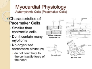
1. Myocardial Physiology
Autorhythmic Cells (Pacemaker Cells)
Characteristics of
Pacemaker Cells
◦ Smaller than
contractile cells
◦ Don’t contain many
myofibrils
◦ No organized
sarcomere structure
do not contribute to
the contractile force of
the heart
normal contractile
myocardial cell
conduction myofibers
SA node cell
AV node cells
2. Myocardial Physiology
Autorhythmic Cells (Pacemaker Cells)
Characteristics of Pacemaker Cells
◦ Unstable membrane potential
“bottoms out” at -60mV
“drifts upward” to -40mV, forming a pacemaker potential
◦ Myogenic
The upward “drift” allows the membrane to reach threshold
potential (-40mV) by itself
This is due to
1. Slow leakage of K+ out & faster leakage Na+ in
Causes slow depolarization
Occurs through If channels (f=funny) that open at negative
membrane potentials and start closing as membrane approaches
threshold potential
2. Ca2+ channels opening as membrane approaches threshold
At threshold additional Ca2+ ion channels open causing more rapid
depolarization
These deactivate shortly after and
3. Slow K+ channels open as membrane depolarizes causing an
efflux of K+ and a repolarization of membrane
4. Myocardial Physiology
Autorhythmic Cells (Pacemaker Cells)
Altering Activity of Pacemaker Cells
◦ Sympathetic activity
NE and E increase If channel activity
Binds to β1 adrenergic receptors which activate cAMP and
increase If channel open time
Causes more rapid pacemaker potential and faster rate of action
potentials
Sympathetic Activity Summary:
increased chronotropic effects
heart rate
increased dromotropic effects
conduction of APs
increased inotropic effects
contractility
5. Myocardial Physiology
Autorhythmic Cells (Pacemaker Cells)
Altering Activity of Pacemaker Cells
◦ Parasympathetic activity
ACh binds to muscarinic receptors
Increases K+ permeability and decreases Ca2+ permeability =
hyperpolarizing the membrane
Longer time to threshold = slower rate of action potentials
Parasympathetic Activity
Summary:
decreased chronotropic effects
heart rate
decreased dromotropic effects
conduction of APs
decreased inotropic effects
contractility
6. Myocardial Physiology
Contractile Cells
Special aspects
◦ Intercalated discs
Highly convoluted and interdigitated
junctions
Joint adjacent cells with
Desmosomes & fascia adherens
Allow for synticial activity
With gap junctions
◦ More mitochondria than skeletal muscle
◦ Less sarcoplasmic reticulum
Ca2+ also influxes from ECF reducing
storage need
◦ Larger t-tubules
Internally branching
◦ Myocardial contractions are graded!
7. Myocardial Physiology
Contractile Cells
Special aspects
◦ The action potential of a contractile cell
Ca2+ plays a major role again
Action potential is longer in duration than a “normal” action potential
due to Ca2+ entry
Phases
4 – resting membrane potential @ -90mV
0 – depolarization
Due to gap junctions or conduction fiber action
Voltage gated Na+ channels open… close at 20mV
1 – temporary repolarization
Open K+ channels allow some K+ to leave the cell
2 – plateau phase
Voltage gated Ca2+ channels are fully open (started during initial
depolarization)
3 – repolarization
Ca2+ channels close and K+ permeability increases as slower activated
K+ channels open, causing a quick repolarization
◦ What is the significance of the plateau phase?
9. Myocardial Physiology
Contractile Cells
Plateau phase prevents summation due
to the elongated refractory period
No summation capacity = no tetanus
◦ Which would be fatal
11. Myocardial Physiology
Contractile Cells
Initiation
◦ Action potential via pacemaker cells to
conduction fibers
Excitation-Contraction Coupling
1. Starts with CICR (Ca2+ induced Ca2+
release)
AP spreads along sarcolemma
T-tubules contain voltage gated L-type Ca2+
channels which open upon depolarization
Ca2+ entrance into myocardial cell and opens
RyR (ryanodine receptors) Ca2+ release
channels
Release of Ca2+ from SR causes a Ca2+
“spark”
Multiple sparks form a Ca2+ signal
Spark Gif
12. Myocardial Physiology
Contractile Cells
Excitation-Contraction Coupling cont…
2. Ca2+ signal (Ca2+ from SR and ECF) binds to troponin to initiate
myosin head attachment to actin
Contraction
◦ Same as skeletal muscle, but…
◦ Strength of contraction varies
Sarcomeres are not “all or none” as it is in skeletal muscle
The response is graded!
Low levels of cytosolic Ca2+ will not activate as many
myosin/actin interactions and the opposite is true
Length tension relationships exist
Strongest contraction generated
when stretched between 80 &
100% of maximum (physiological
range)
What causes stretching?
The filling of chambers
with blood
13. Myocardial Physiology
Contractile Cells
Relaxation
◦ Ca2+ is transported back
into the SR and
◦ Ca2+ is transported out of
the cell by a facilitated
Na+/Ca2+ exchanger (NCX)
◦ As ICF Ca2+ levels drop,
interactions between
myosin/actin are stopped
◦ Sarcomere lengthens
14. Cardiac Cycle
Coordinating the activity
Electrical Conduction Pathway
◦ Initiated by the Sino-Atrial node (SA node) which is myogenic at
70-80 action potentials/minute
◦ Depolarization is spread through the atria via gap junctions and
internodal pathways to the Atrio-Ventricular node (AV node)
The fibrous connective tissue matrix of the heart prevents further
spread of APs to the ventricles
A slight delay at the AV node occurs
Due to slower formation of action potentials
Allows further emptying of the atria
◦ Action potentials travel down the Atrioventricular bundle (Bundle
of His) which splits into left and right atrioventricular bundles
(bundle branches) and then into the conduction myofibers
(Purkinje cells)
Purkinje cells are larger in diameter & conduct impulse very rapidly
Causes the cells at the apex to contract nearly simultaneously
Good for ventricular ejection
61. After atrial contraction is complete
LVEDV typically about 120 ml (preload)
End-diastolic pressures of
LV = 8-12 mmHg and
RV = 3-6 mmHg
AV valves floats upward (pre-position)