SlideShare a Scribd company logo
Introduction to development of
the heart
• It develops early in the middle of 3rd
week , from aggregation of splanchnic
mesodermal cells, in cardiogenic area,
ventral to pericardial coelom, and dorsal
to yolk sac.
• They form 2 angioblastic cords that
canalize to form 2 endocardial heart
tubes.
Development of sinus venosus
• In the middle of the fourth week, the sinus venosus receives
venous blood from the right and left sinus horns .
• Each horn receives blood from three important veins: (1)
the vitelline or the omphalomesenteric vein, (2) the
umbilical vein, and (3) the common cardinal vein.
• Communication between the sinus and the atrium is wide-
initially
• Later the entrance of the sinus shifts to the right
• This shift is caused primarily by left-to-right shunts of blood,
which occur in the venous system during the fourth and fifth
weeks of development.
Development of sinus venosus
• With obliteration of the right umbilical vein and the left
vitelline vein during the fifth week, the left sinus horn rapidly
loses its importance
• When the left common cardinal is obliterated at 10 weeks, all
that remains of the left sinus horn is the oblique vein of the
left atrium and the coronary sinus .
Development of sinus venosus
• As a result of left-to-right shunts of blood, the right sinus horn
and veins enlarge greatly.
• The right horn, which now forms the only communication
between the original sinus venosus and the atrium, is
incorporated into the right atrium to form the smooth-walled
part of the right atrium .
• Its entrance, the sinuatrial orifice, is flanked on each side by a
valvular fold, the right and left venous valves .
• Dorsocranially, the valves fuse, forming a ridge known as the
septum spurium .
Development of sinus venosus
• Initially the valves are large, but when the right sinus horn is
incorporated into the wall of the atrium, the left venous valve
and the septum spurium fuse with the developing atrial
septum .
• The superior portion of the right venous valve disappears
entirely.
• The inferior portion develops into two parts: (1) the valve of
the inferior vena cava and (2) the valve of the coronary
sinus.
• The crista terminalis forms the dividing line between the
original trabeculated part of the right atrium and the smooth-
walled part (sinus venarum), which originates from the right
sinus horn .
Cardiac septa
• The major septa of the heart are formed between the 27th
and 37th days of development, when the embryo grows in
length from 5 mm to approximately 16 to 17 mm.
CARDIAC SEPTA
• The other manner in which a septum is formed does not
involve endocardial cushions.
• a narrow strip of tissue in the wall of the atrium or ventricle
should fail to grow while areas on each side of it expand
rapidly, a narrow ridge forms between the two expanding
portions .
• When growth of the expanding portions continues on either
side of the narrow portion, the two walls approach each other
and eventually merge, forming a septum
CARDIAC SEPTA
Partitioning of the
primitive Heart
• Division of A-V canal , primitive
atrium & primitive ventricle begins
at the middle or end of 4th week and
completed by the end of 5th week.
• These processes occur concurrently.
• At the end of 4th week, 2 endocardial
cushions on dorsal & ventral walls of
atrioventricular canal , develop from
mesenchymal cells of cardiac jelly.
INTERATRIAL SEPTUM
• Begins in 5th week of gestation
• Septum primum begins to develop, growing toward the
endocardial cushions.
• The progressively diminishing space between the
endocardial cushions and the septum primum is known
as the ostium primum.
• Before the septum primum fuses with the endocardial
cushions, small perforations develop and coalesce in the
cephalic portion of the septum primum, which is known
as the ostium secundum.
1-Septum primum :
•A thin crescent-shaped membrane grows
from the roof of common atrium.
•The lower margin of the septum is free and
concave.
•Anterior and posterior horns of the septum
fuse respectively with the ventral and dorsal
endocardial cushions of the primitive
atrioventricular canal.
2-Septum intermedium:
•The ventral and dorsal cushions of the
atrioventricular canal fuse to form a broad
anterioposterior partition, which divide the
canal into right and left atrioventricular
orifices.
• A foramen known as ostium
primum is formed between the
upper surface of septum
intermedium and lower border of
septum primum.
• Later, ostium primum is closed by
the fusion of two septa.
• Associated with the closure of
ostium primum, the upper and
dorsal part of septum disintegrates
forming a foramen known as ostium
secundum.
INTERATRIAL SEPTUM
• To the right of the septum primum, the septum secundum
starts to form as an invagination of the atrial wall.
• The septum secundum stops growing at the end of the
seventh week of gestation, leaving a posterior and inferior gap
known as the fossa ovalis.
• When the left venous valve and the septum spurium fuse with
the right side of the septum secundum, the free concave edge
of the septum secundum begins to overlap the ostium
secundum
3-Septum secundum-
•It arises on the right side of septum
primum from the space of the right
atrium which is interval between
septum primum and septum spurium.
•The septum secundum incorporates
the whole of left venous valve and
extends vertically downwards.
•The lower margin grows sufficiently to
overlap the upper margin of the
septum primum.
•The valvular opening formed between
the lower margin of the septum
secundum and upper margin of the
septum primum is called foramen
ovale.
FORAMEN OVALE
• The opening left by the septum secundum is called the oval
foramen
• When the upper part of the septum primum gradually
disappears, the remaining part becomes the valve of the oval
foramen.
FORAMEN OVALE
• After birth, when lung circulation begins and pressure in
the left atrium increases, the valve of the oval foramen is
pressed against the septum secundum, obliterating the
oval foramen and separating the right and left atria.
• In about 20% of cases, fusion of the septum primum and
septum secundum is incomplete, and a narrow oblique
cleft remains between the two atria.
Features of the
interatrial septum
• On the right side:
1. Fossa ovalis: Oval depression in the lower part of the septum, and the floor is
formed by the septum primum.
2. Limbus fossa ovalis: a sickle shaped fold that surrounds the upper, anterior and
posterior margins of the fossa ovalis. It represents lower free margins of the
septum secundum.
3. Foramina venarium minimarium: Venae cordis minimi open through these
foramina.
4. Atrio-ventricular node: It is situated in the lower part of the septum above the
opening of coronary sinus.
• On the left side:
• 1. Presence of the semilunar fold with the concavity directed upwards; it
is a remnant of the upper margin of the septum primum.
• 2. Lunate impression above the fold is formed by septum secundum.
• 3. Foramina venarium minimarium.
Interatrial septum
Endocardial cushions
• dorsal & ventral swellings
• fuse, dividing the single AV
canal into paired canals
• involved in formation of
interatrial & interventricular
septa
• derived from neural crest
• involved in many CHDs
Septum primum grows from atrial
roof toward endocardial cushions
• Foramen primum: shunt that closes
• Foramen secundum: perforates
septum primum, allowing shunt
• Septum secundum grows down,
Over
lFrom Moore & Persaud 1998apping
foramen secundum
Septum secundum grows down,
overlapping foramen secundum
• Foramen ovale: between septum
primum & septum secundum
• Remaining portion of septum
primum
forms valve of foramen ovale
• Fetus
• • right side high pressure (high pulmonary resistance, etc.)
• • well oxygenated blood streams through foramen ovale
• • valve of foramen ovale closes with left atrial contraction
• • After birth
• • right side low pressure (low pulmonary resistance)
• • valve remains closed (physiological closure)
• • valve eventually fuses (anatomical closure): fossa ovalis
Formation of the Cardiac Septa
• The Atrioventricular (AV) septum
• Atrial septum
• Interventricular septum
• Aorticopulmonary septum
Molecular regulation of septal development
• NKX2.5.-the master gene for heart development
• BMPs 2 and 4
• WNT protein inhibitors-CRESCENT and CERBERUS
• FGF8
• Retinoic acid
• TBX5- DNA- binding motif known as the T-box. Expressed
later than NKX2.5, it plays an important role in septation.
During 5th week, the AV- endocardial
cushions meet and unite in the mid line
to form a septum and divide the
common A-V canal into right & left A-V
canals.
Endocardial cushions also form the
AV- valves + membranous septa of
interventricular septum.
Note in D, coronal section ,begining
of development of interatrial &
intervent. septa.
Formation of the Interatrial septum.
• The septum is developed from three sources:
• 1. Septum primum
• 2. Septum intermedium
• 3. Septum secundum
Development of muscular part of IVS:
Primordial muscular IVS arises in the floor of ventricle , as thick crescentic
fold with concave free edge.
This septum subdivides the original ventricular cavity incompletely into
right & left ventricles that communicate together through IV foramen.
It meets the fused AV cushions (Septum intermedium) and partially fuses
with them
This foramen closes by the end of 7th week as the 2 bulbar ridges fuse
with the endocadial cushion.
• Formation of the membranous part of the IV
septum result from the fusion of tissues from
3 sources:
• The right bulbar ridge
• The left bulbar ridge
• The endocardial cushion
VSD
• Perimembranous(membranous/infracristal)70-
80%
• Muscular- 5-20%
Central- mid muscular
Apical
Marginal- along RV septal junction
Swiss cheese septum – multiple defects
• Inlet/ AV canal type-5-8%
• Supracristal/ subaortic- 5-7%
soto et al
ENDOCARDIAL CUSHION DEFECT
• COMMON AV CANALLEFT AND RIGHT SIDE
BY FUSION OF SUPERIOR AND INFERIOS
ENDOCARDIAL CUSHION
• FAILUREAV SEPTAL DEFECT WITH VARIOUS
COMBINATION
• PARTIAL AV CANAL DEFECTSEPERATE AV
ORIFICE
• COMPLETE AV CANAL DEFECTCOMMON AV
ORIFICE
• ISOLATED INLET VSD
4th week – Formation of septa starts, IVS starts proliferating from
5th w the bulbar ridges & fused endocardial cushions.
 6th w. incorporation of the proximal part of bulbus cordis into the ventricles.
6th w. proliferation of endocardial cushions to diminish I V foramen.
7th w. fusion of bulbar ridges + extensions of endocardial cushions upward with
aortico-pulmonary septum and down with muscular I V septum to close I V foramen ,
so memb. IV septum is formed
DEVELOPMENT OF ARTERY
• First arteries to appear are Right and left primitive
aorta
• They are continous to the endothelial heart tubes
• Each primitive aorta have ventral portion lying in
the first pharygeal arch
• 1st,2nd and 5th arch dissapears
INTERRUPTED AORTIC ARCH
RIGHT AORTIC ARCH
DOUBLE AORTIC ARCH
Embryology of heart
Embryology of heart

More Related Content

What's hot

Development of heart finale copy
Development of heart finale   copyDevelopment of heart finale   copy
Development of heart finale copy
Robin Thomas
 
Embryology of heart
Embryology of heartEmbryology of heart
Embryology of heart
Ramachandra Barik
 
Development of Heart (Embryology)
Development of Heart (Embryology)Development of Heart (Embryology)
Development of Heart (Embryology)
Yukta Wankhede
 
Heart development -I
Heart development -IHeart development -I
Heart development -I
Dr Laxman Khanal
 
Development of venous system
Development of venous systemDevelopment of venous system
Development of venous system
Rohit Paswan
 
EMBRYOLOGY OF CVS
EMBRYOLOGY OF CVSEMBRYOLOGY OF CVS
EMBRYOLOGY OF CVS
profgoodnewszion
 
Fibrous skeleton of the heart
Fibrous  skeleton of the heartFibrous  skeleton of the heart
Fibrous skeleton of the heart
Idris Siddiqui
 
11 development of the heart
11   development of the heart11   development of the heart
11 development of the heart
puneet mahajan
 
Vena cava anatomy and variants
Vena cava anatomy and variantsVena cava anatomy and variants
Vena cava anatomy and variants
Milan Silwal
 
Development of heart ,,embryology,,virbhan
Development of heart ,,embryology,,virbhanDevelopment of heart ,,embryology,,virbhan
Development of heart ,,embryology,,virbhan
Dr Virbhan Balai
 
Mitral valve anatomy - ppt by kunwar sidharth
Mitral valve    anatomy - ppt by kunwar sidharthMitral valve    anatomy - ppt by kunwar sidharth
Mitral valve anatomy - ppt by kunwar sidharth
kunwar sidharth
 
Development of heart ii
Development of heart  iiDevelopment of heart  ii
Development of heart ii
Rohit Paswan
 
Cvs dev
Cvs devCvs dev
Cvs dev
Rohit Paswan
 
Atrial septaum development and
Atrial septaum development andAtrial septaum development and
Atrial septaum development and
Nagendra prasad Kulari
 
Coronary artery anatomy
Coronary artery anatomyCoronary artery anatomy
Coronary artery anatomy
Dr. Yash Kumar Achantani
 
Development of superior venacava and azygous vein
Development of superior venacava and azygous veinDevelopment of superior venacava and azygous vein
Development of superior venacava and azygous vein
anuppslides
 
7. Development of veins.
7. Development of veins.7. Development of veins.
7. Development of veins.
Dr. Mohammad Mahmoud
 
Azygos system
Azygos systemAzygos system
Aortic Arches
Aortic ArchesAortic Arches
Aortic Arches
DRHAFIZ07
 
Embryology of heart and fetal circulation
Embryology of heart and fetal circulationEmbryology of heart and fetal circulation
Embryology of heart and fetal circulation
Archana Koshy
 

What's hot (20)

Development of heart finale copy
Development of heart finale   copyDevelopment of heart finale   copy
Development of heart finale copy
 
Embryology of heart
Embryology of heartEmbryology of heart
Embryology of heart
 
Development of Heart (Embryology)
Development of Heart (Embryology)Development of Heart (Embryology)
Development of Heart (Embryology)
 
Heart development -I
Heart development -IHeart development -I
Heart development -I
 
Development of venous system
Development of venous systemDevelopment of venous system
Development of venous system
 
EMBRYOLOGY OF CVS
EMBRYOLOGY OF CVSEMBRYOLOGY OF CVS
EMBRYOLOGY OF CVS
 
Fibrous skeleton of the heart
Fibrous  skeleton of the heartFibrous  skeleton of the heart
Fibrous skeleton of the heart
 
11 development of the heart
11   development of the heart11   development of the heart
11 development of the heart
 
Vena cava anatomy and variants
Vena cava anatomy and variantsVena cava anatomy and variants
Vena cava anatomy and variants
 
Development of heart ,,embryology,,virbhan
Development of heart ,,embryology,,virbhanDevelopment of heart ,,embryology,,virbhan
Development of heart ,,embryology,,virbhan
 
Mitral valve anatomy - ppt by kunwar sidharth
Mitral valve    anatomy - ppt by kunwar sidharthMitral valve    anatomy - ppt by kunwar sidharth
Mitral valve anatomy - ppt by kunwar sidharth
 
Development of heart ii
Development of heart  iiDevelopment of heart  ii
Development of heart ii
 
Cvs dev
Cvs devCvs dev
Cvs dev
 
Atrial septaum development and
Atrial septaum development andAtrial septaum development and
Atrial septaum development and
 
Coronary artery anatomy
Coronary artery anatomyCoronary artery anatomy
Coronary artery anatomy
 
Development of superior venacava and azygous vein
Development of superior venacava and azygous veinDevelopment of superior venacava and azygous vein
Development of superior venacava and azygous vein
 
7. Development of veins.
7. Development of veins.7. Development of veins.
7. Development of veins.
 
Azygos system
Azygos systemAzygos system
Azygos system
 
Aortic Arches
Aortic ArchesAortic Arches
Aortic Arches
 
Embryology of heart and fetal circulation
Embryology of heart and fetal circulationEmbryology of heart and fetal circulation
Embryology of heart and fetal circulation
 

Similar to Embryology of heart

Embryology of heart
Embryology of heartEmbryology of heart
Embryology of heart
Rengarajan Rajagopal
 
Embryology of heart and lung
Embryology of heart and lungEmbryology of heart and lung
Embryology of heart and lung
Princy Francis M
 
Cardiovascularsystem 110514061056-phpapp02
Cardiovascularsystem 110514061056-phpapp02Cardiovascularsystem 110514061056-phpapp02
Cardiovascularsystem 110514061056-phpapp02
Ifrah Javaid
 
development of heart.pptx
development of heart.pptxdevelopment of heart.pptx
development of heart.pptx
KARTHIKD765407
 
Cardiovascular System Embryology 2024 .pptx
Cardiovascular System Embryology 2024 .pptxCardiovascular System Embryology 2024 .pptx
Cardiovascular System Embryology 2024 .pptx
ZAPPAC1
 
Embryology of the heart
Embryology of the heartEmbryology of the heart
Embryology of the heart
Anand Manjunath
 
Development of heart.pptx
Development of heart.pptxDevelopment of heart.pptx
Development of heart.pptx
Sureshdayalan1
 
Embryology of heart and understand basic heart disease
Embryology of heart  and understand basic heart diseaseEmbryology of heart  and understand basic heart disease
Embryology of heart and understand basic heart disease
VikramChaudhry
 
123.ppt
123.ppt123.ppt
123.ppt
TasnimSPansar
 
Final heart development.pptx
Final heart development.pptxFinal heart development.pptx
Final heart development.pptx
SayanRDey
 
Development of heart
Development of heartDevelopment of heart
Development of heart
Lawrence James
 
Cardiac embryology
Cardiac embryologyCardiac embryology
Cardiac embryology
Rezaul hayat
 
CVS Embryology (Amoud University)
CVS Embryology (Amoud University)CVS Embryology (Amoud University)
CVS Embryology (Amoud University)
moaminnca
 
Development of the heart tube and fetal circulation
Development of the heart tube and fetal circulationDevelopment of the heart tube and fetal circulation
Development of the heart tube and fetal circulation
Mohamed El Fiky
 
Final heart development.pptx
Final heart development.pptxFinal heart development.pptx
Final heart development.pptx
mayank360671
 
Heart development
Heart developmentHeart development
Heart development
SohamKhedekar2
 
Development of Cardiovascular System.pptx
Development of Cardiovascular System.pptxDevelopment of Cardiovascular System.pptx
Development of Cardiovascular System.pptx
Suresh Managutti
 
Cardiac Embryology basics.pptx
Cardiac Embryology basics.pptxCardiac Embryology basics.pptx
Cardiac Embryology basics.pptx
Shivani Rao
 
Manoja_Gullapalli_ug
Manoja_Gullapalli_ugManoja_Gullapalli_ug
Manoja_Gullapalli_ug
Debiprasad Panda
 
Final heart development.ppt
Final heart development.pptFinal heart development.ppt
Final heart development.ppt
mukeshgupta500425
 

Similar to Embryology of heart (20)

Embryology of heart
Embryology of heartEmbryology of heart
Embryology of heart
 
Embryology of heart and lung
Embryology of heart and lungEmbryology of heart and lung
Embryology of heart and lung
 
Cardiovascularsystem 110514061056-phpapp02
Cardiovascularsystem 110514061056-phpapp02Cardiovascularsystem 110514061056-phpapp02
Cardiovascularsystem 110514061056-phpapp02
 
development of heart.pptx
development of heart.pptxdevelopment of heart.pptx
development of heart.pptx
 
Cardiovascular System Embryology 2024 .pptx
Cardiovascular System Embryology 2024 .pptxCardiovascular System Embryology 2024 .pptx
Cardiovascular System Embryology 2024 .pptx
 
Embryology of the heart
Embryology of the heartEmbryology of the heart
Embryology of the heart
 
Development of heart.pptx
Development of heart.pptxDevelopment of heart.pptx
Development of heart.pptx
 
Embryology of heart and understand basic heart disease
Embryology of heart  and understand basic heart diseaseEmbryology of heart  and understand basic heart disease
Embryology of heart and understand basic heart disease
 
123.ppt
123.ppt123.ppt
123.ppt
 
Final heart development.pptx
Final heart development.pptxFinal heart development.pptx
Final heart development.pptx
 
Development of heart
Development of heartDevelopment of heart
Development of heart
 
Cardiac embryology
Cardiac embryologyCardiac embryology
Cardiac embryology
 
CVS Embryology (Amoud University)
CVS Embryology (Amoud University)CVS Embryology (Amoud University)
CVS Embryology (Amoud University)
 
Development of the heart tube and fetal circulation
Development of the heart tube and fetal circulationDevelopment of the heart tube and fetal circulation
Development of the heart tube and fetal circulation
 
Final heart development.pptx
Final heart development.pptxFinal heart development.pptx
Final heart development.pptx
 
Heart development
Heart developmentHeart development
Heart development
 
Development of Cardiovascular System.pptx
Development of Cardiovascular System.pptxDevelopment of Cardiovascular System.pptx
Development of Cardiovascular System.pptx
 
Cardiac Embryology basics.pptx
Cardiac Embryology basics.pptxCardiac Embryology basics.pptx
Cardiac Embryology basics.pptx
 
Manoja_Gullapalli_ug
Manoja_Gullapalli_ugManoja_Gullapalli_ug
Manoja_Gullapalli_ug
 
Final heart development.ppt
Final heart development.pptFinal heart development.ppt
Final heart development.ppt
 

More from nmonty02

Levosimendan
LevosimendanLevosimendan
Levosimendan
nmonty02
 
Early repolarisation syndrome
Early repolarisation syndromeEarly repolarisation syndrome
Early repolarisation syndrome
nmonty02
 
Epicardial fat
Epicardial fatEpicardial fat
Epicardial fat
nmonty02
 
Echo views
Echo viewsEcho views
Echo views
nmonty02
 
Anatomy of heart
Anatomy of heartAnatomy of heart
Anatomy of heart
nmonty02
 
Physiology of heart
Physiology of heartPhysiology of heart
Physiology of heart
nmonty02
 

More from nmonty02 (6)

Levosimendan
LevosimendanLevosimendan
Levosimendan
 
Early repolarisation syndrome
Early repolarisation syndromeEarly repolarisation syndrome
Early repolarisation syndrome
 
Epicardial fat
Epicardial fatEpicardial fat
Epicardial fat
 
Echo views
Echo viewsEcho views
Echo views
 
Anatomy of heart
Anatomy of heartAnatomy of heart
Anatomy of heart
 
Physiology of heart
Physiology of heartPhysiology of heart
Physiology of heart
 

Recently uploaded

Chapter 11 Nutrition and Chronic Diseases.pptx
Chapter 11 Nutrition and Chronic Diseases.pptxChapter 11 Nutrition and Chronic Diseases.pptx
Chapter 11 Nutrition and Chronic Diseases.pptx
Earlene McNair
 
Aortic Association CBL Pilot April 19 – 20 Bern
Aortic Association CBL Pilot April 19 – 20 BernAortic Association CBL Pilot April 19 – 20 Bern
Aortic Association CBL Pilot April 19 – 20 Bern
suvadeepdas911
 
#cALL# #gIRLS# In Dehradun ꧁❤8107221448❤꧂#cALL# #gIRLS# Service In Dehradun W...
#cALL# #gIRLS# In Dehradun ꧁❤8107221448❤꧂#cALL# #gIRLS# Service In Dehradun W...#cALL# #gIRLS# In Dehradun ꧁❤8107221448❤꧂#cALL# #gIRLS# Service In Dehradun W...
#cALL# #gIRLS# In Dehradun ꧁❤8107221448❤꧂#cALL# #gIRLS# Service In Dehradun W...
chandankumarsmartiso
 
Integrating Ayurveda into Parkinson’s Management: A Holistic Approach
Integrating Ayurveda into Parkinson’s Management: A Holistic ApproachIntegrating Ayurveda into Parkinson’s Management: A Holistic Approach
Integrating Ayurveda into Parkinson’s Management: A Holistic Approach
Ayurveda ForAll
 
Basavarajeeyam - Ayurvedic heritage book of Andhra pradesh
Basavarajeeyam - Ayurvedic heritage book of Andhra pradeshBasavarajeeyam - Ayurvedic heritage book of Andhra pradesh
Basavarajeeyam - Ayurvedic heritage book of Andhra pradesh
Dr. Madduru Muni Haritha
 
Local Advanced Lung Cancer: Artificial Intelligence, Synergetics, Complex Sys...
Local Advanced Lung Cancer: Artificial Intelligence, Synergetics, Complex Sys...Local Advanced Lung Cancer: Artificial Intelligence, Synergetics, Complex Sys...
Local Advanced Lung Cancer: Artificial Intelligence, Synergetics, Complex Sys...
Oleg Kshivets
 
The Electrocardiogram - Physiologic Principles
The Electrocardiogram - Physiologic PrinciplesThe Electrocardiogram - Physiologic Principles
The Electrocardiogram - Physiologic Principles
MedicoseAcademics
 
8 Surprising Reasons To Meditate 40 Minutes A Day That Can Change Your Life.pptx
8 Surprising Reasons To Meditate 40 Minutes A Day That Can Change Your Life.pptx8 Surprising Reasons To Meditate 40 Minutes A Day That Can Change Your Life.pptx
8 Surprising Reasons To Meditate 40 Minutes A Day That Can Change Your Life.pptx
Holistified Wellness
 
Journal Article Review on Rasamanikya
Journal Article Review on RasamanikyaJournal Article Review on Rasamanikya
Journal Article Review on Rasamanikya
Dr. Jyothirmai Paindla
 
Top 10 Best Ayurvedic Kidney Stone Syrups in India
Top 10 Best Ayurvedic Kidney Stone Syrups in IndiaTop 10 Best Ayurvedic Kidney Stone Syrups in India
Top 10 Best Ayurvedic Kidney Stone Syrups in India
Swastik Ayurveda
 
Phone Us ❤8107221448❤ #ℂall #gIRLS In Dehradun By Dehradun @ℂall @Girls Hotel...
Phone Us ❤8107221448❤ #ℂall #gIRLS In Dehradun By Dehradun @ℂall @Girls Hotel...Phone Us ❤8107221448❤ #ℂall #gIRLS In Dehradun By Dehradun @ℂall @Girls Hotel...
Phone Us ❤8107221448❤ #ℂall #gIRLS In Dehradun By Dehradun @ℂall @Girls Hotel...
chandankumarsmartiso
 
Tests for analysis of different pharmaceutical.pptx
Tests for analysis of different pharmaceutical.pptxTests for analysis of different pharmaceutical.pptx
Tests for analysis of different pharmaceutical.pptx
taiba qazi
 
Dehradun #ℂall #gIRLS Oyo Hotel 8107221448 #ℂall #gIRL in Dehradun
Dehradun #ℂall #gIRLS Oyo Hotel 8107221448 #ℂall #gIRL in DehradunDehradun #ℂall #gIRLS Oyo Hotel 8107221448 #ℂall #gIRL in Dehradun
Dehradun #ℂall #gIRLS Oyo Hotel 8107221448 #ℂall #gIRL in Dehradun
chandankumarsmartiso
 
CHEMOTHERAPY_RDP_CHAPTER 1_ANTI TB DRUGS.pdf
CHEMOTHERAPY_RDP_CHAPTER 1_ANTI TB DRUGS.pdfCHEMOTHERAPY_RDP_CHAPTER 1_ANTI TB DRUGS.pdf
CHEMOTHERAPY_RDP_CHAPTER 1_ANTI TB DRUGS.pdf
rishi2789
 
Role of Mukta Pishti in the Management of Hyperthyroidism
Role of Mukta Pishti in the Management of HyperthyroidismRole of Mukta Pishti in the Management of Hyperthyroidism
Role of Mukta Pishti in the Management of Hyperthyroidism
Dr. Jyothirmai Paindla
 
share - Lions, tigers, AI and health misinformation, oh my!.pptx
share - Lions, tigers, AI and health misinformation, oh my!.pptxshare - Lions, tigers, AI and health misinformation, oh my!.pptx
share - Lions, tigers, AI and health misinformation, oh my!.pptx
Tina Purnat
 
TEST BANK For Community Health Nursing A Canadian Perspective, 5th Edition by...
TEST BANK For Community Health Nursing A Canadian Perspective, 5th Edition by...TEST BANK For Community Health Nursing A Canadian Perspective, 5th Edition by...
TEST BANK For Community Health Nursing A Canadian Perspective, 5th Edition by...
Donc Test
 
Thyroid Gland- Gross Anatomy by Dr. Rabia Inam Gandapore.pptx
Thyroid Gland- Gross Anatomy by Dr. Rabia Inam Gandapore.pptxThyroid Gland- Gross Anatomy by Dr. Rabia Inam Gandapore.pptx
Thyroid Gland- Gross Anatomy by Dr. Rabia Inam Gandapore.pptx
Dr. Rabia Inam Gandapore
 
THERAPEUTIC ANTISENSE MOLECULES .pptx
THERAPEUTIC ANTISENSE MOLECULES    .pptxTHERAPEUTIC ANTISENSE MOLECULES    .pptx
THERAPEUTIC ANTISENSE MOLECULES .pptx
70KRISHPATEL
 
Part II - Body Grief: Losing parts of ourselves and our identity before, duri...
Part II - Body Grief: Losing parts of ourselves and our identity before, duri...Part II - Body Grief: Losing parts of ourselves and our identity before, duri...
Part II - Body Grief: Losing parts of ourselves and our identity before, duri...
bkling
 

Recently uploaded (20)

Chapter 11 Nutrition and Chronic Diseases.pptx
Chapter 11 Nutrition and Chronic Diseases.pptxChapter 11 Nutrition and Chronic Diseases.pptx
Chapter 11 Nutrition and Chronic Diseases.pptx
 
Aortic Association CBL Pilot April 19 – 20 Bern
Aortic Association CBL Pilot April 19 – 20 BernAortic Association CBL Pilot April 19 – 20 Bern
Aortic Association CBL Pilot April 19 – 20 Bern
 
#cALL# #gIRLS# In Dehradun ꧁❤8107221448❤꧂#cALL# #gIRLS# Service In Dehradun W...
#cALL# #gIRLS# In Dehradun ꧁❤8107221448❤꧂#cALL# #gIRLS# Service In Dehradun W...#cALL# #gIRLS# In Dehradun ꧁❤8107221448❤꧂#cALL# #gIRLS# Service In Dehradun W...
#cALL# #gIRLS# In Dehradun ꧁❤8107221448❤꧂#cALL# #gIRLS# Service In Dehradun W...
 
Integrating Ayurveda into Parkinson’s Management: A Holistic Approach
Integrating Ayurveda into Parkinson’s Management: A Holistic ApproachIntegrating Ayurveda into Parkinson’s Management: A Holistic Approach
Integrating Ayurveda into Parkinson’s Management: A Holistic Approach
 
Basavarajeeyam - Ayurvedic heritage book of Andhra pradesh
Basavarajeeyam - Ayurvedic heritage book of Andhra pradeshBasavarajeeyam - Ayurvedic heritage book of Andhra pradesh
Basavarajeeyam - Ayurvedic heritage book of Andhra pradesh
 
Local Advanced Lung Cancer: Artificial Intelligence, Synergetics, Complex Sys...
Local Advanced Lung Cancer: Artificial Intelligence, Synergetics, Complex Sys...Local Advanced Lung Cancer: Artificial Intelligence, Synergetics, Complex Sys...
Local Advanced Lung Cancer: Artificial Intelligence, Synergetics, Complex Sys...
 
The Electrocardiogram - Physiologic Principles
The Electrocardiogram - Physiologic PrinciplesThe Electrocardiogram - Physiologic Principles
The Electrocardiogram - Physiologic Principles
 
8 Surprising Reasons To Meditate 40 Minutes A Day That Can Change Your Life.pptx
8 Surprising Reasons To Meditate 40 Minutes A Day That Can Change Your Life.pptx8 Surprising Reasons To Meditate 40 Minutes A Day That Can Change Your Life.pptx
8 Surprising Reasons To Meditate 40 Minutes A Day That Can Change Your Life.pptx
 
Journal Article Review on Rasamanikya
Journal Article Review on RasamanikyaJournal Article Review on Rasamanikya
Journal Article Review on Rasamanikya
 
Top 10 Best Ayurvedic Kidney Stone Syrups in India
Top 10 Best Ayurvedic Kidney Stone Syrups in IndiaTop 10 Best Ayurvedic Kidney Stone Syrups in India
Top 10 Best Ayurvedic Kidney Stone Syrups in India
 
Phone Us ❤8107221448❤ #ℂall #gIRLS In Dehradun By Dehradun @ℂall @Girls Hotel...
Phone Us ❤8107221448❤ #ℂall #gIRLS In Dehradun By Dehradun @ℂall @Girls Hotel...Phone Us ❤8107221448❤ #ℂall #gIRLS In Dehradun By Dehradun @ℂall @Girls Hotel...
Phone Us ❤8107221448❤ #ℂall #gIRLS In Dehradun By Dehradun @ℂall @Girls Hotel...
 
Tests for analysis of different pharmaceutical.pptx
Tests for analysis of different pharmaceutical.pptxTests for analysis of different pharmaceutical.pptx
Tests for analysis of different pharmaceutical.pptx
 
Dehradun #ℂall #gIRLS Oyo Hotel 8107221448 #ℂall #gIRL in Dehradun
Dehradun #ℂall #gIRLS Oyo Hotel 8107221448 #ℂall #gIRL in DehradunDehradun #ℂall #gIRLS Oyo Hotel 8107221448 #ℂall #gIRL in Dehradun
Dehradun #ℂall #gIRLS Oyo Hotel 8107221448 #ℂall #gIRL in Dehradun
 
CHEMOTHERAPY_RDP_CHAPTER 1_ANTI TB DRUGS.pdf
CHEMOTHERAPY_RDP_CHAPTER 1_ANTI TB DRUGS.pdfCHEMOTHERAPY_RDP_CHAPTER 1_ANTI TB DRUGS.pdf
CHEMOTHERAPY_RDP_CHAPTER 1_ANTI TB DRUGS.pdf
 
Role of Mukta Pishti in the Management of Hyperthyroidism
Role of Mukta Pishti in the Management of HyperthyroidismRole of Mukta Pishti in the Management of Hyperthyroidism
Role of Mukta Pishti in the Management of Hyperthyroidism
 
share - Lions, tigers, AI and health misinformation, oh my!.pptx
share - Lions, tigers, AI and health misinformation, oh my!.pptxshare - Lions, tigers, AI and health misinformation, oh my!.pptx
share - Lions, tigers, AI and health misinformation, oh my!.pptx
 
TEST BANK For Community Health Nursing A Canadian Perspective, 5th Edition by...
TEST BANK For Community Health Nursing A Canadian Perspective, 5th Edition by...TEST BANK For Community Health Nursing A Canadian Perspective, 5th Edition by...
TEST BANK For Community Health Nursing A Canadian Perspective, 5th Edition by...
 
Thyroid Gland- Gross Anatomy by Dr. Rabia Inam Gandapore.pptx
Thyroid Gland- Gross Anatomy by Dr. Rabia Inam Gandapore.pptxThyroid Gland- Gross Anatomy by Dr. Rabia Inam Gandapore.pptx
Thyroid Gland- Gross Anatomy by Dr. Rabia Inam Gandapore.pptx
 
THERAPEUTIC ANTISENSE MOLECULES .pptx
THERAPEUTIC ANTISENSE MOLECULES    .pptxTHERAPEUTIC ANTISENSE MOLECULES    .pptx
THERAPEUTIC ANTISENSE MOLECULES .pptx
 
Part II - Body Grief: Losing parts of ourselves and our identity before, duri...
Part II - Body Grief: Losing parts of ourselves and our identity before, duri...Part II - Body Grief: Losing parts of ourselves and our identity before, duri...
Part II - Body Grief: Losing parts of ourselves and our identity before, duri...
 

Embryology of heart

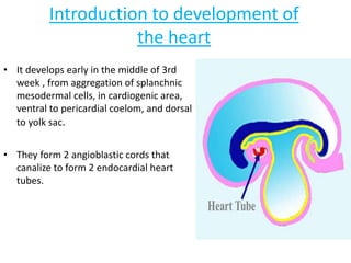
  • 1. Introduction to development of the heart • It develops early in the middle of 3rd week , from aggregation of splanchnic mesodermal cells, in cardiogenic area, ventral to pericardial coelom, and dorsal to yolk sac. • They form 2 angioblastic cords that canalize to form 2 endocardial heart tubes.
  • 2.
  • 3.
  • 4. Development of sinus venosus • In the middle of the fourth week, the sinus venosus receives venous blood from the right and left sinus horns . • Each horn receives blood from three important veins: (1) the vitelline or the omphalomesenteric vein, (2) the umbilical vein, and (3) the common cardinal vein. • Communication between the sinus and the atrium is wide- initially • Later the entrance of the sinus shifts to the right • This shift is caused primarily by left-to-right shunts of blood, which occur in the venous system during the fourth and fifth weeks of development.
  • 5. Development of sinus venosus • With obliteration of the right umbilical vein and the left vitelline vein during the fifth week, the left sinus horn rapidly loses its importance • When the left common cardinal is obliterated at 10 weeks, all that remains of the left sinus horn is the oblique vein of the left atrium and the coronary sinus .
  • 6. Development of sinus venosus • As a result of left-to-right shunts of blood, the right sinus horn and veins enlarge greatly. • The right horn, which now forms the only communication between the original sinus venosus and the atrium, is incorporated into the right atrium to form the smooth-walled part of the right atrium . • Its entrance, the sinuatrial orifice, is flanked on each side by a valvular fold, the right and left venous valves . • Dorsocranially, the valves fuse, forming a ridge known as the septum spurium .
  • 7. Development of sinus venosus • Initially the valves are large, but when the right sinus horn is incorporated into the wall of the atrium, the left venous valve and the septum spurium fuse with the developing atrial septum . • The superior portion of the right venous valve disappears entirely. • The inferior portion develops into two parts: (1) the valve of the inferior vena cava and (2) the valve of the coronary sinus. • The crista terminalis forms the dividing line between the original trabeculated part of the right atrium and the smooth- walled part (sinus venarum), which originates from the right sinus horn .
  • 8. Cardiac septa • The major septa of the heart are formed between the 27th and 37th days of development, when the embryo grows in length from 5 mm to approximately 16 to 17 mm.
  • 9. CARDIAC SEPTA • The other manner in which a septum is formed does not involve endocardial cushions. • a narrow strip of tissue in the wall of the atrium or ventricle should fail to grow while areas on each side of it expand rapidly, a narrow ridge forms between the two expanding portions . • When growth of the expanding portions continues on either side of the narrow portion, the two walls approach each other and eventually merge, forming a septum
  • 11. Partitioning of the primitive Heart • Division of A-V canal , primitive atrium & primitive ventricle begins at the middle or end of 4th week and completed by the end of 5th week. • These processes occur concurrently. • At the end of 4th week, 2 endocardial cushions on dorsal & ventral walls of atrioventricular canal , develop from mesenchymal cells of cardiac jelly.
  • 12. INTERATRIAL SEPTUM • Begins in 5th week of gestation • Septum primum begins to develop, growing toward the endocardial cushions. • The progressively diminishing space between the endocardial cushions and the septum primum is known as the ostium primum. • Before the septum primum fuses with the endocardial cushions, small perforations develop and coalesce in the cephalic portion of the septum primum, which is known as the ostium secundum.
  • 13. 1-Septum primum : •A thin crescent-shaped membrane grows from the roof of common atrium. •The lower margin of the septum is free and concave. •Anterior and posterior horns of the septum fuse respectively with the ventral and dorsal endocardial cushions of the primitive atrioventricular canal. 2-Septum intermedium: •The ventral and dorsal cushions of the atrioventricular canal fuse to form a broad anterioposterior partition, which divide the canal into right and left atrioventricular orifices.
  • 14. • A foramen known as ostium primum is formed between the upper surface of septum intermedium and lower border of septum primum. • Later, ostium primum is closed by the fusion of two septa. • Associated with the closure of ostium primum, the upper and dorsal part of septum disintegrates forming a foramen known as ostium secundum.
  • 15. INTERATRIAL SEPTUM • To the right of the septum primum, the septum secundum starts to form as an invagination of the atrial wall. • The septum secundum stops growing at the end of the seventh week of gestation, leaving a posterior and inferior gap known as the fossa ovalis. • When the left venous valve and the septum spurium fuse with the right side of the septum secundum, the free concave edge of the septum secundum begins to overlap the ostium secundum
  • 16. 3-Septum secundum- •It arises on the right side of septum primum from the space of the right atrium which is interval between septum primum and septum spurium. •The septum secundum incorporates the whole of left venous valve and extends vertically downwards. •The lower margin grows sufficiently to overlap the upper margin of the septum primum. •The valvular opening formed between the lower margin of the septum secundum and upper margin of the septum primum is called foramen ovale.
  • 17. FORAMEN OVALE • The opening left by the septum secundum is called the oval foramen • When the upper part of the septum primum gradually disappears, the remaining part becomes the valve of the oval foramen.
  • 18. FORAMEN OVALE • After birth, when lung circulation begins and pressure in the left atrium increases, the valve of the oval foramen is pressed against the septum secundum, obliterating the oval foramen and separating the right and left atria. • In about 20% of cases, fusion of the septum primum and septum secundum is incomplete, and a narrow oblique cleft remains between the two atria.
  • 19. Features of the interatrial septum • On the right side: 1. Fossa ovalis: Oval depression in the lower part of the septum, and the floor is formed by the septum primum. 2. Limbus fossa ovalis: a sickle shaped fold that surrounds the upper, anterior and posterior margins of the fossa ovalis. It represents lower free margins of the septum secundum. 3. Foramina venarium minimarium: Venae cordis minimi open through these foramina. 4. Atrio-ventricular node: It is situated in the lower part of the septum above the opening of coronary sinus.
  • 20. • On the left side: • 1. Presence of the semilunar fold with the concavity directed upwards; it is a remnant of the upper margin of the septum primum. • 2. Lunate impression above the fold is formed by septum secundum. • 3. Foramina venarium minimarium.
  • 22. Endocardial cushions • dorsal & ventral swellings • fuse, dividing the single AV canal into paired canals • involved in formation of interatrial & interventricular septa • derived from neural crest • involved in many CHDs
  • 23. Septum primum grows from atrial roof toward endocardial cushions • Foramen primum: shunt that closes • Foramen secundum: perforates septum primum, allowing shunt • Septum secundum grows down, Over lFrom Moore & Persaud 1998apping foramen secundum
  • 24. Septum secundum grows down, overlapping foramen secundum • Foramen ovale: between septum primum & septum secundum • Remaining portion of septum primum forms valve of foramen ovale
  • 25.
  • 26. • Fetus • • right side high pressure (high pulmonary resistance, etc.) • • well oxygenated blood streams through foramen ovale • • valve of foramen ovale closes with left atrial contraction • • After birth • • right side low pressure (low pulmonary resistance) • • valve remains closed (physiological closure) • • valve eventually fuses (anatomical closure): fossa ovalis
  • 27.
  • 28. Formation of the Cardiac Septa • The Atrioventricular (AV) septum • Atrial septum • Interventricular septum • Aorticopulmonary septum
  • 29. Molecular regulation of septal development • NKX2.5.-the master gene for heart development • BMPs 2 and 4 • WNT protein inhibitors-CRESCENT and CERBERUS • FGF8 • Retinoic acid • TBX5- DNA- binding motif known as the T-box. Expressed later than NKX2.5, it plays an important role in septation.
  • 30.
  • 31. During 5th week, the AV- endocardial cushions meet and unite in the mid line to form a septum and divide the common A-V canal into right & left A-V canals. Endocardial cushions also form the AV- valves + membranous septa of interventricular septum. Note in D, coronal section ,begining of development of interatrial & intervent. septa.
  • 32. Formation of the Interatrial septum. • The septum is developed from three sources: • 1. Septum primum • 2. Septum intermedium • 3. Septum secundum
  • 33.
  • 34. Development of muscular part of IVS: Primordial muscular IVS arises in the floor of ventricle , as thick crescentic fold with concave free edge. This septum subdivides the original ventricular cavity incompletely into right & left ventricles that communicate together through IV foramen. It meets the fused AV cushions (Septum intermedium) and partially fuses with them This foramen closes by the end of 7th week as the 2 bulbar ridges fuse with the endocadial cushion.
  • 35. • Formation of the membranous part of the IV septum result from the fusion of tissues from 3 sources: • The right bulbar ridge • The left bulbar ridge • The endocardial cushion
  • 36.
  • 37.
  • 38. VSD • Perimembranous(membranous/infracristal)70- 80% • Muscular- 5-20% Central- mid muscular Apical Marginal- along RV septal junction Swiss cheese septum – multiple defects • Inlet/ AV canal type-5-8% • Supracristal/ subaortic- 5-7% soto et al
  • 39. ENDOCARDIAL CUSHION DEFECT • COMMON AV CANALLEFT AND RIGHT SIDE BY FUSION OF SUPERIOR AND INFERIOS ENDOCARDIAL CUSHION • FAILUREAV SEPTAL DEFECT WITH VARIOUS COMBINATION
  • 40. • PARTIAL AV CANAL DEFECTSEPERATE AV ORIFICE • COMPLETE AV CANAL DEFECTCOMMON AV ORIFICE • ISOLATED INLET VSD
  • 41. 4th week – Formation of septa starts, IVS starts proliferating from 5th w the bulbar ridges & fused endocardial cushions.  6th w. incorporation of the proximal part of bulbus cordis into the ventricles. 6th w. proliferation of endocardial cushions to diminish I V foramen. 7th w. fusion of bulbar ridges + extensions of endocardial cushions upward with aortico-pulmonary septum and down with muscular I V septum to close I V foramen , so memb. IV septum is formed
  • 43. • First arteries to appear are Right and left primitive aorta • They are continous to the endothelial heart tubes • Each primitive aorta have ventral portion lying in the first pharygeal arch • 1st,2nd and 5th arch dissapears
  • 44.
  • 45.
  • 46.
  • 47.
  • 48.
  • 49.
  • 50.
  • 51.
  • 52.
  • 54.