Downloaded 143 times

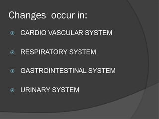
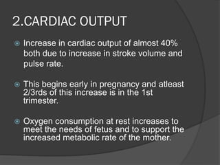
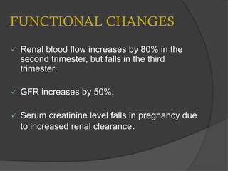
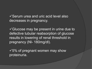


Changes occur in several body systems during pregnancy, including the cardiovascular, respiratory, gastrointestinal, and urinary systems. The cardiovascular system experiences an increased cardiac output of around 40% due to higher stroke volume and heart rate. Respiration increases through a 40% rise in tidal volume without changing breath frequency. The gastrointestinal system exhibits nausea, increased appetite, and constipation. The urinary system shows an enlarged kidney and dilated ureters along with higher glomerular filtration rate and decreased serum creatinine.


![H:\Physiological Changes In Pregnancy[2]](https://cdn.slidesharecdn.com/ss_thumbnails/hphysiologicalchangesinpregnancy2-100305140624-phpapp02-thumbnail.jpg?width=640&height=640&fit=bounds)


![CASE_PRESENTATION_ON_subdural_hematoma(SDH)[1 FINAL PPT]-1.pptx](https://cdn.slidesharecdn.com/ss_thumbnails/casepresentationonsubduralhematomasdh1finalppt-1-260129172522-d405d375-thumbnail.jpg?width=640&height=640&fit=bounds)


