SlideShare a Scribd company logo
Perthes Disease
Credit -
Dr. Qamrul Hoda
M.S(Ortho),D.Ortho.
P.M.C.H
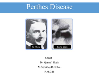
Perthes Bone Scan
Perthes Disease
• Also known as-
o Legg-Calve-Perthes disease.
o Calve Perthes disease.
o Avascular necrosis.
o Coxa plana
o Pseudocoxalgia
• It is an osteochondritis of the epiphysis of the femoral head.
• In this disease the femoral head become partly or wholly avascular and
deformed.
Epidemiology
• Legg-Calve-Perthes disease is the
most common hip disease that
afflicts school-age children.
• The disorder was described by three
independent authors in 1910.
• Arthur Legg
• Jacques Calve
• Georg Perthes
• They might have surprised to see(if
alive)how much and how little have
changed in 100 years
Legg Calve
What is Perthes Disease ?
• Perthes disease is a childhood disorder which affects
the head of the femur.
• It is a vascular problem affect 1 in 10000 child (4 in
10000 in India)
• In Perthes disease the blood supply to the growth
plate of the bone at the end of the femur becomes
inadequate.
• As a result the bone softens and breaks down due to
avascular necrosis.
Incidence
• Legg-Calve-Perthes disease generally occurs in children between 4 to 8
years of age.
• With males being affected about four to five times more frequently than
females.
• It is a unilateral in 90 percent of patients, while the other 10 percent of
patients have bilateral hip.
• Rates as low as 3.8 per 100,000 have been reported in Korea, and very
similar rates of 4.4 per 100,000 have been identified in Southern India.
Normal Blood Supply
Vascular Phenomena
• Up to the age of 4 months, the femoral head is supplied by -
1) Metaphyseal vessels which penetrate the growth disc
2) Lateral epiphyseal vessels running in the retinacula and
3) Scanty vessels in the ligamentum teres
• The metaphyseal blood supply -
1) Gradually declines until, by the age of 4 years.
2) It has virtually disappeared; by the age of 7 years.
3) However, the vessels in the ligamentum teres have developed.
• Between 4 and 7 years of age the femoral head may depend for its blood
supply and venous drainage almost entirely on the Lateral Epiphyseal
Vessels whose situation in the retinacula makes them susceptible to
stretching and pressure from an effusion.
Pathophysiology
• Another factor which needs to be taken into account Is that the vascular
sinusoids which nourish the marrow and bone cells ,unlike arterial
capillaries, have no adventitial layer and their patency is determined by the
volume and pressure of the surrounding marrow tissue ,which itself is
encased in unyielding bone.
• The system functions essentially as a closed compartment within which one
element can expand the presser(marrow presser)
Bones Vulnerable to Ischemia
• Sites which are vulnerable to ischemic necrosis are-
1. Femoral Head,
2. Femoral Condyles
3. Head of the Humerus
4. Capitulum
5. Proximal Part of Scaphoid
6. Talus.
Pathology
• Once a critical portion of blood flow to the femoral head has been
compromised.
• The femoral head will begin to take on a pathologic shape leading to -
• Flatening
• Fragmentation
• Collapse
Flatening Fragmentation Collapse Collapse
Etiology
• It is not clear why this blood vessel
problem occurs in the femoral head.
• Factors which has been implicated are -
1) Heredity
2) Trauma
3) Endocrine
4) Inflammatory
5) Nutritional
6) Altered circulatory hemodynamic
Bilateral -15% Cases
Unilateral - 80% Cases
Clinical Features
• It occur commonly in boys of age group from 5 to 10 years.
• The signs and symptoms of -Perthes disease can be rather slow and may not be
immediately recognized by parents or by treating doctor.
• They may simply perceive a slightly abnormal gait
• Since there is no complaint of pain, little attention on may be paid to it.
• If hip/knee/groin pain is present, it may worsen towards the end of a strenuous day
and is often relieved by rest.
• Gait pattern disturbed-limp
• Often present with a Trendelenburg gait, ( abductor lurch.)
• ROM diminished-- loss of abduction and internal rotation.
Clinical Feature
• Symptoms -
1) Hip, knee or groin pain,
exacerbated by hip/leg
movement, especially internal hip
rotation .
2) Hip pain radiating to knee.
3) There may limping and stiffness.
4) Reduced range of motion –
i. Abduction and
ii. Internal rotation
iii. Patient presents with a limp.
5) Pain is usually mild.
• Signs -
1) Wasting of the muscles in the
upper thigh.
2) Shortening of the leg
3) Stiffness of the Hip
4) Difficulty in Squatting
PAIN IN KNEE- SHOULD BE CONSIDERED AS HIP IN CHILDREN UNLESS OTHERWISE PROVED
Stages of Perthes Disease
• Necrosis: Initial period of ischemia/loss of blood supply to femoral head.
• Fragmentation: Re-absorption of bone with femoral head collapse.
• Re-ossification: New bone re-grows to reshape the femoral head.
• Remodeling: Femoral head reshapes itself into normal spherical shape.
Diagnosis
• X-rays of the hip
• Finding
1) Femoral head broken or damaged.
2) Collapse and sclerosis of epiphysis
of femoral head.
3) Hip joint space increase.
4) Flattened femoral head(Typical).
• Bone Scan
• Done for confirmation of Perthes
Disease , when X-ray picture is normal.
• MRI
• Use to asses the extent of damage.
Differential Diagnosis
• The irritable hip of early Perthes’ disease must be differentiated from
other causes of irritability -
1) Morquio’s disease
2) Cretinism
3) Multiple epiphyseal dysplasia
4) Sickle-cell disease
5) Gaucher’s disease (may resemble Perthes’ disease radiologically, especially if they
are bilateral; however, in bilateral Perthes’ disease the two sides are likely to be
at different stages. )
• In Old Perthes deformities’ in adults, in the 10 per cent of cases with
bilateral involvement, may resemble those of certain bone dysplasias,
especially multiple epiphyseal dysplasia.
What is the Treatment for Perthes Disease ?
• The overall goal of treatment is -
1) To reduce hip irritability and pain
2) Restore and maintain hip mobility
3) To prevent the femoral head from extruding or collapsing
4) To regain a spherical shape of the femoral head
• It can be achieved by-
• Conservative (or)
• Operative Method
Conservative Treatment
• Observation
• Physical
• Therapy/Traction to improve range of motion
• Splints and abduction. Cast in internal rotation
Ptrei Cast
Treatment Related to Age
• Children under 6 years
• No specific form of treatment has much influence on the outcome. Symptomatic
treatment, including activity modification.
• Bone age at or below 6 years
• Lateral pillar group A and B (or Catterall stage I and II) – symptomatic treatment.
• Lateral pillar group C (or Catterall stage III and IV)– abduction brace.
• Bone age over 6 years
• Lateral pillar group A and B (Catterall stage I and II) – abduction brace or
osteotomy.
• Lateral pillar group C (Catterall stage III and IV) – outcome probably unaffected by
treatment, but some would operate.
• Children 9 years and older
• Operative containment is the treatment of choice.
Surgery
• Femoral Varus Osteotomy
• Acetabular Osteototomy( with
internal fixation)
Operative Procedure
• Schematic diagram of surgical options
for containment of an extruded
avascular femoral epiphysis –
1) Proximal femoral varus osteotomy
2) Innominate Osteotomy
3) Shelf Procedure
Total Hip Replacement
• In older cases THR can be done.
Prognostic Feature
• The outlook for children with Perthes’ disease, as a group, is well
summarized by Herring (1994):
• A small % of patients have a very difficult course, with recurrent loss of motion,
pain, and an eventual poor outcome.
• However, most children have moderate problems in the active phase of the
disease and then improve steadily, leading to satisfactory outcome.’

More Related Content

What's hot

Ctev.ppt by krr
Ctev.ppt by krrCtev.ppt by krr
Ctev.ppt by krr
ramachandra reddy
 
Osteochondritis of different bones
Osteochondritis of different bonesOsteochondritis of different bones
Osteochondritis of different bonesPramod Govindraj
 
Tendoachilles rupture and its management
Tendoachilles rupture and its managementTendoachilles rupture and its management
Tendoachilles rupture and its management
Rohan Vakta
 
Congenital pseudoarthrosis tibia
Congenital pseudoarthrosis tibiaCongenital pseudoarthrosis tibia
Congenital pseudoarthrosis tibia
Saikrishna Katragadda
 
Heterotrophic ossification
Heterotrophic ossificationHeterotrophic ossification
Heterotrophic ossification
Annnoble
 
Neck of femur fractures
Neck  of femur fracturesNeck  of femur fractures
Neck of femur fractures
BADAL BALOCH
 
Carries sicca
Carries siccaCarries sicca
Carries sicca
PratikDhabalia
 
Cubitus varus by Dhrumil Patel
Cubitus varus by Dhrumil PatelCubitus varus by Dhrumil Patel
Cubitus varus by Dhrumil Pateldhrumil88
 
Legg calve perthes disease
Legg calve perthes disease Legg calve perthes disease
Legg calve perthes disease Ratan Khuman
 
Dislocation of hip joint
Dislocation of hip jointDislocation of hip joint
Dislocation of hip jointorthoprince
 
Leg Calve Perthes disease
Leg Calve Perthes disease Leg Calve Perthes disease
Leg Calve Perthes disease
darshanck89
 
Spondylolisthesis
Spondylolisthesis Spondylolisthesis
Spondylolisthesis
Dibyendunarayan Bid
 
Cubitus varus deformity
Cubitus varus deformityCubitus varus deformity
Cubitus varus deformity
ramachandra reddy
 
Genu valgus
Genu valgusGenu valgus
Genu valgus
Santosh Batajoo
 
Congenital talipes equinovarus (club foot/ctev) ppt by Dr Pratik
Congenital talipes equinovarus (club foot/ctev) ppt by Dr PratikCongenital talipes equinovarus (club foot/ctev) ppt by Dr Pratik
Congenital talipes equinovarus (club foot/ctev) ppt by Dr Pratik
Dr. Pratik Agarwal
 
Tuberculosis of hip
Tuberculosis of hipTuberculosis of hip
Tuberculosis of hip
Hardik Pawar
 
The pathology and management of blount’s disease
The pathology and management of blount’s diseaseThe pathology and management of blount’s disease
The pathology and management of blount’s disease
Asi-oqua Bassey
 
Avascular necrosis of femoral head
Avascular necrosis of femoral headAvascular necrosis of femoral head
Avascular necrosis of femoral head
sayf aldeen hussam
 
Pelvic fractures classification and management
Pelvic fractures classification and managementPelvic fractures classification and management
Pelvic fractures classification and management
Joydeep Mandal
 

What's hot (20)

Ctev.ppt by krr
Ctev.ppt by krrCtev.ppt by krr
Ctev.ppt by krr
 
Osteochondritis of different bones
Osteochondritis of different bonesOsteochondritis of different bones
Osteochondritis of different bones
 
Tendoachilles rupture and its management
Tendoachilles rupture and its managementTendoachilles rupture and its management
Tendoachilles rupture and its management
 
Congenital pseudoarthrosis tibia
Congenital pseudoarthrosis tibiaCongenital pseudoarthrosis tibia
Congenital pseudoarthrosis tibia
 
Heterotrophic ossification
Heterotrophic ossificationHeterotrophic ossification
Heterotrophic ossification
 
Neck of femur fractures
Neck  of femur fracturesNeck  of femur fractures
Neck of femur fractures
 
Carries sicca
Carries siccaCarries sicca
Carries sicca
 
Subtrochanteric fracture
Subtrochanteric fractureSubtrochanteric fracture
Subtrochanteric fracture
 
Cubitus varus by Dhrumil Patel
Cubitus varus by Dhrumil PatelCubitus varus by Dhrumil Patel
Cubitus varus by Dhrumil Patel
 
Legg calve perthes disease
Legg calve perthes disease Legg calve perthes disease
Legg calve perthes disease
 
Dislocation of hip joint
Dislocation of hip jointDislocation of hip joint
Dislocation of hip joint
 
Leg Calve Perthes disease
Leg Calve Perthes disease Leg Calve Perthes disease
Leg Calve Perthes disease
 
Spondylolisthesis
Spondylolisthesis Spondylolisthesis
Spondylolisthesis
 
Cubitus varus deformity
Cubitus varus deformityCubitus varus deformity
Cubitus varus deformity
 
Genu valgus
Genu valgusGenu valgus
Genu valgus
 
Congenital talipes equinovarus (club foot/ctev) ppt by Dr Pratik
Congenital talipes equinovarus (club foot/ctev) ppt by Dr PratikCongenital talipes equinovarus (club foot/ctev) ppt by Dr Pratik
Congenital talipes equinovarus (club foot/ctev) ppt by Dr Pratik
 
Tuberculosis of hip
Tuberculosis of hipTuberculosis of hip
Tuberculosis of hip
 
The pathology and management of blount’s disease
The pathology and management of blount’s diseaseThe pathology and management of blount’s disease
The pathology and management of blount’s disease
 
Avascular necrosis of femoral head
Avascular necrosis of femoral headAvascular necrosis of femoral head
Avascular necrosis of femoral head
 
Pelvic fractures classification and management
Pelvic fractures classification and managementPelvic fractures classification and management
Pelvic fractures classification and management
 

Similar to Perthes disease

PERTHES DISEASE
PERTHES  DISEASEPERTHES  DISEASE
PERTHES DISEASE
RITESHJAISWAL57
 
Perthes disease
Perthes diseasePerthes disease
Perthes disease
vashisth narayan
 
Skeletal dysplasias and dwarfism
Skeletal dysplasias and dwarfismSkeletal dysplasias and dwarfism
Skeletal dysplasias and dwarfism
Praveen Kumar Reddy Gorantla
 
Legg-Calve-Perthes Disease.pptx
Legg-Calve-Perthes Disease.pptxLegg-Calve-Perthes Disease.pptx
Legg-Calve-Perthes Disease.pptx
NellyPhiri5
 
Legg calve-perthes disease
Legg calve-perthes diseaseLegg calve-perthes disease
Legg calve-perthes disease
Dr Rajeev
 
Perthes disease
Perthes diseasePerthes disease
Perthes disease
Rifhan Kamaruddin
 
Irritable hip and perthe's disease
Irritable hip and perthe's diseaseIrritable hip and perthe's disease
Irritable hip and perthe's disease
Abdul Basit
 
Case discussion of perthes disease-Dr. Siddharth Deshwal PG Orthopaedics
Case discussion of perthes disease-Dr. Siddharth Deshwal PG OrthopaedicsCase discussion of perthes disease-Dr. Siddharth Deshwal PG Orthopaedics
Case discussion of perthes disease-Dr. Siddharth Deshwal PG Orthopaedics
SIDDHARTHDESHWAL3
 
Perthes disease
Perthes diseasePerthes disease
Perthes disease
Ahmed Azim
 
imaging in intrauterine skeletal dysplasia
imaging in intrauterine skeletal dysplasia imaging in intrauterine skeletal dysplasia
imaging in intrauterine skeletal dysplasia Shail Padmani
 
Perthes disease
Perthes diseasePerthes disease
Perthes disease
Fadzlina Zabri
 
Skeletal dysplasia final
Skeletal dysplasia finalSkeletal dysplasia final
Skeletal dysplasia final
Nihit Jain
 
Perthes disease ADOLESCENT COXA VARA
Perthes disease ADOLESCENT COXA VARAPerthes disease ADOLESCENT COXA VARA
Perthes disease ADOLESCENT COXA VARA
Dr ashwani panchal
 
skeletal dysplasia ppt
skeletal dysplasia pptskeletal dysplasia ppt
skeletal dysplasia ppt
Naba Kumar Barman
 
Perthes disease
Perthes diseasePerthes disease
Perthes disease
Ponnilavan Ponz
 
Perthes
PerthesPerthes
Congenital hip disease
Congenital hip disease Congenital hip disease
Congenital hip disease
Vivesh Singh
 
Craniosynostosis
Craniosynostosis Craniosynostosis
Craniosynostosis
Kode Sashanka
 
Referat pedi I-Anggrian-HYPOPLASIA OF FEMUR.pptx
Referat pedi I-Anggrian-HYPOPLASIA OF FEMUR.pptxReferat pedi I-Anggrian-HYPOPLASIA OF FEMUR.pptx
Referat pedi I-Anggrian-HYPOPLASIA OF FEMUR.pptx
Reza Hambali
 
developmental condition of musculoskelatal system
developmental condition of musculoskelatal systemdevelopmental condition of musculoskelatal system
developmental condition of musculoskelatal system
BipulBorthakur
 

Similar to Perthes disease (20)

PERTHES DISEASE
PERTHES  DISEASEPERTHES  DISEASE
PERTHES DISEASE
 
Perthes disease
Perthes diseasePerthes disease
Perthes disease
 
Skeletal dysplasias and dwarfism
Skeletal dysplasias and dwarfismSkeletal dysplasias and dwarfism
Skeletal dysplasias and dwarfism
 
Legg-Calve-Perthes Disease.pptx
Legg-Calve-Perthes Disease.pptxLegg-Calve-Perthes Disease.pptx
Legg-Calve-Perthes Disease.pptx
 
Legg calve-perthes disease
Legg calve-perthes diseaseLegg calve-perthes disease
Legg calve-perthes disease
 
Perthes disease
Perthes diseasePerthes disease
Perthes disease
 
Irritable hip and perthe's disease
Irritable hip and perthe's diseaseIrritable hip and perthe's disease
Irritable hip and perthe's disease
 
Case discussion of perthes disease-Dr. Siddharth Deshwal PG Orthopaedics
Case discussion of perthes disease-Dr. Siddharth Deshwal PG OrthopaedicsCase discussion of perthes disease-Dr. Siddharth Deshwal PG Orthopaedics
Case discussion of perthes disease-Dr. Siddharth Deshwal PG Orthopaedics
 
Perthes disease
Perthes diseasePerthes disease
Perthes disease
 
imaging in intrauterine skeletal dysplasia
imaging in intrauterine skeletal dysplasia imaging in intrauterine skeletal dysplasia
imaging in intrauterine skeletal dysplasia
 
Perthes disease
Perthes diseasePerthes disease
Perthes disease
 
Skeletal dysplasia final
Skeletal dysplasia finalSkeletal dysplasia final
Skeletal dysplasia final
 
Perthes disease ADOLESCENT COXA VARA
Perthes disease ADOLESCENT COXA VARAPerthes disease ADOLESCENT COXA VARA
Perthes disease ADOLESCENT COXA VARA
 
skeletal dysplasia ppt
skeletal dysplasia pptskeletal dysplasia ppt
skeletal dysplasia ppt
 
Perthes disease
Perthes diseasePerthes disease
Perthes disease
 
Perthes
PerthesPerthes
Perthes
 
Congenital hip disease
Congenital hip disease Congenital hip disease
Congenital hip disease
 
Craniosynostosis
Craniosynostosis Craniosynostosis
Craniosynostosis
 
Referat pedi I-Anggrian-HYPOPLASIA OF FEMUR.pptx
Referat pedi I-Anggrian-HYPOPLASIA OF FEMUR.pptxReferat pedi I-Anggrian-HYPOPLASIA OF FEMUR.pptx
Referat pedi I-Anggrian-HYPOPLASIA OF FEMUR.pptx
 
developmental condition of musculoskelatal system
developmental condition of musculoskelatal systemdevelopmental condition of musculoskelatal system
developmental condition of musculoskelatal system
 

Recently uploaded

Maxilla, Mandible & Hyoid Bone & Clinical Correlations by Dr. RIG.pptx
Maxilla, Mandible & Hyoid Bone & Clinical Correlations by Dr. RIG.pptxMaxilla, Mandible & Hyoid Bone & Clinical Correlations by Dr. RIG.pptx
Maxilla, Mandible & Hyoid Bone & Clinical Correlations by Dr. RIG.pptx
Dr. Rabia Inam Gandapore
 
KDIGO 2024 guidelines for diabetologists
KDIGO 2024 guidelines for diabetologistsKDIGO 2024 guidelines for diabetologists
KDIGO 2024 guidelines for diabetologists
د.محمود نجيب
 
How to Give Better Lectures: Some Tips for Doctors
How to Give Better Lectures: Some Tips for DoctorsHow to Give Better Lectures: Some Tips for Doctors
How to Give Better Lectures: Some Tips for Doctors
LanceCatedral
 
Ocular injury ppt Upendra pal optometrist upums saifai etawah
Ocular injury  ppt  Upendra pal  optometrist upums saifai etawahOcular injury  ppt  Upendra pal  optometrist upums saifai etawah
Ocular injury ppt Upendra pal optometrist upums saifai etawah
pal078100
 
24 Upakrama.pptx class ppt useful in all
24 Upakrama.pptx class ppt useful in all24 Upakrama.pptx class ppt useful in all
24 Upakrama.pptx class ppt useful in all
DrSathishMS1
 
Hemodialysis: Chapter 3, Dialysis Water Unit - Dr.Gawad
Hemodialysis: Chapter 3, Dialysis Water Unit - Dr.GawadHemodialysis: Chapter 3, Dialysis Water Unit - Dr.Gawad
Hemodialysis: Chapter 3, Dialysis Water Unit - Dr.Gawad
NephroTube - Dr.Gawad
 
ARTIFICIAL INTELLIGENCE IN HEALTHCARE.pdf
ARTIFICIAL INTELLIGENCE IN  HEALTHCARE.pdfARTIFICIAL INTELLIGENCE IN  HEALTHCARE.pdf
ARTIFICIAL INTELLIGENCE IN HEALTHCARE.pdf
Anujkumaranit
 
Surat @ℂall @Girls ꧁❤8527049040❤꧂@ℂall @Girls Service Vip Top Model Safe
Surat @ℂall @Girls ꧁❤8527049040❤꧂@ℂall @Girls Service Vip Top Model SafeSurat @ℂall @Girls ꧁❤8527049040❤꧂@ℂall @Girls Service Vip Top Model Safe
Surat @ℂall @Girls ꧁❤8527049040❤꧂@ℂall @Girls Service Vip Top Model Safe
Savita Shen $i11
 
POST OPERATIVE OLIGURIA and its management
POST OPERATIVE OLIGURIA and its managementPOST OPERATIVE OLIGURIA and its management
POST OPERATIVE OLIGURIA and its management
touseefaziz1
 
Cervical & Brachial Plexus By Dr. RIG.pptx
Cervical & Brachial Plexus By Dr. RIG.pptxCervical & Brachial Plexus By Dr. RIG.pptx
Cervical & Brachial Plexus By Dr. RIG.pptx
Dr. Rabia Inam Gandapore
 
micro teaching on communication m.sc nursing.pdf
micro teaching on communication m.sc nursing.pdfmicro teaching on communication m.sc nursing.pdf
micro teaching on communication m.sc nursing.pdf
Anurag Sharma
 
Superficial & Deep Fascia of the NECK.pptx
Superficial & Deep Fascia of the NECK.pptxSuperficial & Deep Fascia of the NECK.pptx
Superficial & Deep Fascia of the NECK.pptx
Dr. Rabia Inam Gandapore
 
BENIGN PROSTATIC HYPERPLASIA.BPH. BPHpdf
BENIGN PROSTATIC HYPERPLASIA.BPH. BPHpdfBENIGN PROSTATIC HYPERPLASIA.BPH. BPHpdf
BENIGN PROSTATIC HYPERPLASIA.BPH. BPHpdf
DR SETH JOTHAM
 
heat stroke and heat exhaustion in children
heat stroke and heat exhaustion in childrenheat stroke and heat exhaustion in children
heat stroke and heat exhaustion in children
SumeraAhmad5
 
Factory Supply Best Quality Pmk Oil CAS 28578–16–7 PMK Powder in Stock
Factory Supply Best Quality Pmk Oil CAS 28578–16–7 PMK Powder in StockFactory Supply Best Quality Pmk Oil CAS 28578–16–7 PMK Powder in Stock
Factory Supply Best Quality Pmk Oil CAS 28578–16–7 PMK Powder in Stock
rebeccabio
 
Non-respiratory Functions of the Lungs.pdf
Non-respiratory Functions of the Lungs.pdfNon-respiratory Functions of the Lungs.pdf
Non-respiratory Functions of the Lungs.pdf
MedicoseAcademics
 
Prix Galien International 2024 Forum Program
Prix Galien International 2024 Forum ProgramPrix Galien International 2024 Forum Program
Prix Galien International 2024 Forum Program
Levi Shapiro
 
ARTHROLOGY PPT NCISM SYLLABUS AYURVEDA STUDENTS
ARTHROLOGY PPT NCISM SYLLABUS AYURVEDA STUDENTSARTHROLOGY PPT NCISM SYLLABUS AYURVEDA STUDENTS
ARTHROLOGY PPT NCISM SYLLABUS AYURVEDA STUDENTS
Dr. Vinay Pareek
 
Charaka Samhita Sutra Sthana 9 Chapter khuddakachatuspadadhyaya
Charaka Samhita Sutra Sthana 9 Chapter khuddakachatuspadadhyayaCharaka Samhita Sutra Sthana 9 Chapter khuddakachatuspadadhyaya
Charaka Samhita Sutra Sthana 9 Chapter khuddakachatuspadadhyaya
Dr KHALID B.M
 
Antiulcer drugs Advance Pharmacology .pptx
Antiulcer drugs Advance Pharmacology .pptxAntiulcer drugs Advance Pharmacology .pptx
Antiulcer drugs Advance Pharmacology .pptx
Rohit chaurpagar
 

Recently uploaded (20)

Maxilla, Mandible & Hyoid Bone & Clinical Correlations by Dr. RIG.pptx
Maxilla, Mandible & Hyoid Bone & Clinical Correlations by Dr. RIG.pptxMaxilla, Mandible & Hyoid Bone & Clinical Correlations by Dr. RIG.pptx
Maxilla, Mandible & Hyoid Bone & Clinical Correlations by Dr. RIG.pptx
 
KDIGO 2024 guidelines for diabetologists
KDIGO 2024 guidelines for diabetologistsKDIGO 2024 guidelines for diabetologists
KDIGO 2024 guidelines for diabetologists
 
How to Give Better Lectures: Some Tips for Doctors
How to Give Better Lectures: Some Tips for DoctorsHow to Give Better Lectures: Some Tips for Doctors
How to Give Better Lectures: Some Tips for Doctors
 
Ocular injury ppt Upendra pal optometrist upums saifai etawah
Ocular injury  ppt  Upendra pal  optometrist upums saifai etawahOcular injury  ppt  Upendra pal  optometrist upums saifai etawah
Ocular injury ppt Upendra pal optometrist upums saifai etawah
 
24 Upakrama.pptx class ppt useful in all
24 Upakrama.pptx class ppt useful in all24 Upakrama.pptx class ppt useful in all
24 Upakrama.pptx class ppt useful in all
 
Hemodialysis: Chapter 3, Dialysis Water Unit - Dr.Gawad
Hemodialysis: Chapter 3, Dialysis Water Unit - Dr.GawadHemodialysis: Chapter 3, Dialysis Water Unit - Dr.Gawad
Hemodialysis: Chapter 3, Dialysis Water Unit - Dr.Gawad
 
ARTIFICIAL INTELLIGENCE IN HEALTHCARE.pdf
ARTIFICIAL INTELLIGENCE IN  HEALTHCARE.pdfARTIFICIAL INTELLIGENCE IN  HEALTHCARE.pdf
ARTIFICIAL INTELLIGENCE IN HEALTHCARE.pdf
 
Surat @ℂall @Girls ꧁❤8527049040❤꧂@ℂall @Girls Service Vip Top Model Safe
Surat @ℂall @Girls ꧁❤8527049040❤꧂@ℂall @Girls Service Vip Top Model SafeSurat @ℂall @Girls ꧁❤8527049040❤꧂@ℂall @Girls Service Vip Top Model Safe
Surat @ℂall @Girls ꧁❤8527049040❤꧂@ℂall @Girls Service Vip Top Model Safe
 
POST OPERATIVE OLIGURIA and its management
POST OPERATIVE OLIGURIA and its managementPOST OPERATIVE OLIGURIA and its management
POST OPERATIVE OLIGURIA and its management
 
Cervical & Brachial Plexus By Dr. RIG.pptx
Cervical & Brachial Plexus By Dr. RIG.pptxCervical & Brachial Plexus By Dr. RIG.pptx
Cervical & Brachial Plexus By Dr. RIG.pptx
 
micro teaching on communication m.sc nursing.pdf
micro teaching on communication m.sc nursing.pdfmicro teaching on communication m.sc nursing.pdf
micro teaching on communication m.sc nursing.pdf
 
Superficial & Deep Fascia of the NECK.pptx
Superficial & Deep Fascia of the NECK.pptxSuperficial & Deep Fascia of the NECK.pptx
Superficial & Deep Fascia of the NECK.pptx
 
BENIGN PROSTATIC HYPERPLASIA.BPH. BPHpdf
BENIGN PROSTATIC HYPERPLASIA.BPH. BPHpdfBENIGN PROSTATIC HYPERPLASIA.BPH. BPHpdf
BENIGN PROSTATIC HYPERPLASIA.BPH. BPHpdf
 
heat stroke and heat exhaustion in children
heat stroke and heat exhaustion in childrenheat stroke and heat exhaustion in children
heat stroke and heat exhaustion in children
 
Factory Supply Best Quality Pmk Oil CAS 28578–16–7 PMK Powder in Stock
Factory Supply Best Quality Pmk Oil CAS 28578–16–7 PMK Powder in StockFactory Supply Best Quality Pmk Oil CAS 28578–16–7 PMK Powder in Stock
Factory Supply Best Quality Pmk Oil CAS 28578–16–7 PMK Powder in Stock
 
Non-respiratory Functions of the Lungs.pdf
Non-respiratory Functions of the Lungs.pdfNon-respiratory Functions of the Lungs.pdf
Non-respiratory Functions of the Lungs.pdf
 
Prix Galien International 2024 Forum Program
Prix Galien International 2024 Forum ProgramPrix Galien International 2024 Forum Program
Prix Galien International 2024 Forum Program
 
ARTHROLOGY PPT NCISM SYLLABUS AYURVEDA STUDENTS
ARTHROLOGY PPT NCISM SYLLABUS AYURVEDA STUDENTSARTHROLOGY PPT NCISM SYLLABUS AYURVEDA STUDENTS
ARTHROLOGY PPT NCISM SYLLABUS AYURVEDA STUDENTS
 
Charaka Samhita Sutra Sthana 9 Chapter khuddakachatuspadadhyaya
Charaka Samhita Sutra Sthana 9 Chapter khuddakachatuspadadhyayaCharaka Samhita Sutra Sthana 9 Chapter khuddakachatuspadadhyaya
Charaka Samhita Sutra Sthana 9 Chapter khuddakachatuspadadhyaya
 
Antiulcer drugs Advance Pharmacology .pptx
Antiulcer drugs Advance Pharmacology .pptxAntiulcer drugs Advance Pharmacology .pptx
Antiulcer drugs Advance Pharmacology .pptx
 

Perthes disease

  • 1. Perthes Disease Credit - Dr. Qamrul Hoda M.S(Ortho),D.Ortho. P.M.C.H Perthes Bone Scan
  • 2. Perthes Disease • Also known as- o Legg-Calve-Perthes disease. o Calve Perthes disease. o Avascular necrosis. o Coxa plana o Pseudocoxalgia • It is an osteochondritis of the epiphysis of the femoral head. • In this disease the femoral head become partly or wholly avascular and deformed.
  • 3. Epidemiology • Legg-Calve-Perthes disease is the most common hip disease that afflicts school-age children. • The disorder was described by three independent authors in 1910. • Arthur Legg • Jacques Calve • Georg Perthes • They might have surprised to see(if alive)how much and how little have changed in 100 years Legg Calve
  • 4. What is Perthes Disease ? • Perthes disease is a childhood disorder which affects the head of the femur. • It is a vascular problem affect 1 in 10000 child (4 in 10000 in India) • In Perthes disease the blood supply to the growth plate of the bone at the end of the femur becomes inadequate. • As a result the bone softens and breaks down due to avascular necrosis.
  • 5. Incidence • Legg-Calve-Perthes disease generally occurs in children between 4 to 8 years of age. • With males being affected about four to five times more frequently than females. • It is a unilateral in 90 percent of patients, while the other 10 percent of patients have bilateral hip. • Rates as low as 3.8 per 100,000 have been reported in Korea, and very similar rates of 4.4 per 100,000 have been identified in Southern India.
  • 7. Vascular Phenomena • Up to the age of 4 months, the femoral head is supplied by - 1) Metaphyseal vessels which penetrate the growth disc 2) Lateral epiphyseal vessels running in the retinacula and 3) Scanty vessels in the ligamentum teres • The metaphyseal blood supply - 1) Gradually declines until, by the age of 4 years. 2) It has virtually disappeared; by the age of 7 years. 3) However, the vessels in the ligamentum teres have developed. • Between 4 and 7 years of age the femoral head may depend for its blood supply and venous drainage almost entirely on the Lateral Epiphyseal Vessels whose situation in the retinacula makes them susceptible to stretching and pressure from an effusion.
  • 8. Pathophysiology • Another factor which needs to be taken into account Is that the vascular sinusoids which nourish the marrow and bone cells ,unlike arterial capillaries, have no adventitial layer and their patency is determined by the volume and pressure of the surrounding marrow tissue ,which itself is encased in unyielding bone. • The system functions essentially as a closed compartment within which one element can expand the presser(marrow presser)
  • 9. Bones Vulnerable to Ischemia • Sites which are vulnerable to ischemic necrosis are- 1. Femoral Head, 2. Femoral Condyles 3. Head of the Humerus 4. Capitulum 5. Proximal Part of Scaphoid 6. Talus.
  • 10. Pathology • Once a critical portion of blood flow to the femoral head has been compromised. • The femoral head will begin to take on a pathologic shape leading to - • Flatening • Fragmentation • Collapse Flatening Fragmentation Collapse Collapse
  • 11. Etiology • It is not clear why this blood vessel problem occurs in the femoral head. • Factors which has been implicated are - 1) Heredity 2) Trauma 3) Endocrine 4) Inflammatory 5) Nutritional 6) Altered circulatory hemodynamic Bilateral -15% Cases Unilateral - 80% Cases
  • 12. Clinical Features • It occur commonly in boys of age group from 5 to 10 years. • The signs and symptoms of -Perthes disease can be rather slow and may not be immediately recognized by parents or by treating doctor. • They may simply perceive a slightly abnormal gait • Since there is no complaint of pain, little attention on may be paid to it. • If hip/knee/groin pain is present, it may worsen towards the end of a strenuous day and is often relieved by rest. • Gait pattern disturbed-limp • Often present with a Trendelenburg gait, ( abductor lurch.) • ROM diminished-- loss of abduction and internal rotation.
  • 13. Clinical Feature • Symptoms - 1) Hip, knee or groin pain, exacerbated by hip/leg movement, especially internal hip rotation . 2) Hip pain radiating to knee. 3) There may limping and stiffness. 4) Reduced range of motion – i. Abduction and ii. Internal rotation iii. Patient presents with a limp. 5) Pain is usually mild. • Signs - 1) Wasting of the muscles in the upper thigh. 2) Shortening of the leg 3) Stiffness of the Hip 4) Difficulty in Squatting PAIN IN KNEE- SHOULD BE CONSIDERED AS HIP IN CHILDREN UNLESS OTHERWISE PROVED
  • 14. Stages of Perthes Disease • Necrosis: Initial period of ischemia/loss of blood supply to femoral head. • Fragmentation: Re-absorption of bone with femoral head collapse. • Re-ossification: New bone re-grows to reshape the femoral head. • Remodeling: Femoral head reshapes itself into normal spherical shape.
  • 15. Diagnosis • X-rays of the hip • Finding 1) Femoral head broken or damaged. 2) Collapse and sclerosis of epiphysis of femoral head. 3) Hip joint space increase. 4) Flattened femoral head(Typical). • Bone Scan • Done for confirmation of Perthes Disease , when X-ray picture is normal. • MRI • Use to asses the extent of damage.
  • 16. Differential Diagnosis • The irritable hip of early Perthes’ disease must be differentiated from other causes of irritability - 1) Morquio’s disease 2) Cretinism 3) Multiple epiphyseal dysplasia 4) Sickle-cell disease 5) Gaucher’s disease (may resemble Perthes’ disease radiologically, especially if they are bilateral; however, in bilateral Perthes’ disease the two sides are likely to be at different stages. ) • In Old Perthes deformities’ in adults, in the 10 per cent of cases with bilateral involvement, may resemble those of certain bone dysplasias, especially multiple epiphyseal dysplasia.
  • 17. What is the Treatment for Perthes Disease ? • The overall goal of treatment is - 1) To reduce hip irritability and pain 2) Restore and maintain hip mobility 3) To prevent the femoral head from extruding or collapsing 4) To regain a spherical shape of the femoral head • It can be achieved by- • Conservative (or) • Operative Method
  • 18. Conservative Treatment • Observation • Physical • Therapy/Traction to improve range of motion • Splints and abduction. Cast in internal rotation Ptrei Cast
  • 19. Treatment Related to Age • Children under 6 years • No specific form of treatment has much influence on the outcome. Symptomatic treatment, including activity modification. • Bone age at or below 6 years • Lateral pillar group A and B (or Catterall stage I and II) – symptomatic treatment. • Lateral pillar group C (or Catterall stage III and IV)– abduction brace. • Bone age over 6 years • Lateral pillar group A and B (Catterall stage I and II) – abduction brace or osteotomy. • Lateral pillar group C (Catterall stage III and IV) – outcome probably unaffected by treatment, but some would operate. • Children 9 years and older • Operative containment is the treatment of choice.
  • 20. Surgery • Femoral Varus Osteotomy • Acetabular Osteototomy( with internal fixation)
  • 21. Operative Procedure • Schematic diagram of surgical options for containment of an extruded avascular femoral epiphysis – 1) Proximal femoral varus osteotomy 2) Innominate Osteotomy 3) Shelf Procedure
  • 22. Total Hip Replacement • In older cases THR can be done.
  • 23. Prognostic Feature • The outlook for children with Perthes’ disease, as a group, is well summarized by Herring (1994): • A small % of patients have a very difficult course, with recurrent loss of motion, pain, and an eventual poor outcome. • However, most children have moderate problems in the active phase of the disease and then improve steadily, leading to satisfactory outcome.’