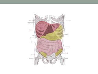
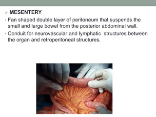
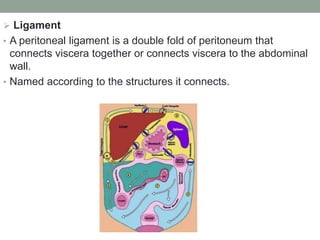
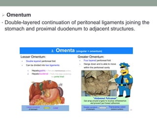
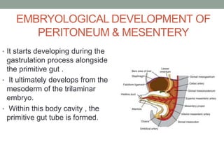
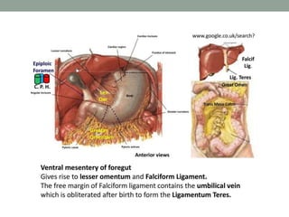
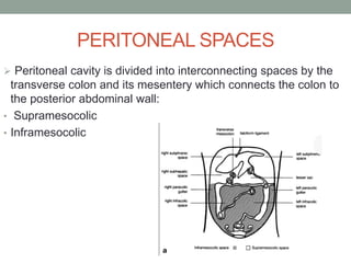
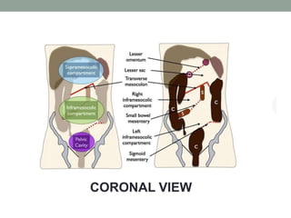
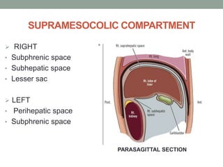
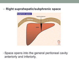
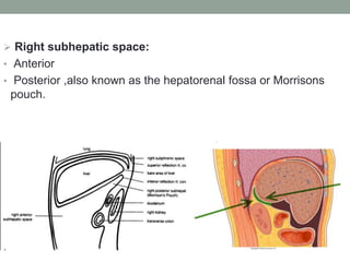
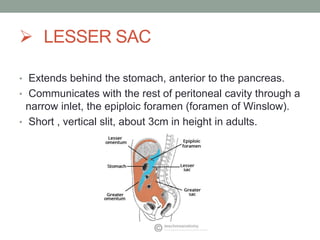
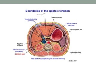
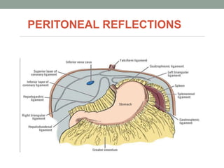
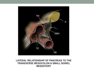
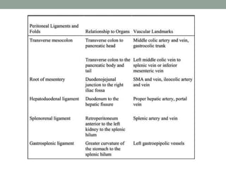
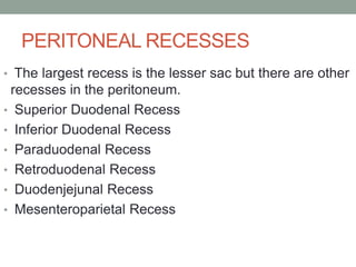
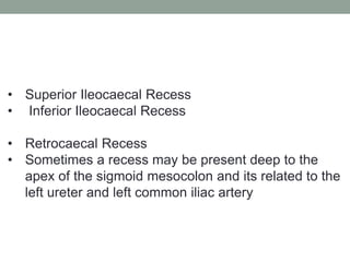
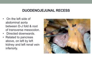
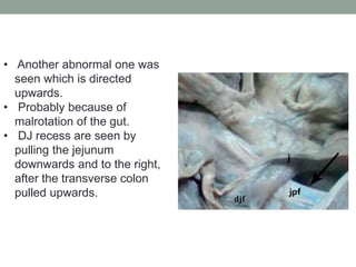
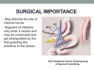
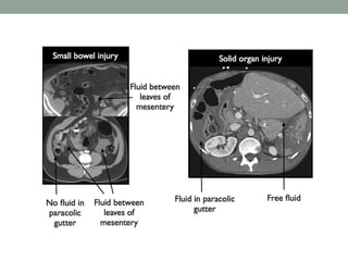
SlideShare a Scribd company logo
SURGICAL ANATOMY OF
PERITONEUM,MESENTERY AND
RETROPERITONEUM
DR.MEENAL
DEPT OF GENERAL SURGERY
INTRODUCTION
PERITONEUM
• Serous membrane lining the abdominal cavity.
• Comprises of 2 layers i.e. parietal peritoneum and
visceral peritoneum.
• Surface area is 1.0 to 1.7 mt sq.
• In males, the peritoneal cavity is sealed.
• In females, it is open to the exterior through the ostia of
the fallopian tubes.
 MESENTERY
• Fan shaped double layer of peritoneum that suspends the
small and large bowel from the posterior abdominal wall.
• Conduit for neurovascular and lymphatic structures between
the organ and retroperitoneal structures.
BLOOD SUPPLY
INNERVATION
• The superior mesenteric plexus (a continuation of the
celiac plexus) accompanies the superior mesenteric
artery into the mesentery.
• The superior mesenteric plexus then divides into many
secondary plexuses which
contain parasympathetic and sympathetic
innervation to the mesentery associated with a particular
organ, the organs themselves and their related blood
vessels.
LYMPHATICS
• The mesentery contains both lymph nodes and lymphatic
vessels. There are several groups of lymph nodes found
within the mesentery:
• Inferior mesenteric lymph nodes – receives lymph from
the hindgut organs, and drains into the superior
mesenteric lymph nodes.
• Superior mesenteric lymph nodes – receives lymph
from the midgut organs (and from the inferior mesenteric
nodes), and drains into the pre-aortic lymph nodes.
 Ligament
• A peritoneal ligament is a double fold of peritoneum that
connects viscera together or connects viscera to the abdominal
wall.
• Named according to the structures it connects.
 Omentum
• Double-layered continuation of peritoneal ligaments joining the
stomach and proximal duodenum to adjacent structures.
EMBRYOLOGICAL DEVELOPMENT OF
PERITONEUM & MESENTERY
• It starts developing during the
gastrulation process alongside
the primitive gut .
• It ultimately develops from the
mesoderm of the trilaminar
embryo.
• Within this body cavity , the
primitive gut tube is formed.
• The layer covering the gut is later known as the visceral
peritoneum.
• One covering the wall is known as the parietal
peritoneum.
• Primitive foregut separates upper part of the body cavity
into right & left body cavities by the virtue of its dorsal and
ventral mesentries.
• Rotation of the foregut
occurs separately from
the midgut and the
hindgut.
• Gives rise to the liver in
the ventral mesentery
and the spleen within the
dorsal mesentery.
• Rotation occurs 90
degrees to the stomach
such that ventral
mesentery comes to lie
to the right and dorsal
mesentery to the left.
LATERAL VIEW OF
THE PRIMITIVE GUT
TUBE
• In the region of the stomach it forms- dorsal
mesogastrium or greater omentum.
Region of duodenum -dorsal mesoduodenum
Region of colon-mesocolon
Dorsal mesentery of jejunal and ileal loops-mesentery
proper.
• Ventral mesentery, which exists only in the region of the
terminal part of the esophagus, the stomach, and the upper part
of the duodenum is derived from the septum transversum.
• Growth of the liver into the mesenchyme of the septum
transversum divides the ventral mesentery into:
i. the lesser omentum,
ii. the falciform ligament
PERITONEAL SPACES
 Peritoneal cavity is divided into interconnecting spaces by the
transverse colon and its mesentery which connects the colon to
the posterior abdominal wall:
• Supramesocolic
• Inframesocolic
CORONAL VIEW
SUPRAMESOCOLIC COMPARTMENT
 RIGHT
• Subphrenic space
• Subhepatic space
• Lesser sac
 LEFT
• Perihepatic space
• Subphrenic space
•
PARASAGITTAL SECTION
 Right suprahepatic/subphrenic space
• Space opens into the general peritoneal cavity
anteriorly and inferiorly.
 Right subhepatic space:
• Anterior
• Posterior ,also known as the hepatorenal fossa or Morrisons
pouch.
 LESSER SAC
• Extends behind the stomach, anterior to the pancreas.
• Communicates with the rest of peritoneal cavity through a
narrow inlet, the epiploic foramen (foramen of Winslow).
• Short , vertical slit, about 3cm in height in adults.
THE LESSER OMENTUM REMOVED TO SHOW
THE EPIPLOIC FORAMEN OF WINSLOW.
CLINICAL RELEVANCE
A posterior gastric ulcer may
perforate into the lesser sac.
• Gastric content and pus
accumulates in the lesser sac,
forming an abcess.
• Through foramen of Winslow this
passes into the peritoneal cavity,
leading to generalized peritonitis.
 Usually a loop of small
intestine can pass through the
epiploic foramen and become
twisted and strangulated in
the lesser sac.
• Pre-disposing factors include:
i. Large epiploic foramen
ii. Reduntant or mobile
mesentery
iii. Elongated right liver
iv. Defect in the lesser
omentum.
SURGICALAPPROACH OF THE
LESSER SAC
 During minimally invasive upper GI surgery, it can
be approached in 3 ways:
i. By opening the hepatogastric ligament (pars
flaccida of the lesser omentum)
ii. Through the gastrocolic and gastrosplenic
ligament, and;
iii. By opening the transverse mesocolon at the level
of the pancreas.
• First two approaches are used in upper GI
surgery, being the approach through the
gastrosplenic ligament used to create any type of
fundoplication during surgery for GERD.
• The last option is used in colorectal surgery for
mobilization of the splenic flexure.
INTRA-ABDOMINAL STRUCTURES OF
LESSER SAC
 LEFT SUPRAMESOCOLIC SPACE
• Four arbitrary communicating subspaces
i. Left anterior perihepatic space
ii. Left posterior perihepatic space
iii. Left anterior subphrenic space
iv. Posterior subphrenic(perisplenic) space
PARASAGITTAL SECTION
THROUGH THE TRUNK
SAGITTAL SECTION THROUGH THE LEFT
LOBE OF THE LIVER AND LESSER SAC
CLINICAL RELEVANCE:SUBPHRENIC
ABCESSES
• Refer to accumulation of pus in the right or left
subphrenic space.
• More common on the right side due to the increased
frequency of appendicitis and ruptured duodenal ulcers
(pus from the appendix can track up to the subphrenic
space via the right paracolic gutter).
RELATIONS OF AN ABCESS IN THE ANTERIOR
AND POSTERIOR PORTION OF RIGHT
SUBPHRENIC SPACE
• Phrenicocolic ligament forms a partial barrier to the spread of
fluid from left paracolic gutter into the left subphrenic space
hence left subphrenic collections are less common than right
sided.
 SURGICAL CONSIDERATIONS
• Drain a right anterior subphrenic abcess using a small right
subcostal incision to establish an extraperitoneal route.
• Posteriorly, the approach must be by an incision at the level of
the spinous process of L1. This avoids the pleura.
• The pleura and the 12th rib are related at the vertebral
spine thus care should be taken to avoid traversing the
bed of the 12th rib.
INFRAMESOCOLIC SPACE
 Is divided into 2 unequal spaces by the root of the small
bowel mesentery running from the DJ flexure in the left
upper quadrant to the ileocaecal valve in the right lower
quadrant:
• The smaller right infracolic space
• The larger left infracolic space
PARACOLIC SPACES
• Located lateral to the peritoneal reflections of the left and
right sides of the colon.
• Right is larger than the left and communicates freely with
the right subphrenic space.
• The connection between the left paracolic gutter an
subphrenic space is partially limited by the phrenicocolic
ligament.
• Both the right and left paracolic gutters communicate with
the pelvic space.
PELVIC SPACES
• Inferiorly the peritoneum is reflected over the fundus of
the bladder , the anterior & posterior surface of the uterus
in females, and on to the superior part of the rectum.
• Divided into the right & left paravesical spaces by the
urinary bladder.
PERITONEAL REFLECTIONS
GREATER OMENTUM
• Can be divided into 2 parts from a surgical point of view:
i. An upper part , the gastrocolic ligament
ii. Lower part the true greater omentum or fat apron
• Formed by 4 peritoneal layers, 2 anterior and 2 posterior-
fuse together during development & growth.
BLOOD SUPPLY OF GREATER
OMENTUM
SURGICAL APPLICATIONS
• The two-layer gastrocolic ligament, is the best pathway
for lesser sac exploration, especially closer to the greater
curvature.
• All the 6 layers of gastrocolic ligament are fused and the
middle colic artery as well as the other omental vessels
may be the cause of complications.
 For mobilization of the right side of the omentum:
• transect the gastrocolic ligament inferior to the right
gastroepiploic artery.
• ligate the anterior epiploics or the right gastroepiploic
artery distal to the origin of the right epiploic vessels.
 For mobilization of left side:
• ligate only the gastric branches of the left gastroepiploic
arches.
• do not ligate the left epiploic arteries
 LESSER OMENTUM
• Posterior component of the ventral mesentery.
CONTENTS
SURGICAL CONSIDERATIONS
 Falciform ligament
• Cut should be made between proximal and distal ligations
to avoid bleeding from a patent round ligament.
 Hepatogastric ligament
• Routinely divided during mobilization of the liver or
stomach.
• Care must be taken to preserve a dominant left hepatic
artery that travels through the ligament if a right hepatic
lobectomy, or if the patient has had prior interruption of
the right hepatic artery.
 GASTROSPLENIC LIGAMENT
• Extends from the posterolateral wall of the fundus and greater
curvature of the stomach to the splenic hilum.
• Continuous with the gastrocolic ligament.
• Short gastric arteries, veins and lymph nodes in the upper part.
• Left GE artery and vein, terminal branches of the splenic artery
and lymph nodes in the lower part.
• It’s a frequent route for sub-peritoneal spread of pancreatitis
related fluid.
 TRANSVERSE MESOCOLON
• Suspends the transverse colon in the abdominal
cavity.
• Diseases of the pancreas, such as pancreatic
cancer & acute pancreatitis, can spread via the
subperitoneal route along the transverse
mesocolon anteriorly to the colon, the gastrocolic
ligament, and the omentum.
• Colonic involvement is caused by direct spread
of pancreatic enzymes through the
extraperitoneal fat planes along the mesocolon,
causing pericolitis.
 SMALL BOWEL MESENTERY
• Voluminous, fat laden peritoneal reflection.
• Suspends the ileal & jejunal loops in the abdomen.
• Short base which is continuous with the right mesocolon.
• Root of the mesentery is approximately 15cm.
• It’s root is a bare area continuous with the anterior pararenal
space of the retroperitoneum.
• The root also contains 2 major blood vessels, the superior
mesenteric artery and the superior mesenteric vein.
• One of the most likely structure to be involved in metastatic
disease.
• Inflammation and tumour may involve the mesentery directly
e.g,from the pancreatic body or jejunum or by way of
neurovascular plexus or lymphatic channels that run within it.
• Rarely rotational and fusion anomalies of the mesentery may
lead to volvulus or internal hernia.
• Fat necrosis due to pancreatitis and occasionally pancreatic
cancer spread subperitoneally into the small bowel mesentery.
SIGMOID MESOCOLON
• Most common pathologic process involving this structure is
acute diverticulitis.
LATERAL RELATIONSHIP OF PANCREAS TO THE
TRANSVERSE MESOCOLON & SMALL BOWEL
MESENTERY
PERITONEAL RECESSES
• The largest recess is the lesser sac but there are other
recesses in the peritoneum.
• Superior Duodenal Recess
• Inferior Duodenal Recess
• Paraduodenal Recess
• Retroduodenal Recess
• Duodenjejunal Recess
• Mesenteroparietal Recess
• Superior Ileocaecal Recess
• Inferior Ileocaecal Recess
• Retrocaecal Recess
• Sometimes a recess may be present deep to the
apex of the sigmoid mesocolon and its related to the
left ureter and left common iliac artery
SUPERIOR DUODENAL RECESS
• Behind the D-J fold, left
of 4th part of duodenum.
• Directed downwards.
• Depth-1-3.5cm
• Width-0.5-2.0cm.
INFERIOR DUODENAL RECESS
• Fossa duodeno-jejunalis
of Treitz.
• Situated on the left of
the ascending(4th) part of
the duodenum.
• Directed upwards.
• Max depth-5.0cm.
• Max width-2.9cm
• Common opening for
both Superior and
inferior duodenal
recess.
• Due to vertical growth
of the two folds, and
these folds grow
extensively towards
each other and fused
laterally.
PARADUODENAL RECESS
• Recess venosus, fossa of
Landzert.
• Originates as congenital
peritoneal anomaly owing to
failure of mesentric fusion
with the parietal peritoneum.
• Situated on the left side of
the 4th part of the duodenum.
• Directed downwards and to
the right.
• Normally related to the IMV.
• Anomalous, related to
ascending branch of left colic
artery.
DUODENOJEJUNAL RECESS
• On the left side of
abdominal aorta
between D-J fold & root
of transverse mesocolon.
• Directed downwards.
• Related to pancreas
above, on left by left
kidney and left renal vein
inferiorly.
• Another abnormal one was
seen which is directed
upwards.
• Probably because of
malrotation of the gut.
• DJ recess are seen by
pulling the jejunum
downwards and to the right,
after the transverse colon
pulled upwards.
SURGICAL IMPORTANCE
• May become the site of
internal hernia.
• Segment of intestine
may enter a recess and
may be constricted and
get strangulated by the
fold guarding the
entrance to the recess.
Para duodenal hernia showing loop
of jejunum herniating.
OVERVIEW OF DISEASE SPREAD
• Potential routes are along the mesenteries and ligaments, via
visceral lymphatics to nodes. And by periarterial, perineural or
transvenous routes and as well as along ducts.
• For organs which are covered by peritoneum, there is an
additional pathway for spread:
I. Transperitoneal
II. Subperitoneal
APPLIED ANATOMY
 Intraperitoneal spread of infection & malignancy
• Spread is bi-directional, allowing disease spread via
ligaments & mesenteries to the extraperitoneal region &
vice versa.
• Lesser sac is not a common site for intraperitoneal
spread of infection as the epiploic foramen is easily
closed off by adhesions.
• It also provides a route for the spread of disease to the
anterior pararenal space, mesenteric root & transverse
colon.
• For example, gastric neoplasms can spread to involve
the superior border of the transverse colon & vice-versa.
• Rectro-uterine pouch(female)/rectovesical pouch (male)
is the most dependent region of the pelvis in both erect
and supine patient and hence the site of maximum fluid
stasis.
• Therefore,is a common site for abcesses, fluid,
haemorrhage & mestastasis.
 Perforation
• Most caused by a perforated duodenal ulcer or gastric
ulcer or perforated diverticulitis.
• With a perforated duodenal ulcer , free gas will pass
along the hepatoduodenal ligament to collect in the
fissure for ligamentum venosum.
• Colonic perforation is usually secondary to diverticulitis
and initally results in free gas passing along the sigmoid
mesentery. It then extends into the peritoneal spaces.
•
 Trauma
• Following a small bowel injury, fluid/blood collects
between the leaves of the small bowel mesentery
(extraperitoneal).
• It initially extends between these mesenteric folds
forming triangular pockets of fluid.
• Fluid is not seen in the paracolic gutters or pelvis.
• Following a solid organ injury, fluid/blood will initially be
seen around the injured organ.
• This extends down the paracolic gutters & into the pelvis.
• Once these are filled up there will be extension between
the leaves of the mesentery.
RETROPERITONEUM
RETROPERITONEAL SPACES
 Anterior pararenal space:
• Extends from the posterior parietal peritoneum to the
anterior renal fascia.
• Includes the ascending and descending colon, the
duodenal loop, and the pancreas.
• Continuous across the midline.
 Posterior pararenal space
• Extends from the posterior renal fascia to the
transversalis fascia.
• It’s a thin layer of fat, also known as the preperitoneal fat.
 Perirenal space:
• Within this space, the kidney and the perirenal fat reside
within the confines of Gerotas fascia.
CLINICAL IMPORTANCE
 Extravasation of Pancreatic
Fluid
• Track of pathological
peripancreatic fluid collections
depends on the involved part of
the organ, however the chest &
peritoneal cavity are not
immune.
• Primarily, the spaces around
the kidneys are the first to be
occupied by the pancreatic
fluid.
• In patients with acute pancreatitis,the Grey Turners sign
is caused by spread of disease from the anterior pararenal
space to the area between the leaves of the posterior renal
fascia and subsequently the lateral edge of quadratus
lamborum muscle.
• The superior and inferior lumbar triangles, sites of anatomic
weakness in the flank wall, structurally predisposes this area
to development of lumbar hernias.
SURGICAL APPROACH
• Can be approached and explored by several routes
including the transperitoneal route and the extraperitoneal
route.
• RETROPERITONEAL HEMATOMA- Produced by blunt
or penetrating injuries
• For practical purposes, retroperitoneum is an areolar
space without any geographic limitation.
• Hematoma may be localised or spread rapidly.
 OPERATIVE MANAGEMENT OF RETROPERITONEAL
HEMATOMA :
MECHANISM
OF INJURY
BLUNT PENETRATING
Zone I(centromedial) Explore Explore
Zone II(lateral) Observe Explore
Zone III(pelvic) Observe Explore
 2 accepted procedures used for diagnosis of
retroperitoneal injuries and for exploration of
clinicopathological entities:
• Cattell maneuver for right sided structures
• Mattox maneuver for left sided structures
• Cattell maneuver:
• Step 1:Incise the lateral
peritoneum along the caecum,
ascending colon and hepatic
flexure.
• Step 2: Divide the white line of
Toldt
• Step 3: Perform duodenal
mobilization(Kocherization)
• Step 4: Mobilize all right sided
anatomic structures
anteromedially.
• Mattox maneuver:
• Step 1:Incise the lateral peritoneum
along the splenic flexure, descending
colon and upper sigmoid.
• Step 2: Divide the white line of Toldt
• Step 3: Carefully mobilise the spleen,
including the pancreatic tail, stomach
and left colon.
• Step 4: Gently push all left sided
anatomic structures anteromedially.
REFERENCES
• Langmans Medical Embryology.
• Sabiston Textbook of Surgery.
• Skandalaki's Book of Surgical Anatomy.
• Applied peritoneal anatomy. Clin Radiol. 2013 May
• The peritoneum, mesenteries and omenta: normal anatomy
and pathological processes. Eur Radiol. 1998 Jul
• Surgical anatomy of the omental bursa and the stomach based
on a minimally invasive approach: different approaches and
technical steps to resection and lymphadenectomy. J Thorac
Dis. 2017.
• Anatomy & Physiology of the peritoneum. Seminars in
Pediatric Surgery, Southhampton, 2014.
• Clinical importance of duodenal recesses with special
reference to internal hernias. Arch Med Sci AMS. 2017 Feb
1;13(1):148–56.
• Pancreas:Peritoneal reflections,Ligamentous connections, and
pathways of disease spread. RSNA, 2009 March.
• The subperitoneal space and peritoneal cavity: basic
concepts.
• Department of Radiology, Sloan Cancer center,New York, May
2015.
THANK YOU

More Related Content

What's hot

Laparoscopic inguinal hernia repair (TAPP)
Laparoscopic inguinal hernia repair (TAPP)Laparoscopic inguinal hernia repair (TAPP)
Laparoscopic inguinal hernia repair (TAPP)
Dr-Maryam Khan
 
Surgical Anatomy of Esophagus
Surgical Anatomy of EsophagusSurgical Anatomy of Esophagus
Surgical Anatomy of Esophagus
John Thanakumar
 
Peritoneum , Intraperitoneal Spaces
Peritoneum , Intraperitoneal SpacesPeritoneum , Intraperitoneal Spaces
Peritoneum , Intraperitoneal Spaces
Jisa Rafael Rodriguez
 
Open right hemicolectomy/ step by step/ operative surgery
Open right hemicolectomy/ step by step/ operative surgeryOpen right hemicolectomy/ step by step/ operative surgery
Open right hemicolectomy/ step by step/ operative surgery
Selvaraj Balasubramani
 
Resectional Anatomy of Pancreas
Resectional Anatomy of PancreasResectional Anatomy of Pancreas
Resectional Anatomy of Pancreas
Happykumar Kagathara
 
Surgical anatomy of hepatobiliary system by biswajit deka
Surgical    anatomy   of hepatobiliary   system by biswajit dekaSurgical    anatomy   of hepatobiliary   system by biswajit deka
Surgical anatomy of hepatobiliary system by biswajit deka
Biswajit Deka
 
Surgical Anatomy of anterior abdominal wall
Surgical Anatomy of anterior abdominal wallSurgical Anatomy of anterior abdominal wall
Surgical Anatomy of anterior abdominal wall
Abdul Rahim Shaan
 
Esophagectomy : APPROACHES, CONTROVERSIES AND CURRENT EVIDENCE
Esophagectomy : APPROACHES, CONTROVERSIES AND CURRENT EVIDENCEEsophagectomy : APPROACHES, CONTROVERSIES AND CURRENT EVIDENCE
Esophagectomy : APPROACHES, CONTROVERSIES AND CURRENT EVIDENCE
Dr Amit Dangi
 
Surgical anatomy of liver
Surgical anatomy of liverSurgical anatomy of liver
Surgical anatomy of liver
Ashish Tripathi
 
Laparoscopic ipom plus
Laparoscopic ipom plusLaparoscopic ipom plus
Laparoscopic ipom plus
John Thanakumar
 
Laparoscopic Ventral Hernia Repair Ppt. DR DILIP S.RAJPAL
Laparoscopic Ventral Hernia Repair Ppt. DR DILIP S.RAJPALLaparoscopic Ventral Hernia Repair Ppt. DR DILIP S.RAJPAL
Laparoscopic Ventral Hernia Repair Ppt. DR DILIP S.RAJPALdiliprajpal
 
Whipple's procedure - Indications, Steps, Complications
Whipple's procedure - Indications, Steps, ComplicationsWhipple's procedure - Indications, Steps, Complications
Whipple's procedure - Indications, Steps, Complications
Vikas V
 
Diseases of Peritoneum Mesentry and Omentum
Diseases of Peritoneum Mesentry and OmentumDiseases of Peritoneum Mesentry and Omentum
Diseases of Peritoneum Mesentry and OmentumRanjeet Patil
 
Lap inguinal hernia repair/ operative surgery
Lap inguinal hernia repair/  operative surgeryLap inguinal hernia repair/  operative surgery
Lap inguinal hernia repair/ operative surgery
Selvaraj Balasubramani
 
anatomy of Peritoneal spaces
 anatomy of Peritoneal spaces anatomy of Peritoneal spaces
anatomy of Peritoneal spaces
reddyvjm
 
SURGERY OF THE COLON
SURGERY OF THE COLONSURGERY OF THE COLON
SURGERY OF THE COLONshabeel pn
 
Presentation
PresentationPresentation
Presentation
Dr-Maryam Khan
 
Laparoscopic Herniorrhaphy: TEP
Laparoscopic Herniorrhaphy: TEPLaparoscopic Herniorrhaphy: TEP
Laparoscopic Herniorrhaphy: TEPGeorge S. Ferzli
 
TAPP : tips,tricks & technique
TAPP : tips,tricks & techniqueTAPP : tips,tricks & technique
TAPP : tips,tricks & technique
piyushpatwa
 

What's hot (20)

Laparoscopic inguinal hernia repair (TAPP)
Laparoscopic inguinal hernia repair (TAPP)Laparoscopic inguinal hernia repair (TAPP)
Laparoscopic inguinal hernia repair (TAPP)
 
Surgical Anatomy of Esophagus
Surgical Anatomy of EsophagusSurgical Anatomy of Esophagus
Surgical Anatomy of Esophagus
 
Peritoneum , Intraperitoneal Spaces
Peritoneum , Intraperitoneal SpacesPeritoneum , Intraperitoneal Spaces
Peritoneum , Intraperitoneal Spaces
 
Open right hemicolectomy/ step by step/ operative surgery
Open right hemicolectomy/ step by step/ operative surgeryOpen right hemicolectomy/ step by step/ operative surgery
Open right hemicolectomy/ step by step/ operative surgery
 
Resectional Anatomy of Pancreas
Resectional Anatomy of PancreasResectional Anatomy of Pancreas
Resectional Anatomy of Pancreas
 
Surgical anatomy of hepatobiliary system by biswajit deka
Surgical    anatomy   of hepatobiliary   system by biswajit dekaSurgical    anatomy   of hepatobiliary   system by biswajit deka
Surgical anatomy of hepatobiliary system by biswajit deka
 
Surgical Anatomy of anterior abdominal wall
Surgical Anatomy of anterior abdominal wallSurgical Anatomy of anterior abdominal wall
Surgical Anatomy of anterior abdominal wall
 
Laparoscopic Splenectomy
Laparoscopic SplenectomyLaparoscopic Splenectomy
Laparoscopic Splenectomy
 
Esophagectomy : APPROACHES, CONTROVERSIES AND CURRENT EVIDENCE
Esophagectomy : APPROACHES, CONTROVERSIES AND CURRENT EVIDENCEEsophagectomy : APPROACHES, CONTROVERSIES AND CURRENT EVIDENCE
Esophagectomy : APPROACHES, CONTROVERSIES AND CURRENT EVIDENCE
 
Surgical anatomy of liver
Surgical anatomy of liverSurgical anatomy of liver
Surgical anatomy of liver
 
Laparoscopic ipom plus
Laparoscopic ipom plusLaparoscopic ipom plus
Laparoscopic ipom plus
 
Laparoscopic Ventral Hernia Repair Ppt. DR DILIP S.RAJPAL
Laparoscopic Ventral Hernia Repair Ppt. DR DILIP S.RAJPALLaparoscopic Ventral Hernia Repair Ppt. DR DILIP S.RAJPAL
Laparoscopic Ventral Hernia Repair Ppt. DR DILIP S.RAJPAL
 
Whipple's procedure - Indications, Steps, Complications
Whipple's procedure - Indications, Steps, ComplicationsWhipple's procedure - Indications, Steps, Complications
Whipple's procedure - Indications, Steps, Complications
 
Diseases of Peritoneum Mesentry and Omentum
Diseases of Peritoneum Mesentry and OmentumDiseases of Peritoneum Mesentry and Omentum
Diseases of Peritoneum Mesentry and Omentum
 
Lap inguinal hernia repair/ operative surgery
Lap inguinal hernia repair/  operative surgeryLap inguinal hernia repair/  operative surgery
Lap inguinal hernia repair/ operative surgery
 
anatomy of Peritoneal spaces
 anatomy of Peritoneal spaces anatomy of Peritoneal spaces
anatomy of Peritoneal spaces
 
SURGERY OF THE COLON
SURGERY OF THE COLONSURGERY OF THE COLON
SURGERY OF THE COLON
 
Presentation
PresentationPresentation
Presentation
 
Laparoscopic Herniorrhaphy: TEP
Laparoscopic Herniorrhaphy: TEPLaparoscopic Herniorrhaphy: TEP
Laparoscopic Herniorrhaphy: TEP
 
TAPP : tips,tricks & technique
TAPP : tips,tricks & techniqueTAPP : tips,tricks & technique
TAPP : tips,tricks & technique
 

Similar to SURGICAL ANATOMY

SURGICAL ANATOMY OF STOMACH [Autosaved].pptx
SURGICAL ANATOMY OF STOMACH [Autosaved].pptxSURGICAL ANATOMY OF STOMACH [Autosaved].pptx
SURGICAL ANATOMY OF STOMACH [Autosaved].pptx
dhanu23610
 
Abdomen Presentation anat.ppt
Abdomen Presentation anat.pptAbdomen Presentation anat.ppt
Abdomen Presentation anat.ppt
WinstonM3
 
Peritoneum & Abdominal cavity .pptx
Peritoneum & Abdominal cavity .pptxPeritoneum & Abdominal cavity .pptx
Peritoneum & Abdominal cavity .pptx
ABHIJIT BHOYAR
 
Stomach anatomy
Stomach anatomy Stomach anatomy
Stomach anatomy
Mawara Ali
 
Stomach.pptx
Stomach.pptxStomach.pptx
Stomach.pptx
OtemaJames
 
Imaging anatomy of peritoneum
Imaging  anatomy of peritoneumImaging  anatomy of peritoneum
Imaging anatomy of peritoneum
Bajanagaraju
 
Peritoneal cavity and relations
Peritoneal cavity and relationsPeritoneal cavity and relations
Peritoneal cavity and relations
AmarSaleh1
 
Development of-WPS Office.pptxHVDSUVHDSUIVDSVDF
Development of-WPS Office.pptxHVDSUVHDSUIVDSVDFDevelopment of-WPS Office.pptxHVDSUVHDSUIVDSVDF
Development of-WPS Office.pptxHVDSUVHDSUIVDSVDF
sgaur356
 
Peritoneum and peritoneal cavity
Peritoneum and peritoneal cavityPeritoneum and peritoneal cavity
Peritoneum and peritoneal cavity
Dr. Noura El Tahawy
 
SURGICAL ANATOMY OF STOMACH AND DUODENUM dr. bruhath^final. .pdf
SURGICAL ANATOMY OF STOMACH AND DUODENUM dr. bruhath^final. .pdfSURGICAL ANATOMY OF STOMACH AND DUODENUM dr. bruhath^final. .pdf
SURGICAL ANATOMY OF STOMACH AND DUODENUM dr. bruhath^final. .pdf
madhurikakarnati
 
Prolapse rectum
Prolapse rectumProlapse rectum
Prolapse rectum
Dr KAMBLE
 
Omentum – anatomy, pathological conditions and surgical importance
Omentum – anatomy, pathological conditions and surgical importanceOmentum – anatomy, pathological conditions and surgical importance
Omentum – anatomy, pathological conditions and surgical importanceAravind Endamu
 
The abdominal wall
The abdominal wallThe abdominal wall
The abdominal wall
RajeevPandit10
 
Abdomen and Perinium.powerpoint -marrah001
Abdomen and Perinium.powerpoint -marrah001Abdomen and Perinium.powerpoint -marrah001
Abdomen and Perinium.powerpoint -marrah001
marrahmohamed33
 
Anatomy of abdomen to medicine and health student
Anatomy of abdomen to medicine and health studentAnatomy of abdomen to medicine and health student
Anatomy of abdomen to medicine and health student
osamaessa10
 
Large intestine
Large intestineLarge intestine
Large intestine
Y Alsfah
 
Abdomen & pelvis part I
Abdomen & pelvis part IAbdomen & pelvis part I
Abdomen & pelvis part I
SaruGosain
 
LARGE INTESTINE in the human body of a person
LARGE INTESTINE in the human  body of a personLARGE INTESTINE in the human  body of a person
LARGE INTESTINE in the human body of a person
AtemJoshua
 
Peritonium
Peritonium  Peritonium
Peritonium
Hitesh Kumar Godra
 

Similar to SURGICAL ANATOMY (20)

SURGICAL ANATOMY OF STOMACH [Autosaved].pptx
SURGICAL ANATOMY OF STOMACH [Autosaved].pptxSURGICAL ANATOMY OF STOMACH [Autosaved].pptx
SURGICAL ANATOMY OF STOMACH [Autosaved].pptx
 
Abdomen Presentation anat.ppt
Abdomen Presentation anat.pptAbdomen Presentation anat.ppt
Abdomen Presentation anat.ppt
 
Intraperitoneal anatomy
Intraperitoneal anatomyIntraperitoneal anatomy
Intraperitoneal anatomy
 
Peritoneum & Abdominal cavity .pptx
Peritoneum & Abdominal cavity .pptxPeritoneum & Abdominal cavity .pptx
Peritoneum & Abdominal cavity .pptx
 
Stomach anatomy
Stomach anatomy Stomach anatomy
Stomach anatomy
 
Stomach.pptx
Stomach.pptxStomach.pptx
Stomach.pptx
 
Imaging anatomy of peritoneum
Imaging  anatomy of peritoneumImaging  anatomy of peritoneum
Imaging anatomy of peritoneum
 
Peritoneal cavity and relations
Peritoneal cavity and relationsPeritoneal cavity and relations
Peritoneal cavity and relations
 
Development of-WPS Office.pptxHVDSUVHDSUIVDSVDF
Development of-WPS Office.pptxHVDSUVHDSUIVDSVDFDevelopment of-WPS Office.pptxHVDSUVHDSUIVDSVDF
Development of-WPS Office.pptxHVDSUVHDSUIVDSVDF
 
Peritoneum and peritoneal cavity
Peritoneum and peritoneal cavityPeritoneum and peritoneal cavity
Peritoneum and peritoneal cavity
 
SURGICAL ANATOMY OF STOMACH AND DUODENUM dr. bruhath^final. .pdf
SURGICAL ANATOMY OF STOMACH AND DUODENUM dr. bruhath^final. .pdfSURGICAL ANATOMY OF STOMACH AND DUODENUM dr. bruhath^final. .pdf
SURGICAL ANATOMY OF STOMACH AND DUODENUM dr. bruhath^final. .pdf
 
Prolapse rectum
Prolapse rectumProlapse rectum
Prolapse rectum
 
Omentum – anatomy, pathological conditions and surgical importance
Omentum – anatomy, pathological conditions and surgical importanceOmentum – anatomy, pathological conditions and surgical importance
Omentum – anatomy, pathological conditions and surgical importance
 
The abdominal wall
The abdominal wallThe abdominal wall
The abdominal wall
 
Abdomen and Perinium.powerpoint -marrah001
Abdomen and Perinium.powerpoint -marrah001Abdomen and Perinium.powerpoint -marrah001
Abdomen and Perinium.powerpoint -marrah001
 
Anatomy of abdomen to medicine and health student
Anatomy of abdomen to medicine and health studentAnatomy of abdomen to medicine and health student
Anatomy of abdomen to medicine and health student
 
Large intestine
Large intestineLarge intestine
Large intestine
 
Abdomen & pelvis part I
Abdomen & pelvis part IAbdomen & pelvis part I
Abdomen & pelvis part I
 
LARGE INTESTINE in the human body of a person
LARGE INTESTINE in the human  body of a personLARGE INTESTINE in the human  body of a person
LARGE INTESTINE in the human body of a person
 
Peritonium
Peritonium  Peritonium
Peritonium
 

Recently uploaded

The Electrocardiogram - Physiologic Principles
The Electrocardiogram - Physiologic PrinciplesThe Electrocardiogram - Physiologic Principles
The Electrocardiogram - Physiologic Principles
MedicoseAcademics
 
Pictures of Superficial & Deep Fascia.ppt.pdf
Pictures of Superficial & Deep Fascia.ppt.pdfPictures of Superficial & Deep Fascia.ppt.pdf
Pictures of Superficial & Deep Fascia.ppt.pdf
Dr. Rabia Inam Gandapore
 
Physiology of Special Chemical Sensation of Taste
Physiology of Special Chemical Sensation of TastePhysiology of Special Chemical Sensation of Taste
Physiology of Special Chemical Sensation of Taste
MedicoseAcademics
 
ABDOMINAL TRAUMA in pediatrics part one.
ABDOMINAL TRAUMA in pediatrics part one.ABDOMINAL TRAUMA in pediatrics part one.
ABDOMINAL TRAUMA in pediatrics part one.
drhasanrajab
 
Vision-1.pptx, Eye structure, basics of optics
Vision-1.pptx, Eye structure, basics of opticsVision-1.pptx, Eye structure, basics of optics
Vision-1.pptx, Eye structure, basics of optics
Sai Sailesh Kumar Goothy
 
Knee anatomy and clinical tests 2024.pdf
Knee anatomy and clinical tests 2024.pdfKnee anatomy and clinical tests 2024.pdf
Knee anatomy and clinical tests 2024.pdf
vimalpl1234
 
Local Advanced Lung Cancer: Artificial Intelligence, Synergetics, Complex Sys...
Local Advanced Lung Cancer: Artificial Intelligence, Synergetics, Complex Sys...Local Advanced Lung Cancer: Artificial Intelligence, Synergetics, Complex Sys...
Local Advanced Lung Cancer: Artificial Intelligence, Synergetics, Complex Sys...
Oleg Kshivets
 
Physiology of Chemical Sensation of smell.pdf
Physiology of Chemical Sensation of smell.pdfPhysiology of Chemical Sensation of smell.pdf
Physiology of Chemical Sensation of smell.pdf
MedicoseAcademics
 
Aortic Association CBL Pilot April 19 – 20 Bern
Aortic Association CBL Pilot April 19 – 20 BernAortic Association CBL Pilot April 19 – 20 Bern
Aortic Association CBL Pilot April 19 – 20 Bern
suvadeepdas911
 
NVBDCP.pptx Nation vector borne disease control program
NVBDCP.pptx Nation vector borne disease control programNVBDCP.pptx Nation vector borne disease control program
NVBDCP.pptx Nation vector borne disease control program
Sapna Thakur
 
How STIs Influence the Development of Pelvic Inflammatory Disease.pptx
How STIs Influence the Development of Pelvic Inflammatory Disease.pptxHow STIs Influence the Development of Pelvic Inflammatory Disease.pptx
How STIs Influence the Development of Pelvic Inflammatory Disease.pptx
FFragrant
 
Dehradun #ℂall #gIRLS Oyo Hotel 8107221448 #ℂall #gIRL in Dehradun
Dehradun #ℂall #gIRLS Oyo Hotel 8107221448 #ℂall #gIRL in DehradunDehradun #ℂall #gIRLS Oyo Hotel 8107221448 #ℂall #gIRL in Dehradun
Dehradun #ℂall #gIRLS Oyo Hotel 8107221448 #ℂall #gIRL in Dehradun
chandankumarsmartiso
 
Thyroid Gland- Gross Anatomy by Dr. Rabia Inam Gandapore.pptx
Thyroid Gland- Gross Anatomy by Dr. Rabia Inam Gandapore.pptxThyroid Gland- Gross Anatomy by Dr. Rabia Inam Gandapore.pptx
Thyroid Gland- Gross Anatomy by Dr. Rabia Inam Gandapore.pptx
Dr. Rabia Inam Gandapore
 
263778731218 Abortion Clinic /Pills In Harare ,
263778731218 Abortion Clinic /Pills In Harare ,263778731218 Abortion Clinic /Pills In Harare ,
263778731218 Abortion Clinic /Pills In Harare ,
sisternakatoto
 
Journal Article Review on Rasamanikya
Journal Article Review on RasamanikyaJournal Article Review on Rasamanikya
Journal Article Review on Rasamanikya
Dr. Jyothirmai Paindla
 
Cardiac Assessment for B.sc Nursing Student.pdf
Cardiac Assessment for B.sc Nursing Student.pdfCardiac Assessment for B.sc Nursing Student.pdf
Cardiac Assessment for B.sc Nursing Student.pdf
shivalingatalekar1
 
Ophthalmology Clinical Tests for OSCE exam
Ophthalmology Clinical Tests for OSCE examOphthalmology Clinical Tests for OSCE exam
Ophthalmology Clinical Tests for OSCE exam
KafrELShiekh University
 
Best Ayurvedic medicine for Gas and Indigestion
Best Ayurvedic medicine for Gas and IndigestionBest Ayurvedic medicine for Gas and Indigestion
Best Ayurvedic medicine for Gas and Indigestion
Swastik Ayurveda
 
Effective-Soaps-for-Fungal-Skin-Infections.pptx
Effective-Soaps-for-Fungal-Skin-Infections.pptxEffective-Soaps-for-Fungal-Skin-Infections.pptx
Effective-Soaps-for-Fungal-Skin-Infections.pptx
SwisschemDerma
 
Top-Vitamin-Supplement-Brands-in-India.pptx
Top-Vitamin-Supplement-Brands-in-India.pptxTop-Vitamin-Supplement-Brands-in-India.pptx
Top-Vitamin-Supplement-Brands-in-India.pptx
SwisschemDerma
 

Recently uploaded (20)

The Electrocardiogram - Physiologic Principles
The Electrocardiogram - Physiologic PrinciplesThe Electrocardiogram - Physiologic Principles
The Electrocardiogram - Physiologic Principles
 
Pictures of Superficial & Deep Fascia.ppt.pdf
Pictures of Superficial & Deep Fascia.ppt.pdfPictures of Superficial & Deep Fascia.ppt.pdf
Pictures of Superficial & Deep Fascia.ppt.pdf
 
Physiology of Special Chemical Sensation of Taste
Physiology of Special Chemical Sensation of TastePhysiology of Special Chemical Sensation of Taste
Physiology of Special Chemical Sensation of Taste
 
ABDOMINAL TRAUMA in pediatrics part one.
ABDOMINAL TRAUMA in pediatrics part one.ABDOMINAL TRAUMA in pediatrics part one.
ABDOMINAL TRAUMA in pediatrics part one.
 
Vision-1.pptx, Eye structure, basics of optics
Vision-1.pptx, Eye structure, basics of opticsVision-1.pptx, Eye structure, basics of optics
Vision-1.pptx, Eye structure, basics of optics
 
Knee anatomy and clinical tests 2024.pdf
Knee anatomy and clinical tests 2024.pdfKnee anatomy and clinical tests 2024.pdf
Knee anatomy and clinical tests 2024.pdf
 
Local Advanced Lung Cancer: Artificial Intelligence, Synergetics, Complex Sys...
Local Advanced Lung Cancer: Artificial Intelligence, Synergetics, Complex Sys...Local Advanced Lung Cancer: Artificial Intelligence, Synergetics, Complex Sys...
Local Advanced Lung Cancer: Artificial Intelligence, Synergetics, Complex Sys...
 
Physiology of Chemical Sensation of smell.pdf
Physiology of Chemical Sensation of smell.pdfPhysiology of Chemical Sensation of smell.pdf
Physiology of Chemical Sensation of smell.pdf
 
Aortic Association CBL Pilot April 19 – 20 Bern
Aortic Association CBL Pilot April 19 – 20 BernAortic Association CBL Pilot April 19 – 20 Bern
Aortic Association CBL Pilot April 19 – 20 Bern
 
NVBDCP.pptx Nation vector borne disease control program
NVBDCP.pptx Nation vector borne disease control programNVBDCP.pptx Nation vector borne disease control program
NVBDCP.pptx Nation vector borne disease control program
 
How STIs Influence the Development of Pelvic Inflammatory Disease.pptx
How STIs Influence the Development of Pelvic Inflammatory Disease.pptxHow STIs Influence the Development of Pelvic Inflammatory Disease.pptx
How STIs Influence the Development of Pelvic Inflammatory Disease.pptx
 
Dehradun #ℂall #gIRLS Oyo Hotel 8107221448 #ℂall #gIRL in Dehradun
Dehradun #ℂall #gIRLS Oyo Hotel 8107221448 #ℂall #gIRL in DehradunDehradun #ℂall #gIRLS Oyo Hotel 8107221448 #ℂall #gIRL in Dehradun
Dehradun #ℂall #gIRLS Oyo Hotel 8107221448 #ℂall #gIRL in Dehradun
 
Thyroid Gland- Gross Anatomy by Dr. Rabia Inam Gandapore.pptx
Thyroid Gland- Gross Anatomy by Dr. Rabia Inam Gandapore.pptxThyroid Gland- Gross Anatomy by Dr. Rabia Inam Gandapore.pptx
Thyroid Gland- Gross Anatomy by Dr. Rabia Inam Gandapore.pptx
 
263778731218 Abortion Clinic /Pills In Harare ,
263778731218 Abortion Clinic /Pills In Harare ,263778731218 Abortion Clinic /Pills In Harare ,
263778731218 Abortion Clinic /Pills In Harare ,
 
Journal Article Review on Rasamanikya
Journal Article Review on RasamanikyaJournal Article Review on Rasamanikya
Journal Article Review on Rasamanikya
 
Cardiac Assessment for B.sc Nursing Student.pdf
Cardiac Assessment for B.sc Nursing Student.pdfCardiac Assessment for B.sc Nursing Student.pdf
Cardiac Assessment for B.sc Nursing Student.pdf
 
Ophthalmology Clinical Tests for OSCE exam
Ophthalmology Clinical Tests for OSCE examOphthalmology Clinical Tests for OSCE exam
Ophthalmology Clinical Tests for OSCE exam
 
Best Ayurvedic medicine for Gas and Indigestion
Best Ayurvedic medicine for Gas and IndigestionBest Ayurvedic medicine for Gas and Indigestion
Best Ayurvedic medicine for Gas and Indigestion
 
Effective-Soaps-for-Fungal-Skin-Infections.pptx
Effective-Soaps-for-Fungal-Skin-Infections.pptxEffective-Soaps-for-Fungal-Skin-Infections.pptx
Effective-Soaps-for-Fungal-Skin-Infections.pptx
 
Top-Vitamin-Supplement-Brands-in-India.pptx
Top-Vitamin-Supplement-Brands-in-India.pptxTop-Vitamin-Supplement-Brands-in-India.pptx
Top-Vitamin-Supplement-Brands-in-India.pptx
 

SURGICAL ANATOMY

  • 1. SURGICAL ANATOMY OF PERITONEUM,MESENTERY AND RETROPERITONEUM DR.MEENAL DEPT OF GENERAL SURGERY
  • 2. INTRODUCTION PERITONEUM • Serous membrane lining the abdominal cavity. • Comprises of 2 layers i.e. parietal peritoneum and visceral peritoneum. • Surface area is 1.0 to 1.7 mt sq. • In males, the peritoneal cavity is sealed. • In females, it is open to the exterior through the ostia of the fallopian tubes.
  • 3.
  • 4.  MESENTERY • Fan shaped double layer of peritoneum that suspends the small and large bowel from the posterior abdominal wall. • Conduit for neurovascular and lymphatic structures between the organ and retroperitoneal structures.
  • 6. INNERVATION • The superior mesenteric plexus (a continuation of the celiac plexus) accompanies the superior mesenteric artery into the mesentery. • The superior mesenteric plexus then divides into many secondary plexuses which contain parasympathetic and sympathetic innervation to the mesentery associated with a particular organ, the organs themselves and their related blood vessels.
  • 7. LYMPHATICS • The mesentery contains both lymph nodes and lymphatic vessels. There are several groups of lymph nodes found within the mesentery: • Inferior mesenteric lymph nodes – receives lymph from the hindgut organs, and drains into the superior mesenteric lymph nodes. • Superior mesenteric lymph nodes – receives lymph from the midgut organs (and from the inferior mesenteric nodes), and drains into the pre-aortic lymph nodes.
  • 8.
  • 9.  Ligament • A peritoneal ligament is a double fold of peritoneum that connects viscera together or connects viscera to the abdominal wall. • Named according to the structures it connects.
  • 10.  Omentum • Double-layered continuation of peritoneal ligaments joining the stomach and proximal duodenum to adjacent structures.
  • 11. EMBRYOLOGICAL DEVELOPMENT OF PERITONEUM & MESENTERY • It starts developing during the gastrulation process alongside the primitive gut . • It ultimately develops from the mesoderm of the trilaminar embryo. • Within this body cavity , the primitive gut tube is formed.
  • 12. • The layer covering the gut is later known as the visceral peritoneum. • One covering the wall is known as the parietal peritoneum. • Primitive foregut separates upper part of the body cavity into right & left body cavities by the virtue of its dorsal and ventral mesentries.
  • 13. • Rotation of the foregut occurs separately from the midgut and the hindgut. • Gives rise to the liver in the ventral mesentery and the spleen within the dorsal mesentery. • Rotation occurs 90 degrees to the stomach such that ventral mesentery comes to lie to the right and dorsal mesentery to the left. LATERAL VIEW OF THE PRIMITIVE GUT TUBE
  • 14.
  • 15. • In the region of the stomach it forms- dorsal mesogastrium or greater omentum. Region of duodenum -dorsal mesoduodenum Region of colon-mesocolon Dorsal mesentery of jejunal and ileal loops-mesentery proper.
  • 16. • Ventral mesentery, which exists only in the region of the terminal part of the esophagus, the stomach, and the upper part of the duodenum is derived from the septum transversum. • Growth of the liver into the mesenchyme of the septum transversum divides the ventral mesentery into: i. the lesser omentum, ii. the falciform ligament
  • 17.
  • 18. PERITONEAL SPACES  Peritoneal cavity is divided into interconnecting spaces by the transverse colon and its mesentery which connects the colon to the posterior abdominal wall: • Supramesocolic • Inframesocolic
  • 20. SUPRAMESOCOLIC COMPARTMENT  RIGHT • Subphrenic space • Subhepatic space • Lesser sac  LEFT • Perihepatic space • Subphrenic space • PARASAGITTAL SECTION
  • 21.  Right suprahepatic/subphrenic space • Space opens into the general peritoneal cavity anteriorly and inferiorly.
  • 22.  Right subhepatic space: • Anterior • Posterior ,also known as the hepatorenal fossa or Morrisons pouch.
  • 23.  LESSER SAC • Extends behind the stomach, anterior to the pancreas. • Communicates with the rest of peritoneal cavity through a narrow inlet, the epiploic foramen (foramen of Winslow). • Short , vertical slit, about 3cm in height in adults.
  • 24.
  • 25. THE LESSER OMENTUM REMOVED TO SHOW THE EPIPLOIC FORAMEN OF WINSLOW.
  • 26. CLINICAL RELEVANCE A posterior gastric ulcer may perforate into the lesser sac. • Gastric content and pus accumulates in the lesser sac, forming an abcess. • Through foramen of Winslow this passes into the peritoneal cavity, leading to generalized peritonitis.
  • 27.  Usually a loop of small intestine can pass through the epiploic foramen and become twisted and strangulated in the lesser sac. • Pre-disposing factors include: i. Large epiploic foramen ii. Reduntant or mobile mesentery iii. Elongated right liver iv. Defect in the lesser omentum.
  • 28. SURGICALAPPROACH OF THE LESSER SAC  During minimally invasive upper GI surgery, it can be approached in 3 ways: i. By opening the hepatogastric ligament (pars flaccida of the lesser omentum) ii. Through the gastrocolic and gastrosplenic ligament, and; iii. By opening the transverse mesocolon at the level of the pancreas.
  • 29. • First two approaches are used in upper GI surgery, being the approach through the gastrosplenic ligament used to create any type of fundoplication during surgery for GERD. • The last option is used in colorectal surgery for mobilization of the splenic flexure.
  • 31.  LEFT SUPRAMESOCOLIC SPACE • Four arbitrary communicating subspaces i. Left anterior perihepatic space ii. Left posterior perihepatic space iii. Left anterior subphrenic space iv. Posterior subphrenic(perisplenic) space PARASAGITTAL SECTION THROUGH THE TRUNK
  • 32. SAGITTAL SECTION THROUGH THE LEFT LOBE OF THE LIVER AND LESSER SAC
  • 33. CLINICAL RELEVANCE:SUBPHRENIC ABCESSES • Refer to accumulation of pus in the right or left subphrenic space. • More common on the right side due to the increased frequency of appendicitis and ruptured duodenal ulcers (pus from the appendix can track up to the subphrenic space via the right paracolic gutter).
  • 34. RELATIONS OF AN ABCESS IN THE ANTERIOR AND POSTERIOR PORTION OF RIGHT SUBPHRENIC SPACE
  • 35. • Phrenicocolic ligament forms a partial barrier to the spread of fluid from left paracolic gutter into the left subphrenic space hence left subphrenic collections are less common than right sided.
  • 36.  SURGICAL CONSIDERATIONS • Drain a right anterior subphrenic abcess using a small right subcostal incision to establish an extraperitoneal route. • Posteriorly, the approach must be by an incision at the level of the spinous process of L1. This avoids the pleura. • The pleura and the 12th rib are related at the vertebral spine thus care should be taken to avoid traversing the bed of the 12th rib.
  • 37. INFRAMESOCOLIC SPACE  Is divided into 2 unequal spaces by the root of the small bowel mesentery running from the DJ flexure in the left upper quadrant to the ileocaecal valve in the right lower quadrant: • The smaller right infracolic space • The larger left infracolic space
  • 38. PARACOLIC SPACES • Located lateral to the peritoneal reflections of the left and right sides of the colon. • Right is larger than the left and communicates freely with the right subphrenic space. • The connection between the left paracolic gutter an subphrenic space is partially limited by the phrenicocolic ligament. • Both the right and left paracolic gutters communicate with the pelvic space.
  • 39.
  • 40. PELVIC SPACES • Inferiorly the peritoneum is reflected over the fundus of the bladder , the anterior & posterior surface of the uterus in females, and on to the superior part of the rectum. • Divided into the right & left paravesical spaces by the urinary bladder.
  • 42. GREATER OMENTUM • Can be divided into 2 parts from a surgical point of view: i. An upper part , the gastrocolic ligament ii. Lower part the true greater omentum or fat apron • Formed by 4 peritoneal layers, 2 anterior and 2 posterior- fuse together during development & growth.
  • 43. BLOOD SUPPLY OF GREATER OMENTUM
  • 44. SURGICAL APPLICATIONS • The two-layer gastrocolic ligament, is the best pathway for lesser sac exploration, especially closer to the greater curvature. • All the 6 layers of gastrocolic ligament are fused and the middle colic artery as well as the other omental vessels may be the cause of complications.
  • 45.  For mobilization of the right side of the omentum: • transect the gastrocolic ligament inferior to the right gastroepiploic artery. • ligate the anterior epiploics or the right gastroepiploic artery distal to the origin of the right epiploic vessels.  For mobilization of left side: • ligate only the gastric branches of the left gastroepiploic arches. • do not ligate the left epiploic arteries
  • 46.  LESSER OMENTUM • Posterior component of the ventral mesentery.
  • 48. SURGICAL CONSIDERATIONS  Falciform ligament • Cut should be made between proximal and distal ligations to avoid bleeding from a patent round ligament.  Hepatogastric ligament • Routinely divided during mobilization of the liver or stomach. • Care must be taken to preserve a dominant left hepatic artery that travels through the ligament if a right hepatic lobectomy, or if the patient has had prior interruption of the right hepatic artery.
  • 49.  GASTROSPLENIC LIGAMENT • Extends from the posterolateral wall of the fundus and greater curvature of the stomach to the splenic hilum. • Continuous with the gastrocolic ligament. • Short gastric arteries, veins and lymph nodes in the upper part. • Left GE artery and vein, terminal branches of the splenic artery and lymph nodes in the lower part. • It’s a frequent route for sub-peritoneal spread of pancreatitis related fluid.
  • 50.  TRANSVERSE MESOCOLON • Suspends the transverse colon in the abdominal cavity. • Diseases of the pancreas, such as pancreatic cancer & acute pancreatitis, can spread via the subperitoneal route along the transverse mesocolon anteriorly to the colon, the gastrocolic ligament, and the omentum. • Colonic involvement is caused by direct spread of pancreatic enzymes through the extraperitoneal fat planes along the mesocolon, causing pericolitis.
  • 51.  SMALL BOWEL MESENTERY • Voluminous, fat laden peritoneal reflection. • Suspends the ileal & jejunal loops in the abdomen. • Short base which is continuous with the right mesocolon. • Root of the mesentery is approximately 15cm. • It’s root is a bare area continuous with the anterior pararenal space of the retroperitoneum. • The root also contains 2 major blood vessels, the superior mesenteric artery and the superior mesenteric vein.
  • 52. • One of the most likely structure to be involved in metastatic disease. • Inflammation and tumour may involve the mesentery directly e.g,from the pancreatic body or jejunum or by way of neurovascular plexus or lymphatic channels that run within it. • Rarely rotational and fusion anomalies of the mesentery may lead to volvulus or internal hernia. • Fat necrosis due to pancreatitis and occasionally pancreatic cancer spread subperitoneally into the small bowel mesentery. SIGMOID MESOCOLON • Most common pathologic process involving this structure is acute diverticulitis.
  • 53. LATERAL RELATIONSHIP OF PANCREAS TO THE TRANSVERSE MESOCOLON & SMALL BOWEL MESENTERY
  • 54.
  • 55. PERITONEAL RECESSES • The largest recess is the lesser sac but there are other recesses in the peritoneum. • Superior Duodenal Recess • Inferior Duodenal Recess • Paraduodenal Recess • Retroduodenal Recess • Duodenjejunal Recess • Mesenteroparietal Recess
  • 56. • Superior Ileocaecal Recess • Inferior Ileocaecal Recess • Retrocaecal Recess • Sometimes a recess may be present deep to the apex of the sigmoid mesocolon and its related to the left ureter and left common iliac artery
  • 57. SUPERIOR DUODENAL RECESS • Behind the D-J fold, left of 4th part of duodenum. • Directed downwards. • Depth-1-3.5cm • Width-0.5-2.0cm.
  • 58. INFERIOR DUODENAL RECESS • Fossa duodeno-jejunalis of Treitz. • Situated on the left of the ascending(4th) part of the duodenum. • Directed upwards. • Max depth-5.0cm. • Max width-2.9cm
  • 59. • Common opening for both Superior and inferior duodenal recess. • Due to vertical growth of the two folds, and these folds grow extensively towards each other and fused laterally.
  • 60. PARADUODENAL RECESS • Recess venosus, fossa of Landzert. • Originates as congenital peritoneal anomaly owing to failure of mesentric fusion with the parietal peritoneum. • Situated on the left side of the 4th part of the duodenum. • Directed downwards and to the right. • Normally related to the IMV. • Anomalous, related to ascending branch of left colic artery.
  • 61. DUODENOJEJUNAL RECESS • On the left side of abdominal aorta between D-J fold & root of transverse mesocolon. • Directed downwards. • Related to pancreas above, on left by left kidney and left renal vein inferiorly.
  • 62. • Another abnormal one was seen which is directed upwards. • Probably because of malrotation of the gut. • DJ recess are seen by pulling the jejunum downwards and to the right, after the transverse colon pulled upwards.
  • 63. SURGICAL IMPORTANCE • May become the site of internal hernia. • Segment of intestine may enter a recess and may be constricted and get strangulated by the fold guarding the entrance to the recess. Para duodenal hernia showing loop of jejunum herniating.
  • 64. OVERVIEW OF DISEASE SPREAD • Potential routes are along the mesenteries and ligaments, via visceral lymphatics to nodes. And by periarterial, perineural or transvenous routes and as well as along ducts. • For organs which are covered by peritoneum, there is an additional pathway for spread: I. Transperitoneal II. Subperitoneal
  • 65. APPLIED ANATOMY  Intraperitoneal spread of infection & malignancy • Spread is bi-directional, allowing disease spread via ligaments & mesenteries to the extraperitoneal region & vice versa. • Lesser sac is not a common site for intraperitoneal spread of infection as the epiploic foramen is easily closed off by adhesions. • It also provides a route for the spread of disease to the anterior pararenal space, mesenteric root & transverse colon. • For example, gastric neoplasms can spread to involve the superior border of the transverse colon & vice-versa.
  • 66. • Rectro-uterine pouch(female)/rectovesical pouch (male) is the most dependent region of the pelvis in both erect and supine patient and hence the site of maximum fluid stasis. • Therefore,is a common site for abcesses, fluid, haemorrhage & mestastasis.  Perforation • Most caused by a perforated duodenal ulcer or gastric ulcer or perforated diverticulitis. • With a perforated duodenal ulcer , free gas will pass along the hepatoduodenal ligament to collect in the fissure for ligamentum venosum.
  • 67. • Colonic perforation is usually secondary to diverticulitis and initally results in free gas passing along the sigmoid mesentery. It then extends into the peritoneal spaces. •
  • 68.  Trauma • Following a small bowel injury, fluid/blood collects between the leaves of the small bowel mesentery (extraperitoneal). • It initially extends between these mesenteric folds forming triangular pockets of fluid. • Fluid is not seen in the paracolic gutters or pelvis. • Following a solid organ injury, fluid/blood will initially be seen around the injured organ. • This extends down the paracolic gutters & into the pelvis. • Once these are filled up there will be extension between the leaves of the mesentery.
  • 69.
  • 71. RETROPERITONEAL SPACES  Anterior pararenal space: • Extends from the posterior parietal peritoneum to the anterior renal fascia. • Includes the ascending and descending colon, the duodenal loop, and the pancreas. • Continuous across the midline.  Posterior pararenal space • Extends from the posterior renal fascia to the transversalis fascia. • It’s a thin layer of fat, also known as the preperitoneal fat.
  • 72.  Perirenal space: • Within this space, the kidney and the perirenal fat reside within the confines of Gerotas fascia.
  • 73. CLINICAL IMPORTANCE  Extravasation of Pancreatic Fluid • Track of pathological peripancreatic fluid collections depends on the involved part of the organ, however the chest & peritoneal cavity are not immune. • Primarily, the spaces around the kidneys are the first to be occupied by the pancreatic fluid.
  • 74. • In patients with acute pancreatitis,the Grey Turners sign is caused by spread of disease from the anterior pararenal space to the area between the leaves of the posterior renal fascia and subsequently the lateral edge of quadratus lamborum muscle. • The superior and inferior lumbar triangles, sites of anatomic weakness in the flank wall, structurally predisposes this area to development of lumbar hernias.
  • 75. SURGICAL APPROACH • Can be approached and explored by several routes including the transperitoneal route and the extraperitoneal route. • RETROPERITONEAL HEMATOMA- Produced by blunt or penetrating injuries • For practical purposes, retroperitoneum is an areolar space without any geographic limitation. • Hematoma may be localised or spread rapidly.
  • 76.  OPERATIVE MANAGEMENT OF RETROPERITONEAL HEMATOMA : MECHANISM OF INJURY BLUNT PENETRATING Zone I(centromedial) Explore Explore Zone II(lateral) Observe Explore Zone III(pelvic) Observe Explore
  • 77.  2 accepted procedures used for diagnosis of retroperitoneal injuries and for exploration of clinicopathological entities: • Cattell maneuver for right sided structures • Mattox maneuver for left sided structures
  • 78. • Cattell maneuver: • Step 1:Incise the lateral peritoneum along the caecum, ascending colon and hepatic flexure. • Step 2: Divide the white line of Toldt • Step 3: Perform duodenal mobilization(Kocherization) • Step 4: Mobilize all right sided anatomic structures anteromedially.
  • 79. • Mattox maneuver: • Step 1:Incise the lateral peritoneum along the splenic flexure, descending colon and upper sigmoid. • Step 2: Divide the white line of Toldt • Step 3: Carefully mobilise the spleen, including the pancreatic tail, stomach and left colon. • Step 4: Gently push all left sided anatomic structures anteromedially.
  • 80. REFERENCES • Langmans Medical Embryology. • Sabiston Textbook of Surgery. • Skandalaki's Book of Surgical Anatomy. • Applied peritoneal anatomy. Clin Radiol. 2013 May • The peritoneum, mesenteries and omenta: normal anatomy and pathological processes. Eur Radiol. 1998 Jul • Surgical anatomy of the omental bursa and the stomach based on a minimally invasive approach: different approaches and technical steps to resection and lymphadenectomy. J Thorac Dis. 2017. • Anatomy & Physiology of the peritoneum. Seminars in Pediatric Surgery, Southhampton, 2014.
  • 81. • Clinical importance of duodenal recesses with special reference to internal hernias. Arch Med Sci AMS. 2017 Feb 1;13(1):148–56. • Pancreas:Peritoneal reflections,Ligamentous connections, and pathways of disease spread. RSNA, 2009 March. • The subperitoneal space and peritoneal cavity: basic concepts. • Department of Radiology, Sloan Cancer center,New York, May 2015.