Chapter 8
Supporting Structures
Copyright © 2014 by Mosby, an imprint of Elsevier Inc.
Copyright © 2003 by Mosby, Inc., an affiliate of Elsevier Inc.
Previous editions copyrighted 1977, 1982, 1986, 1990, 1994, 1998.
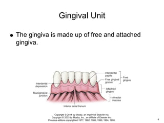
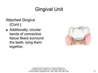
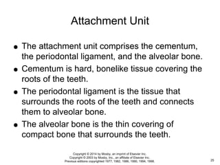
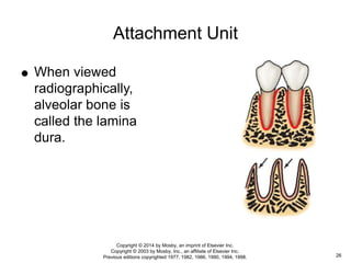
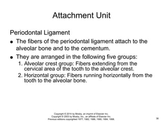
 The periodontium consists of tissues that
support the teeth.
 It is divided into a gingival unit and an
attachment unit or attachment apparatus.
The Periodontium
2
Copyright © 2014 by Mosby, an imprint of Elsevier Inc.
Copyright © 2003 by Mosby, Inc., an affiliate of Elsevier Inc.
Previous editions copyrighted 1977, 1982, 1986, 1990, 1994, 1998.
 The various parts of the gingival unit include:
Gingiva
Free gingiva
Attached gingiva
Alveolar mucosa
Attachment unit
Cementum
Alveolar bone
Periodontal ligament
The Periodontium
3
Copyright © 2014 by Mosby, an imprint of Elsevier Inc.
Copyright © 2003 by Mosby, Inc., an affiliate of Elsevier Inc.
Previous editions copyrighted 1977, 1982, 1986, 1990, 1994, 1998.
Gingival Unit
 The gingiva is made up of free and attached
gingiva.
4
Copyright © 2014 by Mosby, an imprint of Elsevier Inc.
Copyright © 2003 by Mosby, Inc., an affiliate of Elsevier Inc.
Previous editions copyrighted 1977, 1982, 1986, 1990, 1994, 1998.
 Gingiva is composed of very dense mucosa
called masticatory mucosa and has a thick
epithelial covering and keratinized cells.
 The underlying mucosa is composed of dense
collagen fibers.
Gingival Unit
5
Copyright © 2014 by Mosby, an imprint of Elsevier Inc.
Copyright © 2003 by Mosby, Inc., an affiliate of Elsevier Inc.
Previous editions copyrighted 1977, 1982, 1986, 1990, 1994, 1998.
Gingival Unit
6
Copyright © 2014 by Mosby, an imprint of Elsevier Inc.
Copyright © 2003 by Mosby, Inc., an affiliate of Elsevier Inc.
Previous editions copyrighted 1977, 1982, 1986, 1990, 1994, 1998.
 Masticatory mucosa is also found on the hard
palate and is designed to withstand the trauma
to which it is subjected in grinding food.
Gingival Unit
7
Copyright © 2014 by Mosby, an imprint of Elsevier Inc.
Copyright © 2003 by Mosby, Inc., an affiliate of Elsevier Inc.
Previous editions copyrighted 1977, 1982, 1986, 1990, 1994, 1998.
 The rest of the mouth is lined with a type of
mucosa called lining mucosa.
Alveolar mucosa is lining mucosa.
 The epithelium covering lining mucosa is thin,
nonkeratinized, and freely movable.
 Its mucosa is composed of loose connective
tissue and muscle fibers.
Gingival Unit
8
Copyright © 2014 by Mosby, an imprint of Elsevier Inc.
Copyright © 2003 by Mosby, Inc., an affiliate of Elsevier Inc.
Previous editions copyrighted 1977, 1982, 1986, 1990, 1994, 1998.
Free Gingiva
 Free gingiva is the gum tissue that extends from the
gingival margin to the base of the gingival sulcus.
It is usually light pink in color and averages between
0.5 to 2 mm in depth.
Gingival Unit
9
Copyright © 2014 by Mosby, an imprint of Elsevier Inc.
Copyright © 2003 by Mosby, Inc., an affiliate of Elsevier Inc.
Previous editions copyrighted 1977, 1982, 1986, 1990, 1994, 1998.
Free Gingiva (Cont.)
 The free gingival margin around a fully erupted tooth
is about 0.5 to 2 mm coronal to the cementoenamel
junction (CEJ).
 It forms a collar, which is separated from the tooth
by the gingival sulcus.
 A healthy gingival sulcus is approximately 2.5 mm in
depth.
Gingival Unit
10
Copyright © 2014 by Mosby, an imprint of Elsevier Inc.
Copyright © 2003 by Mosby, Inc., an affiliate of Elsevier Inc.
Previous editions copyrighted 1977, 1982, 1986, 1990, 1994, 1998.
Free Gingiva (Cont.)
 The gingival papilla is the free gingiva located in the
triangular interdental spaces.
 The shape of gingival papilla is affected by the
location of the contact area of the adjacent teeth,
the shape of the interproximal surfaces of the
adjacent teeth, and the CEJ of the adjacent teeth.
Gingival Unit
11
Copyright © 2014 by Mosby, an imprint of Elsevier Inc.
Copyright © 2003 by Mosby, Inc., an affiliate of Elsevier Inc.
Previous editions copyrighted 1977, 1982, 1986, 1990, 1994, 1998.
Free Gingiva (Cont.)
 The inner portion of the gingival sulcus is lined with
nonkeratinized epithelium; the outer portion is the
free gingiva, which is covered with keratinized
epithelium.
 A gingival groove often occurs on the outside of the
free gingiva and corresponds to the base of the
sulcus.
Gingival Unit
12
Copyright © 2014 by Mosby, an imprint of Elsevier Inc.
Copyright © 2003 by Mosby, Inc., an affiliate of Elsevier Inc.
Previous editions copyrighted 1977, 1982, 1986, 1990, 1994, 1998.
Attached Gingiva
 The attached gingiva begins at the base of the
gingival sulcus and is attached to the bone and the
cementum by collagenous fibers.
 It often has a stippled texture, resembling the
dimpled surface of an orange.
Gingival Unit
13
Copyright © 2014 by Mosby, an imprint of Elsevier Inc.
Copyright © 2003 by Mosby, Inc., an affiliate of Elsevier Inc.
Previous editions copyrighted 1977, 1982, 1986, 1990, 1994, 1998.
Attached Gingiva (Cont.)
 The attached gingiva is keratinized and is covered
by stratified squamous epithelium in which rete peg
formation is evident.
 The dimpling effect is caused by the rete peg
formation, which is simply the irregular binding of the
epithelium to the bone by collagen fibers.
 This causes depressions or dimples where the
epithelium is pulled tight to the bone.
Gingival Unit
14
Copyright © 2014 by Mosby, an imprint of Elsevier Inc.
Copyright © 2003 by Mosby, Inc., an affiliate of Elsevier Inc.
Previous editions copyrighted 1977, 1982, 1986, 1990, 1994, 1998.
Attached Gingiva (Cont.)
 The color of the gingiva varies from light to dark pink
and may contain pigment, correlating to the skin
pigmentation of the person.
 The darker a person’s skin color, then the more
likely it is that the gingiva is darker and contains
melanin.
Gingival Unit
15
Copyright © 2014 by Mosby, an imprint of Elsevier Inc.
Copyright © 2003 by Mosby, Inc., an affiliate of Elsevier Inc.
Previous editions copyrighted 1977, 1982, 1986, 1990, 1994, 1998.
Attached Gingiva (Cont.)
 The meshwork of collagenous fibers connecting the
gingiva to bone are formed by fibroblasts.
 All fibers embedded in the cementum are known as
Sharpey’s fibers; they extend from the cementum to
the papillary area of the gingiva.
 These fibers pass out from the cementum in small
bundles.
Gingival Unit
16
Copyright © 2014 by Mosby, an imprint of Elsevier Inc.
Copyright © 2003 by Mosby, Inc., an affiliate of Elsevier Inc.
Previous editions copyrighted 1977, 1982, 1986, 1990, 1994, 1998.
Gingival Unit
Attached Gingiva
(Cont.)
 Some of the fibers (A)
curve toward the
mucosa of the free
gingiva and interlace
with one another.
 Other fibers (B) pass
directly across from the
cementum to the
gingiva.
17
Copyright © 2014 by Mosby, an imprint of Elsevier Inc.
Copyright © 2003 by Mosby, Inc., an affiliate of Elsevier Inc.
Previous editions copyrighted 1977, 1982, 1986, 1990, 1994, 1998.
Attached Gingiva (Cont.)
 Still further apically, other fibers pass from the
cementum over the alveolar crest and turn apically
between the outer periosteum of the alveolar
process and the outer epithelial covering of the
attached gingiva.
Gingival Unit
18
Copyright © 2014 by Mosby, an imprint of Elsevier Inc.
Copyright © 2003 by Mosby, Inc., an affiliate of Elsevier Inc.
Previous editions copyrighted 1977, 1982, 1986, 1990, 1994, 1998.
Attached Gingiva (Cont.)
 On the proximal side, the connective tissue fibers
are at a higher level on the cementum because of
the curvature of the CEJ.
 This curvature allows more room for the cementum to
attach to the gingiva.
Gingival Unit
19
Copyright © 2014 by Mosby, an imprint of Elsevier Inc.
Copyright © 2003 by Mosby, Inc., an affiliate of Elsevier Inc.
Previous editions copyrighted 1977, 1982, 1986, 1990, 1994, 1998.
Attached Gingiva (Cont.)
 Attachment is made possible by groups of
connective tissue fibers.
 The most occlusal group travels to the papillary
layer of the epithelium of the interdental papilla.
Gingival Unit
20
Copyright © 2014 by Mosby, an imprint of Elsevier Inc.
Copyright © 2003 by Mosby, Inc., an affiliate of Elsevier Inc.
Previous editions copyrighted 1977, 1982, 1986, 1990, 1994, 1998.
Attached Gingiva (Cont.)
 The next layer of fibers passes occlusally into the
interproximal gingiva.
 The transseptal (TS) group, the next layer of fibers,
travels completely across the interproximal space
and attaches to the adjacent tooth.
These transseptal fiber bundles bind one tooth to
another.
Gingival Unit
21
Copyright © 2014 by Mosby, an imprint of Elsevier Inc.
Copyright © 2003 by Mosby, Inc., an affiliate of Elsevier Inc.
Previous editions copyrighted 1977, 1982, 1986, 1990, 1994, 1998.
Gingival Unit
Attached Gingiva
(Cont.)
 Additionally, circular
bands of connective
tissue fibers surround
the teeth, tying them
together.
22
Copyright © 2014 by Mosby, an imprint of Elsevier Inc.
Copyright © 2003 by Mosby, Inc., an affiliate of Elsevier Inc.
Previous editions copyrighted 1977, 1982, 1986, 1990, 1994, 1998.
Attached Gingiva (Cont.)
 The function of these fibers is to keep the gingiva
closely attached to the tooth surface.
 They also prevent the apical migration of the
epithelial attachment and resist gingival recession.
 Blood supply of the gingival tissue comes from the
supraperiosteal vessels originating from the lingual,
mental, buccal, infraorbital, and palatine arteries.
Gingival Unit
23
Copyright © 2014 by Mosby, an imprint of Elsevier Inc.
Copyright © 2003 by Mosby, Inc., an affiliate of Elsevier Inc.
Previous editions copyrighted 1977, 1982, 1986, 1990, 1994, 1998.
Alveolar Mucosa
 The alveolar mucosa joins the attached gingiva at
the mucogingival junction and is continuous with the
rest of the tissues of the vestibule.
 Alveolar mucosa is composed of lining mucosa, and
its submucosa contains loose connective tissue and
fat.
 The epithelium is smooth, thin, and nonkeratinized.
Gingival Unit
24
Copyright © 2014 by Mosby, an imprint of Elsevier Inc.
Copyright © 2003 by Mosby, Inc., an affiliate of Elsevier Inc.
Previous editions copyrighted 1977, 1982, 1986, 1990, 1994, 1998.
 The attachment unit comprises the cementum,
the periodontal ligament, and the alveolar bone.
 Cementum is hard, bonelike tissue covering the
roots of the teeth.
 The periodontal ligament is the tissue that
surrounds the roots of the teeth and connects
them to alveolar bone.
 The alveolar bone is the thin covering of
compact bone that surrounds the teeth.
Attachment Unit
25
Copyright © 2014 by Mosby, an imprint of Elsevier Inc.
Copyright © 2003 by Mosby, Inc., an affiliate of Elsevier Inc.
Previous editions copyrighted 1977, 1982, 1986, 1990, 1994, 1998.
Attachment Unit
 When viewed
radiographically,
alveolar bone is
called the lamina
dura.
26
Copyright © 2014 by Mosby, an imprint of Elsevier Inc.
Copyright © 2003 by Mosby, Inc., an affiliate of Elsevier Inc.
Previous editions copyrighted 1977, 1982, 1986, 1990, 1994, 1998.
 The function of the attachment apparatus is not
only supportive but also nutritive, formative, and
sensory.
 The supportive function is to maintain the support
for the tooth in the bone and to prevent its
movement.
 The nutritive and sensory functions are fulfilled by
the blood vessels and nerves.
 The nerves act as indicators of pressure or pain
around the tooth.
Attachment Unit
27
Copyright © 2014 by Mosby, an imprint of Elsevier Inc.
Copyright © 2003 by Mosby, Inc., an affiliate of Elsevier Inc.
Previous editions copyrighted 1977, 1982, 1986, 1990, 1994, 1998.
 The formative function is to replace cementum,
periodontal ligament, and alveolar bone.
This is accomplished by specialized cells called
cementoblasts, fibroblasts, and osteoblasts.
Attachment Unit
28
Copyright © 2014 by Mosby, an imprint of Elsevier Inc.
Copyright © 2003 by Mosby, Inc., an affiliate of Elsevier Inc.
Previous editions copyrighted 1977, 1982, 1986, 1990, 1994, 1998.
 In addition to these functions, the periodontal
ligament keeps the root and bone from abrading
each other.
 The periodontal ligament also acts as a cushion
between tooth and bone on the exertion of
pressure.
Attachment Unit
29
Copyright © 2014 by Mosby, an imprint of Elsevier Inc.
Copyright © 2003 by Mosby, Inc., an affiliate of Elsevier Inc.
Previous editions copyrighted 1977, 1982, 1986, 1990, 1994, 1998.
Cementum
 Cementum can be cellular or acellular.
 Both types are formed by cementoblasts.
In the cellular type, the cementoblasts become
embedded in the cementum.
The acellular type is free of embedded cementoblasts
and is clear and without structure.
Attachment Unit
30
Copyright © 2014 by Mosby, an imprint of Elsevier Inc.
Copyright © 2003 by Mosby, Inc., an affiliate of Elsevier Inc.
Previous editions copyrighted 1977, 1982, 1986, 1990, 1994, 1998.
Cementum (Cont.)
 Acellular cementum always covers the cervical third
of the root and sometimes extends over almost all of
the root except the apical portion.
 Cellular cementum covers the apical portion of the
root and sometimes may form over acellular
cementum.
Attachment Unit
31
Copyright © 2014 by Mosby, an imprint of Elsevier Inc.
Copyright © 2003 by Mosby, Inc., an affiliate of Elsevier Inc.
Previous editions copyrighted 1977, 1982, 1986, 1990, 1994, 1998.
Cementum (Cont.)
 Cellular cementum grows by the apposition
(addition) of new layers, one on top of another.
Changes in function and pressure influence the
growth activity of cementum.
Attachment Unit
32
Copyright © 2014 by Mosby, an imprint of Elsevier Inc.
Copyright © 2003 by Mosby, Inc., an affiliate of Elsevier Inc.
Previous editions copyrighted 1977, 1982, 1986, 1990, 1994, 1998.
Cementum (Cont.)
 Both cellular and acellular cementum have collagen
fibers embedded within them.
 These fibers, known as Sharpey’s fibers, are the
embedded ends of connective tissue fibers of the
periodontal membrane.
Some are embedded in cementum, and some are in
bone.
Attachment Unit
33
Copyright © 2014 by Mosby, an imprint of Elsevier Inc.
Copyright © 2003 by Mosby, Inc., an affiliate of Elsevier Inc.
Previous editions copyrighted 1977, 1982, 1986, 1990, 1994, 1998.
Alveolar Bone
 The type of bone that lines the sockets in which the
roots of teeth are held is called alveolar bone
proper.
It is thin and compact, with many small openings
through which blood vessels, nerves, and lymphatic
vessels pass.
Attachment Unit
34
Copyright © 2014 by Mosby, an imprint of Elsevier Inc.
Copyright © 2003 by Mosby, Inc., an affiliate of Elsevier Inc.
Previous editions copyrighted 1977, 1982, 1986, 1990, 1994, 1998.
Alveolar Bone (Cont.)
 The socket in which the tooth rests is called an
alveolus.
 These alveoli are a part of the alveolar process that
surrounds and supports the teeth in the maxilla and
mandible.
Attachment Unit
35
Copyright © 2014 by Mosby, an imprint of Elsevier Inc.
Copyright © 2003 by Mosby, Inc., an affiliate of Elsevier Inc.
Previous editions copyrighted 1977, 1982, 1986, 1990, 1994, 1998.
Alveolar Bone (Cont.)
 Bone consists of an organic matrix and inorganic
matter.
The organic matrix comprises collagen and
intercellular substance.
The inorganic matter comprises apatite crystals of
calcium, phosphate, and carbonates.
Attachment Unit
36
Copyright © 2014 by Mosby, an imprint of Elsevier Inc.
Copyright © 2003 by Mosby, Inc., an affiliate of Elsevier Inc.
Previous editions copyrighted 1977, 1982, 1986, 1990, 1994, 1998.
Alveolar Bone (Cont.)
 The bone forming the alveoli undergoes resorption
when pressure is exerted on it and opposition when
tension is placed on it.
 The term bundle bone applies when numerous
layers of bone are added to the socket wall.
Bundle bone forms the immediate attachment of the
periodontal ligament.
Attachment Unit
37
Copyright © 2014 by Mosby, an imprint of Elsevier Inc.
Copyright © 2003 by Mosby, Inc., an affiliate of Elsevier Inc.
Previous editions copyrighted 1977, 1982, 1986, 1990, 1994, 1998.
Periodontal Ligament
 The fibers of the periodontal ligament attach to the
alveolar bone and to the cementum.
 They are arranged in the following five groups:
1. Alveolar crest group: Fibers extending from the
cervical area of the tooth to the alveolar crest.
2. Horizontal group: Fibers running horizontally from the
tooth to the alveolar bone.
Attachment Unit
38
Copyright © 2014 by Mosby, an imprint of Elsevier Inc.
Copyright © 2003 by Mosby, Inc., an affiliate of Elsevier Inc.
Previous editions copyrighted 1977, 1982, 1986, 1990, 1994, 1998.
Periodontal Ligament (Cont.)
3. Oblique group: Fibers running obliquely from the
cementum to the bone.
4. Apical group: Fibers radiating apically from the tooth
to the bone.
5. Interradicular group: Periodontal fibers between
roots of multirooted teeth.
Attachment Unit
39
Copyright © 2014 by Mosby, an imprint of Elsevier Inc.
Copyright © 2003 by Mosby, Inc., an affiliate of Elsevier Inc.
Previous editions copyrighted 1977, 1982, 1986, 1990, 1994, 1998.
Periodontal Ligament (Cont.)
 The arrangement of fibers ties the tooth to the bone
and prevents it from being pushed into the bone.
 This function is helped by the roots of the teeth and
by fluids within the periodontal ligament.
 The shapes and sizes of the roots of the teeth help
dissipate occlusal stresses in both a lateral and
apical direction.
Attachment Unit
40
Copyright © 2014 by Mosby, an imprint of Elsevier Inc.
Copyright © 2003 by Mosby, Inc., an affiliate of Elsevier Inc.
Previous editions copyrighted 1977, 1982, 1986, 1990, 1994, 1998.
Periodontal Ligament (Cont.)
 The main portion of the periodontal ligament is
composed of bundles of white collagenous
connective tissue fibers.
 These fibers extend from and are embedded in the
cementum of the tooth and alveolar bone.
They are bundled together like the many strands of a
rope.
Attachment Unit
41
Copyright © 2014 by Mosby, an imprint of Elsevier Inc.
Copyright © 2003 by Mosby, Inc., an affiliate of Elsevier Inc.
Previous editions copyrighted 1977, 1982, 1986, 1990, 1994, 1998.
Periodontal Ligament (Cont.)
 The periodontal ligament also contains fibroblasts,
small blood and lymph vessels, and nerves.
Attachment Unit
42
Copyright © 2014 by Mosby, an imprint of Elsevier Inc.
Copyright © 2003 by Mosby, Inc., an affiliate of Elsevier Inc.
Previous editions copyrighted 1977, 1982, 1986, 1990, 1994, 1998.
Periodontal Ligament (Cont.)
 The tooth itself is suspended by the periodontal
ligament and allows movement within the bony
cavity.
 Two forces allow for movement of the tooth.
1. Mesial drift allows the tooth to move forward.
2. Active eruption allows the tooth to migrate occlusally
until it occludes with an antagonist.
Attachment Unit
43
Copyright © 2014 by Mosby, an imprint of Elsevier Inc.
Copyright © 2003 by Mosby, Inc., an affiliate of Elsevier Inc.
Previous editions copyrighted 1977, 1982, 1986, 1990, 1994, 1998.
 For a tooth to move through bone, a force of
some type must be acting on it.
 This force causes pressure on the root,
periodontal ligament, and bone.
 It compresses the periodontal ligament and the
alveolar bone on one side of the root.
 If this force continues for long enough, a
biomechanical system of osteoclastic cells
(bone-destroying cells) is initiated.
Clinical Considerations
44
Copyright © 2014 by Mosby, an imprint of Elsevier Inc.
Copyright © 2003 by Mosby, Inc., an affiliate of Elsevier Inc.
Previous editions copyrighted 1977, 1982, 1986, 1990, 1994, 1998.
 On the other side of this same tooth root, the
periodontal ligament is pulled taut.
 This results in a pulling force on the alveolar
bone on that side of the socket.
 This constant tension causes an opposite
biomechanical system.
 Osteoblastic cells (bone-forming cells) start
laying down bone.
Clinical Considerations
45
Copyright © 2014 by Mosby, an imprint of Elsevier Inc.
Copyright © 2003 by Mosby, Inc., an affiliate of Elsevier Inc.
Previous editions copyrighted 1977, 1982, 1986, 1990, 1994, 1998.
 Some of the forces that exert pressure on the
teeth:
1. Active eruption.
2. Mesial drift.
3. Masticatory occlusal forces.
4. Orthodontic corrective forces.
5. Traumatic occlusal forces.
Clinical Considerations
46
Copyright © 2014 by Mosby, an imprint of Elsevier Inc.
Copyright © 2003 by Mosby, Inc., an affiliate of Elsevier Inc.
Previous editions copyrighted 1977, 1982, 1986, 1990, 1994, 1998.
 The first four of these forces can tip, rotate, or
move the tooth because of the bone-remodeling
system.
 The last of these forces, occlusal trauma, is
usually intermittent and consequently does not
result in tooth movement as much as tooth
mobility.
Clinical Considerations
47
Copyright © 2014 by Mosby, an imprint of Elsevier Inc.
Copyright © 2003 by Mosby, Inc., an affiliate of Elsevier Inc.
Previous editions copyrighted 1977, 1982, 1986, 1990, 1994, 1998.
Clinical Considerations
 The periodontal
ligament becomes
enlarged and
widened, the lamina
dura is compressed,
and the tooth
becomes loose and
mobile within the
socket.
48
Copyright © 2014 by Mosby, an imprint of Elsevier Inc.
Copyright © 2003 by Mosby, Inc., an affiliate of Elsevier Inc.
Previous editions copyrighted 1977, 1982, 1986, 1990, 1994, 1998.

Periodontium: Supporting Structures

  • 1.
    Chapter 8 Supporting Structures Copyright© 2014 by Mosby, an imprint of Elsevier Inc. Copyright © 2003 by Mosby, Inc., an affiliate of Elsevier Inc. Previous editions copyrighted 1977, 1982, 1986, 1990, 1994, 1998.
  • 2.
     The periodontiumconsists of tissues that support the teeth.  It is divided into a gingival unit and an attachment unit or attachment apparatus. The Periodontium 2 Copyright © 2014 by Mosby, an imprint of Elsevier Inc. Copyright © 2003 by Mosby, Inc., an affiliate of Elsevier Inc. Previous editions copyrighted 1977, 1982, 1986, 1990, 1994, 1998.
  • 3.
     The variousparts of the gingival unit include: Gingiva Free gingiva Attached gingiva Alveolar mucosa Attachment unit Cementum Alveolar bone Periodontal ligament The Periodontium 3 Copyright © 2014 by Mosby, an imprint of Elsevier Inc. Copyright © 2003 by Mosby, Inc., an affiliate of Elsevier Inc. Previous editions copyrighted 1977, 1982, 1986, 1990, 1994, 1998.
  • 4.
    Gingival Unit  Thegingiva is made up of free and attached gingiva. 4 Copyright © 2014 by Mosby, an imprint of Elsevier Inc. Copyright © 2003 by Mosby, Inc., an affiliate of Elsevier Inc. Previous editions copyrighted 1977, 1982, 1986, 1990, 1994, 1998.
  • 5.
     Gingiva iscomposed of very dense mucosa called masticatory mucosa and has a thick epithelial covering and keratinized cells.  The underlying mucosa is composed of dense collagen fibers. Gingival Unit 5 Copyright © 2014 by Mosby, an imprint of Elsevier Inc. Copyright © 2003 by Mosby, Inc., an affiliate of Elsevier Inc. Previous editions copyrighted 1977, 1982, 1986, 1990, 1994, 1998.
  • 6.
    Gingival Unit 6 Copyright ©2014 by Mosby, an imprint of Elsevier Inc. Copyright © 2003 by Mosby, Inc., an affiliate of Elsevier Inc. Previous editions copyrighted 1977, 1982, 1986, 1990, 1994, 1998.
  • 7.
     Masticatory mucosais also found on the hard palate and is designed to withstand the trauma to which it is subjected in grinding food. Gingival Unit 7 Copyright © 2014 by Mosby, an imprint of Elsevier Inc. Copyright © 2003 by Mosby, Inc., an affiliate of Elsevier Inc. Previous editions copyrighted 1977, 1982, 1986, 1990, 1994, 1998.
  • 8.
     The restof the mouth is lined with a type of mucosa called lining mucosa. Alveolar mucosa is lining mucosa.  The epithelium covering lining mucosa is thin, nonkeratinized, and freely movable.  Its mucosa is composed of loose connective tissue and muscle fibers. Gingival Unit 8 Copyright © 2014 by Mosby, an imprint of Elsevier Inc. Copyright © 2003 by Mosby, Inc., an affiliate of Elsevier Inc. Previous editions copyrighted 1977, 1982, 1986, 1990, 1994, 1998.
  • 9.
    Free Gingiva  Freegingiva is the gum tissue that extends from the gingival margin to the base of the gingival sulcus. It is usually light pink in color and averages between 0.5 to 2 mm in depth. Gingival Unit 9 Copyright © 2014 by Mosby, an imprint of Elsevier Inc. Copyright © 2003 by Mosby, Inc., an affiliate of Elsevier Inc. Previous editions copyrighted 1977, 1982, 1986, 1990, 1994, 1998.
  • 10.
    Free Gingiva (Cont.) The free gingival margin around a fully erupted tooth is about 0.5 to 2 mm coronal to the cementoenamel junction (CEJ).  It forms a collar, which is separated from the tooth by the gingival sulcus.  A healthy gingival sulcus is approximately 2.5 mm in depth. Gingival Unit 10 Copyright © 2014 by Mosby, an imprint of Elsevier Inc. Copyright © 2003 by Mosby, Inc., an affiliate of Elsevier Inc. Previous editions copyrighted 1977, 1982, 1986, 1990, 1994, 1998.
  • 11.
    Free Gingiva (Cont.) The gingival papilla is the free gingiva located in the triangular interdental spaces.  The shape of gingival papilla is affected by the location of the contact area of the adjacent teeth, the shape of the interproximal surfaces of the adjacent teeth, and the CEJ of the adjacent teeth. Gingival Unit 11 Copyright © 2014 by Mosby, an imprint of Elsevier Inc. Copyright © 2003 by Mosby, Inc., an affiliate of Elsevier Inc. Previous editions copyrighted 1977, 1982, 1986, 1990, 1994, 1998.
  • 12.
    Free Gingiva (Cont.) The inner portion of the gingival sulcus is lined with nonkeratinized epithelium; the outer portion is the free gingiva, which is covered with keratinized epithelium.  A gingival groove often occurs on the outside of the free gingiva and corresponds to the base of the sulcus. Gingival Unit 12 Copyright © 2014 by Mosby, an imprint of Elsevier Inc. Copyright © 2003 by Mosby, Inc., an affiliate of Elsevier Inc. Previous editions copyrighted 1977, 1982, 1986, 1990, 1994, 1998.
  • 13.
    Attached Gingiva  Theattached gingiva begins at the base of the gingival sulcus and is attached to the bone and the cementum by collagenous fibers.  It often has a stippled texture, resembling the dimpled surface of an orange. Gingival Unit 13 Copyright © 2014 by Mosby, an imprint of Elsevier Inc. Copyright © 2003 by Mosby, Inc., an affiliate of Elsevier Inc. Previous editions copyrighted 1977, 1982, 1986, 1990, 1994, 1998.
  • 14.
    Attached Gingiva (Cont.) The attached gingiva is keratinized and is covered by stratified squamous epithelium in which rete peg formation is evident.  The dimpling effect is caused by the rete peg formation, which is simply the irregular binding of the epithelium to the bone by collagen fibers.  This causes depressions or dimples where the epithelium is pulled tight to the bone. Gingival Unit 14 Copyright © 2014 by Mosby, an imprint of Elsevier Inc. Copyright © 2003 by Mosby, Inc., an affiliate of Elsevier Inc. Previous editions copyrighted 1977, 1982, 1986, 1990, 1994, 1998.
  • 15.
    Attached Gingiva (Cont.) The color of the gingiva varies from light to dark pink and may contain pigment, correlating to the skin pigmentation of the person.  The darker a person’s skin color, then the more likely it is that the gingiva is darker and contains melanin. Gingival Unit 15 Copyright © 2014 by Mosby, an imprint of Elsevier Inc. Copyright © 2003 by Mosby, Inc., an affiliate of Elsevier Inc. Previous editions copyrighted 1977, 1982, 1986, 1990, 1994, 1998.
  • 16.
    Attached Gingiva (Cont.) The meshwork of collagenous fibers connecting the gingiva to bone are formed by fibroblasts.  All fibers embedded in the cementum are known as Sharpey’s fibers; they extend from the cementum to the papillary area of the gingiva.  These fibers pass out from the cementum in small bundles. Gingival Unit 16 Copyright © 2014 by Mosby, an imprint of Elsevier Inc. Copyright © 2003 by Mosby, Inc., an affiliate of Elsevier Inc. Previous editions copyrighted 1977, 1982, 1986, 1990, 1994, 1998.
  • 17.
    Gingival Unit Attached Gingiva (Cont.) Some of the fibers (A) curve toward the mucosa of the free gingiva and interlace with one another.  Other fibers (B) pass directly across from the cementum to the gingiva. 17 Copyright © 2014 by Mosby, an imprint of Elsevier Inc. Copyright © 2003 by Mosby, Inc., an affiliate of Elsevier Inc. Previous editions copyrighted 1977, 1982, 1986, 1990, 1994, 1998.
  • 18.
    Attached Gingiva (Cont.) Still further apically, other fibers pass from the cementum over the alveolar crest and turn apically between the outer periosteum of the alveolar process and the outer epithelial covering of the attached gingiva. Gingival Unit 18 Copyright © 2014 by Mosby, an imprint of Elsevier Inc. Copyright © 2003 by Mosby, Inc., an affiliate of Elsevier Inc. Previous editions copyrighted 1977, 1982, 1986, 1990, 1994, 1998.
  • 19.
    Attached Gingiva (Cont.) On the proximal side, the connective tissue fibers are at a higher level on the cementum because of the curvature of the CEJ.  This curvature allows more room for the cementum to attach to the gingiva. Gingival Unit 19 Copyright © 2014 by Mosby, an imprint of Elsevier Inc. Copyright © 2003 by Mosby, Inc., an affiliate of Elsevier Inc. Previous editions copyrighted 1977, 1982, 1986, 1990, 1994, 1998.
  • 20.
    Attached Gingiva (Cont.) Attachment is made possible by groups of connective tissue fibers.  The most occlusal group travels to the papillary layer of the epithelium of the interdental papilla. Gingival Unit 20 Copyright © 2014 by Mosby, an imprint of Elsevier Inc. Copyright © 2003 by Mosby, Inc., an affiliate of Elsevier Inc. Previous editions copyrighted 1977, 1982, 1986, 1990, 1994, 1998.
  • 21.
    Attached Gingiva (Cont.) The next layer of fibers passes occlusally into the interproximal gingiva.  The transseptal (TS) group, the next layer of fibers, travels completely across the interproximal space and attaches to the adjacent tooth. These transseptal fiber bundles bind one tooth to another. Gingival Unit 21 Copyright © 2014 by Mosby, an imprint of Elsevier Inc. Copyright © 2003 by Mosby, Inc., an affiliate of Elsevier Inc. Previous editions copyrighted 1977, 1982, 1986, 1990, 1994, 1998.
  • 22.
    Gingival Unit Attached Gingiva (Cont.) Additionally, circular bands of connective tissue fibers surround the teeth, tying them together. 22 Copyright © 2014 by Mosby, an imprint of Elsevier Inc. Copyright © 2003 by Mosby, Inc., an affiliate of Elsevier Inc. Previous editions copyrighted 1977, 1982, 1986, 1990, 1994, 1998.
  • 23.
    Attached Gingiva (Cont.) The function of these fibers is to keep the gingiva closely attached to the tooth surface.  They also prevent the apical migration of the epithelial attachment and resist gingival recession.  Blood supply of the gingival tissue comes from the supraperiosteal vessels originating from the lingual, mental, buccal, infraorbital, and palatine arteries. Gingival Unit 23 Copyright © 2014 by Mosby, an imprint of Elsevier Inc. Copyright © 2003 by Mosby, Inc., an affiliate of Elsevier Inc. Previous editions copyrighted 1977, 1982, 1986, 1990, 1994, 1998.
  • 24.
    Alveolar Mucosa  Thealveolar mucosa joins the attached gingiva at the mucogingival junction and is continuous with the rest of the tissues of the vestibule.  Alveolar mucosa is composed of lining mucosa, and its submucosa contains loose connective tissue and fat.  The epithelium is smooth, thin, and nonkeratinized. Gingival Unit 24 Copyright © 2014 by Mosby, an imprint of Elsevier Inc. Copyright © 2003 by Mosby, Inc., an affiliate of Elsevier Inc. Previous editions copyrighted 1977, 1982, 1986, 1990, 1994, 1998.
  • 25.
     The attachmentunit comprises the cementum, the periodontal ligament, and the alveolar bone.  Cementum is hard, bonelike tissue covering the roots of the teeth.  The periodontal ligament is the tissue that surrounds the roots of the teeth and connects them to alveolar bone.  The alveolar bone is the thin covering of compact bone that surrounds the teeth. Attachment Unit 25 Copyright © 2014 by Mosby, an imprint of Elsevier Inc. Copyright © 2003 by Mosby, Inc., an affiliate of Elsevier Inc. Previous editions copyrighted 1977, 1982, 1986, 1990, 1994, 1998.
  • 26.
    Attachment Unit  Whenviewed radiographically, alveolar bone is called the lamina dura. 26 Copyright © 2014 by Mosby, an imprint of Elsevier Inc. Copyright © 2003 by Mosby, Inc., an affiliate of Elsevier Inc. Previous editions copyrighted 1977, 1982, 1986, 1990, 1994, 1998.
  • 27.
     The functionof the attachment apparatus is not only supportive but also nutritive, formative, and sensory.  The supportive function is to maintain the support for the tooth in the bone and to prevent its movement.  The nutritive and sensory functions are fulfilled by the blood vessels and nerves.  The nerves act as indicators of pressure or pain around the tooth. Attachment Unit 27 Copyright © 2014 by Mosby, an imprint of Elsevier Inc. Copyright © 2003 by Mosby, Inc., an affiliate of Elsevier Inc. Previous editions copyrighted 1977, 1982, 1986, 1990, 1994, 1998.
  • 28.
     The formativefunction is to replace cementum, periodontal ligament, and alveolar bone. This is accomplished by specialized cells called cementoblasts, fibroblasts, and osteoblasts. Attachment Unit 28 Copyright © 2014 by Mosby, an imprint of Elsevier Inc. Copyright © 2003 by Mosby, Inc., an affiliate of Elsevier Inc. Previous editions copyrighted 1977, 1982, 1986, 1990, 1994, 1998.
  • 29.
     In additionto these functions, the periodontal ligament keeps the root and bone from abrading each other.  The periodontal ligament also acts as a cushion between tooth and bone on the exertion of pressure. Attachment Unit 29 Copyright © 2014 by Mosby, an imprint of Elsevier Inc. Copyright © 2003 by Mosby, Inc., an affiliate of Elsevier Inc. Previous editions copyrighted 1977, 1982, 1986, 1990, 1994, 1998.
  • 30.
    Cementum  Cementum canbe cellular or acellular.  Both types are formed by cementoblasts. In the cellular type, the cementoblasts become embedded in the cementum. The acellular type is free of embedded cementoblasts and is clear and without structure. Attachment Unit 30 Copyright © 2014 by Mosby, an imprint of Elsevier Inc. Copyright © 2003 by Mosby, Inc., an affiliate of Elsevier Inc. Previous editions copyrighted 1977, 1982, 1986, 1990, 1994, 1998.
  • 31.
    Cementum (Cont.)  Acellularcementum always covers the cervical third of the root and sometimes extends over almost all of the root except the apical portion.  Cellular cementum covers the apical portion of the root and sometimes may form over acellular cementum. Attachment Unit 31 Copyright © 2014 by Mosby, an imprint of Elsevier Inc. Copyright © 2003 by Mosby, Inc., an affiliate of Elsevier Inc. Previous editions copyrighted 1977, 1982, 1986, 1990, 1994, 1998.
  • 32.
    Cementum (Cont.)  Cellularcementum grows by the apposition (addition) of new layers, one on top of another. Changes in function and pressure influence the growth activity of cementum. Attachment Unit 32 Copyright © 2014 by Mosby, an imprint of Elsevier Inc. Copyright © 2003 by Mosby, Inc., an affiliate of Elsevier Inc. Previous editions copyrighted 1977, 1982, 1986, 1990, 1994, 1998.
  • 33.
    Cementum (Cont.)  Bothcellular and acellular cementum have collagen fibers embedded within them.  These fibers, known as Sharpey’s fibers, are the embedded ends of connective tissue fibers of the periodontal membrane. Some are embedded in cementum, and some are in bone. Attachment Unit 33 Copyright © 2014 by Mosby, an imprint of Elsevier Inc. Copyright © 2003 by Mosby, Inc., an affiliate of Elsevier Inc. Previous editions copyrighted 1977, 1982, 1986, 1990, 1994, 1998.
  • 34.
    Alveolar Bone  Thetype of bone that lines the sockets in which the roots of teeth are held is called alveolar bone proper. It is thin and compact, with many small openings through which blood vessels, nerves, and lymphatic vessels pass. Attachment Unit 34 Copyright © 2014 by Mosby, an imprint of Elsevier Inc. Copyright © 2003 by Mosby, Inc., an affiliate of Elsevier Inc. Previous editions copyrighted 1977, 1982, 1986, 1990, 1994, 1998.
  • 35.
    Alveolar Bone (Cont.) The socket in which the tooth rests is called an alveolus.  These alveoli are a part of the alveolar process that surrounds and supports the teeth in the maxilla and mandible. Attachment Unit 35 Copyright © 2014 by Mosby, an imprint of Elsevier Inc. Copyright © 2003 by Mosby, Inc., an affiliate of Elsevier Inc. Previous editions copyrighted 1977, 1982, 1986, 1990, 1994, 1998.
  • 36.
    Alveolar Bone (Cont.) Bone consists of an organic matrix and inorganic matter. The organic matrix comprises collagen and intercellular substance. The inorganic matter comprises apatite crystals of calcium, phosphate, and carbonates. Attachment Unit 36 Copyright © 2014 by Mosby, an imprint of Elsevier Inc. Copyright © 2003 by Mosby, Inc., an affiliate of Elsevier Inc. Previous editions copyrighted 1977, 1982, 1986, 1990, 1994, 1998.
  • 37.
    Alveolar Bone (Cont.) The bone forming the alveoli undergoes resorption when pressure is exerted on it and opposition when tension is placed on it.  The term bundle bone applies when numerous layers of bone are added to the socket wall. Bundle bone forms the immediate attachment of the periodontal ligament. Attachment Unit 37 Copyright © 2014 by Mosby, an imprint of Elsevier Inc. Copyright © 2003 by Mosby, Inc., an affiliate of Elsevier Inc. Previous editions copyrighted 1977, 1982, 1986, 1990, 1994, 1998.
  • 38.
    Periodontal Ligament  Thefibers of the periodontal ligament attach to the alveolar bone and to the cementum.  They are arranged in the following five groups: 1. Alveolar crest group: Fibers extending from the cervical area of the tooth to the alveolar crest. 2. Horizontal group: Fibers running horizontally from the tooth to the alveolar bone. Attachment Unit 38 Copyright © 2014 by Mosby, an imprint of Elsevier Inc. Copyright © 2003 by Mosby, Inc., an affiliate of Elsevier Inc. Previous editions copyrighted 1977, 1982, 1986, 1990, 1994, 1998.
  • 39.
    Periodontal Ligament (Cont.) 3.Oblique group: Fibers running obliquely from the cementum to the bone. 4. Apical group: Fibers radiating apically from the tooth to the bone. 5. Interradicular group: Periodontal fibers between roots of multirooted teeth. Attachment Unit 39 Copyright © 2014 by Mosby, an imprint of Elsevier Inc. Copyright © 2003 by Mosby, Inc., an affiliate of Elsevier Inc. Previous editions copyrighted 1977, 1982, 1986, 1990, 1994, 1998.
  • 40.
    Periodontal Ligament (Cont.) The arrangement of fibers ties the tooth to the bone and prevents it from being pushed into the bone.  This function is helped by the roots of the teeth and by fluids within the periodontal ligament.  The shapes and sizes of the roots of the teeth help dissipate occlusal stresses in both a lateral and apical direction. Attachment Unit 40 Copyright © 2014 by Mosby, an imprint of Elsevier Inc. Copyright © 2003 by Mosby, Inc., an affiliate of Elsevier Inc. Previous editions copyrighted 1977, 1982, 1986, 1990, 1994, 1998.
  • 41.
    Periodontal Ligament (Cont.) The main portion of the periodontal ligament is composed of bundles of white collagenous connective tissue fibers.  These fibers extend from and are embedded in the cementum of the tooth and alveolar bone. They are bundled together like the many strands of a rope. Attachment Unit 41 Copyright © 2014 by Mosby, an imprint of Elsevier Inc. Copyright © 2003 by Mosby, Inc., an affiliate of Elsevier Inc. Previous editions copyrighted 1977, 1982, 1986, 1990, 1994, 1998.
  • 42.
    Periodontal Ligament (Cont.) The periodontal ligament also contains fibroblasts, small blood and lymph vessels, and nerves. Attachment Unit 42 Copyright © 2014 by Mosby, an imprint of Elsevier Inc. Copyright © 2003 by Mosby, Inc., an affiliate of Elsevier Inc. Previous editions copyrighted 1977, 1982, 1986, 1990, 1994, 1998.
  • 43.
    Periodontal Ligament (Cont.) The tooth itself is suspended by the periodontal ligament and allows movement within the bony cavity.  Two forces allow for movement of the tooth. 1. Mesial drift allows the tooth to move forward. 2. Active eruption allows the tooth to migrate occlusally until it occludes with an antagonist. Attachment Unit 43 Copyright © 2014 by Mosby, an imprint of Elsevier Inc. Copyright © 2003 by Mosby, Inc., an affiliate of Elsevier Inc. Previous editions copyrighted 1977, 1982, 1986, 1990, 1994, 1998.
  • 44.
     For atooth to move through bone, a force of some type must be acting on it.  This force causes pressure on the root, periodontal ligament, and bone.  It compresses the periodontal ligament and the alveolar bone on one side of the root.  If this force continues for long enough, a biomechanical system of osteoclastic cells (bone-destroying cells) is initiated. Clinical Considerations 44 Copyright © 2014 by Mosby, an imprint of Elsevier Inc. Copyright © 2003 by Mosby, Inc., an affiliate of Elsevier Inc. Previous editions copyrighted 1977, 1982, 1986, 1990, 1994, 1998.
  • 45.
     On theother side of this same tooth root, the periodontal ligament is pulled taut.  This results in a pulling force on the alveolar bone on that side of the socket.  This constant tension causes an opposite biomechanical system.  Osteoblastic cells (bone-forming cells) start laying down bone. Clinical Considerations 45 Copyright © 2014 by Mosby, an imprint of Elsevier Inc. Copyright © 2003 by Mosby, Inc., an affiliate of Elsevier Inc. Previous editions copyrighted 1977, 1982, 1986, 1990, 1994, 1998.
  • 46.
     Some ofthe forces that exert pressure on the teeth: 1. Active eruption. 2. Mesial drift. 3. Masticatory occlusal forces. 4. Orthodontic corrective forces. 5. Traumatic occlusal forces. Clinical Considerations 46 Copyright © 2014 by Mosby, an imprint of Elsevier Inc. Copyright © 2003 by Mosby, Inc., an affiliate of Elsevier Inc. Previous editions copyrighted 1977, 1982, 1986, 1990, 1994, 1998.
  • 47.
     The firstfour of these forces can tip, rotate, or move the tooth because of the bone-remodeling system.  The last of these forces, occlusal trauma, is usually intermittent and consequently does not result in tooth movement as much as tooth mobility. Clinical Considerations 47 Copyright © 2014 by Mosby, an imprint of Elsevier Inc. Copyright © 2003 by Mosby, Inc., an affiliate of Elsevier Inc. Previous editions copyrighted 1977, 1982, 1986, 1990, 1994, 1998.
  • 48.
    Clinical Considerations  Theperiodontal ligament becomes enlarged and widened, the lamina dura is compressed, and the tooth becomes loose and mobile within the socket. 48 Copyright © 2014 by Mosby, an imprint of Elsevier Inc. Copyright © 2003 by Mosby, Inc., an affiliate of Elsevier Inc. Previous editions copyrighted 1977, 1982, 1986, 1990, 1994, 1998.