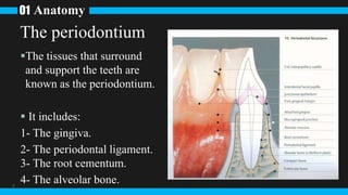
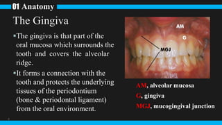
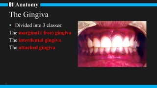
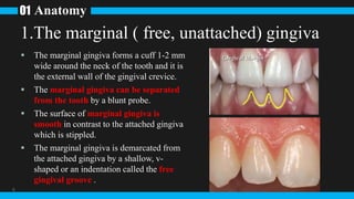
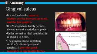
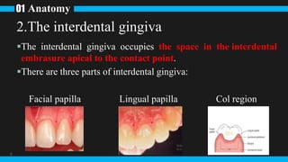
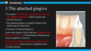
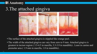
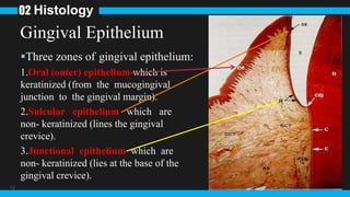
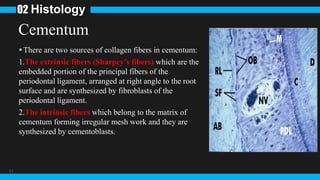
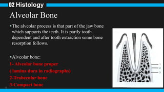
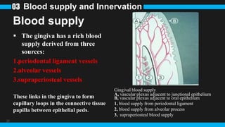
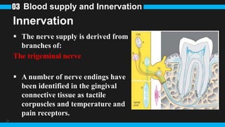
Periodontium: anatomy, histology,
blood supply and innervation
Speaker: Shao-Wei Chao 07/03/2023
1 Anatomy
2
Anatomy
01
3
The tissues that surround
and support the teeth are
known as the periodontium.
 It includes:
1- The gingiva.
2- The periodontal ligament.
3- The root cementum.
4- The alveolar bone.
The periodontium
Anatomy
01
4
The gingiva is that part of the
oral mucosa which surrounds the
tooth and covers the alveolar
ridge.
It forms a connection with the
tooth and protects the underlying
tissues of the periodontium
(bone & periodontal ligament)
from the oral environment.
The Gingiva
AM, alveolar mucosa
G, gingiva
MGJ, mucogingival junction
Anatomy
01
5
 Divided into 3 classes:
The marginal ( free) gingiva
The interdental gingiva
The attached gingiva
The Gingiva
Anatomy
01
6
 The marginal gingiva forms a cuff 1-2 mm
wide around the neck of the tooth and it is
the external wall of the gingival crevice.
 The marginal gingiva can be separated
from the tooth by a blunt probe.
 The surface of marginal gingiva is
smooth in contrast to the attached gingiva
which is stippled.
 The marginal gingiva is demarcated from
the attached gingiva by a shallow, v-
shaped or an indentation called the free
gingival groove .
1.The marginal ( free, unattached) gingiva
Anatomy
01
7
It is defined as the space or
shallow crevice between the tooth
and the free gingiva.
It is V-shaped and barely permits
the entrance of a periodontal probe.
Under normal or ideal conditions it
is about 2 to 3 mm.
The gingival sulcus is probing
depth of a clinically-normal
gingival. It provides good
resistance to mechanical forces.
Gingival sulcus
Anatomy
01
8
The interdental gingiva occupies the space in the interdental
embrasure apical to the contact point.
There are three parts of interdental gingiva:
Facial papilla Lingual papilla Col region
2.The interdental gingiva
Anatomy
01
9
It extends from the free gingival groove to the
mucogingival junction where it meets the
alveolar mucosa.
The attached gingiva is tightly bound to the
underlying alveolar bone.
Function of attached gingiva:
It provides gingival tissue that can withstand the
mechanical forces of mastication, brushing and
tension applied on mucosa.
Prevent free gingiva from being pulled away
from the tooth when tension is applied to the
alveolar mucosa.
3.The attached gingiva
Anatomy
01
10
The surface of the attached gingiva is stippled like orange peel.
The width of the attached gingiva can vary from zero to 9 mm. Attached gingiva is
greatest in incisor region ( 3.5-4.5 in maxilla, 3.3-3.9 in mandible) . Least in canine and
premolar area ( 1.9 mm in maxilla, 1.8 in mandible) .
3.The attached gingiva
2 Histology
11
12
Gingival Epithelium
Three zones of gingival epithelium:
1.Oral (outer) epithelium which is
keratinized (from the mucogingival
junction to the gingival margin).
2.Sulcular epithelium which are
non- keratinized (lines the gingival
crevice).
3.Junctional epithelium which are
non- keratinized (lies at the base of the
gingival crevice).
Histology
02
13
1.Oral (outer) epithelium
Keratinized or parakeratinized consist of four layers:
1.Basal or formative cells layers of columnar cells (The
deepest cells:CT); it is characterized by active mitosis
(stratum basal or stratum grrminativum:SB).
2.Prickle or spinous cell layer of polygonal cells (stratum
spinosum:SS).
3.Granular cell layer in which the cells are flatter and contain
many particles of keratohyaline (stratum granulosum:SG).
4.Cornified or keratinous cell layer in which cells have become
flat, shrunken and keratinized (stratum corneum:SC).
Histology
02
14
2.Sulcular epithelium
It lines the gingival sulcus
facing the tooth similar to
oral epithelium except the
2nd layer (it lacks granular
cell layer)
It acts as semipermeable
membrane allows bacteria to
pass into gingiva.
Histology
02
GS, gingival sulcus
ICS, intercellular spaces
PKC, parakeratinized epithelium
15
3.Junctional epithelium
Lies at the base of the crevice, it mediate the
connection of the gingiva with the tooth.
In health the junctional epithelium lies against
enamel and extends to the cemento-enamel
junction without wavy course at the junction
between the epithelium and connective tissue.
Histology
02
16
Fibers
The fiber bundles are
classified into:
1)Alveolar crest fibers
2)Oblique fibers
3)Transseptal fibers
4)Horizontal fibers
5)Interradicular fibers
6)Apical fibers
Histology
02
Principal periodontal ligament fiber groups:
(1) alveolar crest fibers; (2)oblique fibers;
(3) transseptal fibers; (4) horizontal fibers; (5)
interradicular fibers; (6) apical fibers.
17
Cementum
There are two sources of collagen fibers in cementum:
1.The extrinsic fibers (Sharpey’s fibers) which are the
embedded portion of the principal fibers of the
periodontal ligament, arranged at right angle to the root
surface and are synthesized by fibroblasts of the
periodontal ligament.
2.The intrinsic fibers which belong to the matrix of
cementum forming irregular mesh work and they are
synthesized by cementoblasts.
Histology
02
18
Alveolar Bone
The alveolar process is that part of the jaw bone
which supports the teeth. It is partly tooth
dependent and after tooth extraction some bone
resorption follows.
Alveolar bone:
1- Alveolar bone proper
( lamina dura in radiographs)
2-Trabecular bone
3-Compact bone
Histology
02
3Blood supply and Innervation
19
20
Blood supply
 The gingiva has a rich blood
supply derived from three
sources:
1.periodontal ligament vessels
2.alveolar vessels
3.supraperiosteal vessels
These links in the gingiva to form
capillary loops in the connective tissue
papilla between epithelial peds.
Blood supply and Innervation
03
Gingival blood supply
A, vascular plexus adjacent to junctional epithelium
B, vascular plexus adjacent to oral epithelium
1, blood supply from periodontal ligament
2, blood supply from alveolar process
3, supraperiosteal blood supply
21
Innervation
 The nerve supply is derived from
branches of:
The trigeminal nerve
 A number of nerve endings have
been identified in the gingival
connective tissue as tactile
corpuscles and temperature and
pain receptors.
Blood supply and Innervation
03
Thanks for your participation
22

Periodontium presentation.pptx

  • 1.
    Periodontium: anatomy, histology, bloodsupply and innervation Speaker: Shao-Wei Chao 07/03/2023
  • 2.
  • 3.
    Anatomy 01 3 The tissues thatsurround and support the teeth are known as the periodontium.  It includes: 1- The gingiva. 2- The periodontal ligament. 3- The root cementum. 4- The alveolar bone. The periodontium
  • 4.
    Anatomy 01 4 The gingiva isthat part of the oral mucosa which surrounds the tooth and covers the alveolar ridge. It forms a connection with the tooth and protects the underlying tissues of the periodontium (bone & periodontal ligament) from the oral environment. The Gingiva AM, alveolar mucosa G, gingiva MGJ, mucogingival junction
  • 5.
    Anatomy 01 5  Divided into3 classes: The marginal ( free) gingiva The interdental gingiva The attached gingiva The Gingiva
  • 6.
    Anatomy 01 6  The marginalgingiva forms a cuff 1-2 mm wide around the neck of the tooth and it is the external wall of the gingival crevice.  The marginal gingiva can be separated from the tooth by a blunt probe.  The surface of marginal gingiva is smooth in contrast to the attached gingiva which is stippled.  The marginal gingiva is demarcated from the attached gingiva by a shallow, v- shaped or an indentation called the free gingival groove . 1.The marginal ( free, unattached) gingiva
  • 7.
    Anatomy 01 7 It is definedas the space or shallow crevice between the tooth and the free gingiva. It is V-shaped and barely permits the entrance of a periodontal probe. Under normal or ideal conditions it is about 2 to 3 mm. The gingival sulcus is probing depth of a clinically-normal gingival. It provides good resistance to mechanical forces. Gingival sulcus
  • 8.
    Anatomy 01 8 The interdental gingivaoccupies the space in the interdental embrasure apical to the contact point. There are three parts of interdental gingiva: Facial papilla Lingual papilla Col region 2.The interdental gingiva
  • 9.
    Anatomy 01 9 It extends fromthe free gingival groove to the mucogingival junction where it meets the alveolar mucosa. The attached gingiva is tightly bound to the underlying alveolar bone. Function of attached gingiva: It provides gingival tissue that can withstand the mechanical forces of mastication, brushing and tension applied on mucosa. Prevent free gingiva from being pulled away from the tooth when tension is applied to the alveolar mucosa. 3.The attached gingiva
  • 10.
    Anatomy 01 10 The surface ofthe attached gingiva is stippled like orange peel. The width of the attached gingiva can vary from zero to 9 mm. Attached gingiva is greatest in incisor region ( 3.5-4.5 in maxilla, 3.3-3.9 in mandible) . Least in canine and premolar area ( 1.9 mm in maxilla, 1.8 in mandible) . 3.The attached gingiva
  • 11.
  • 12.
    12 Gingival Epithelium Three zonesof gingival epithelium: 1.Oral (outer) epithelium which is keratinized (from the mucogingival junction to the gingival margin). 2.Sulcular epithelium which are non- keratinized (lines the gingival crevice). 3.Junctional epithelium which are non- keratinized (lies at the base of the gingival crevice). Histology 02
  • 13.
    13 1.Oral (outer) epithelium Keratinizedor parakeratinized consist of four layers: 1.Basal or formative cells layers of columnar cells (The deepest cells:CT); it is characterized by active mitosis (stratum basal or stratum grrminativum:SB). 2.Prickle or spinous cell layer of polygonal cells (stratum spinosum:SS). 3.Granular cell layer in which the cells are flatter and contain many particles of keratohyaline (stratum granulosum:SG). 4.Cornified or keratinous cell layer in which cells have become flat, shrunken and keratinized (stratum corneum:SC). Histology 02
  • 14.
    14 2.Sulcular epithelium It linesthe gingival sulcus facing the tooth similar to oral epithelium except the 2nd layer (it lacks granular cell layer) It acts as semipermeable membrane allows bacteria to pass into gingiva. Histology 02 GS, gingival sulcus ICS, intercellular spaces PKC, parakeratinized epithelium
  • 15.
    15 3.Junctional epithelium Lies atthe base of the crevice, it mediate the connection of the gingiva with the tooth. In health the junctional epithelium lies against enamel and extends to the cemento-enamel junction without wavy course at the junction between the epithelium and connective tissue. Histology 02
  • 16.
    16 Fibers The fiber bundlesare classified into: 1)Alveolar crest fibers 2)Oblique fibers 3)Transseptal fibers 4)Horizontal fibers 5)Interradicular fibers 6)Apical fibers Histology 02 Principal periodontal ligament fiber groups: (1) alveolar crest fibers; (2)oblique fibers; (3) transseptal fibers; (4) horizontal fibers; (5) interradicular fibers; (6) apical fibers.
  • 17.
    17 Cementum There are twosources of collagen fibers in cementum: 1.The extrinsic fibers (Sharpey’s fibers) which are the embedded portion of the principal fibers of the periodontal ligament, arranged at right angle to the root surface and are synthesized by fibroblasts of the periodontal ligament. 2.The intrinsic fibers which belong to the matrix of cementum forming irregular mesh work and they are synthesized by cementoblasts. Histology 02
  • 18.
    18 Alveolar Bone The alveolarprocess is that part of the jaw bone which supports the teeth. It is partly tooth dependent and after tooth extraction some bone resorption follows. Alveolar bone: 1- Alveolar bone proper ( lamina dura in radiographs) 2-Trabecular bone 3-Compact bone Histology 02
  • 19.
    3Blood supply andInnervation 19
  • 20.
    20 Blood supply  Thegingiva has a rich blood supply derived from three sources: 1.periodontal ligament vessels 2.alveolar vessels 3.supraperiosteal vessels These links in the gingiva to form capillary loops in the connective tissue papilla between epithelial peds. Blood supply and Innervation 03 Gingival blood supply A, vascular plexus adjacent to junctional epithelium B, vascular plexus adjacent to oral epithelium 1, blood supply from periodontal ligament 2, blood supply from alveolar process 3, supraperiosteal blood supply
  • 21.
    21 Innervation  The nervesupply is derived from branches of: The trigeminal nerve  A number of nerve endings have been identified in the gingival connective tissue as tactile corpuscles and temperature and pain receptors. Blood supply and Innervation 03
  • 22.
    Thanks for yourparticipation 22

Editor's Notes

  • #4 Disaccharides (such as maltose, lactose, and sucrose) consist of two monosaccharides joined covalently by an O-glycosidic bond, which is formed when a hydroxyl group of one sugar molecule, typically in its cyclic form, reacts with the anomeric carbon of the other An anomer is an epimer at the hemiacetal/hemiketal carbon in a cyclic saccharide, an atom called the anomeric carbon.
  • #5 Disaccharides (such as maltose, lactose, and sucrose) consist of two monosaccharides joined covalently by an O-glycosidic bond, which is formed when a hydroxyl group of one sugar molecule, typically in its cyclic form, reacts with the anomeric carbon of the other An anomer is an epimer at the hemiacetal/hemiketal carbon in a cyclic saccharide, an atom called the anomeric carbon.
  • #6 Disaccharides (such as maltose, lactose, and sucrose) consist of two monosaccharides joined covalently by an O-glycosidic bond, which is formed when a hydroxyl group of one sugar molecule, typically in its cyclic form, reacts with the anomeric carbon of the other An anomer is an epimer at the hemiacetal/hemiketal carbon in a cyclic saccharide, an atom called the anomeric carbon.
  • #7 Disaccharides (such as maltose, lactose, and sucrose) consist of two monosaccharides joined covalently by an O-glycosidic bond, which is formed when a hydroxyl group of one sugar molecule, typically in its cyclic form, reacts with the anomeric carbon of the other An anomer is an epimer at the hemiacetal/hemiketal carbon in a cyclic saccharide, an atom called the anomeric carbon.
  • #8 Disaccharides (such as maltose, lactose, and sucrose) consist of two monosaccharides joined covalently by an O-glycosidic bond, which is formed when a hydroxyl group of one sugar molecule, typically in its cyclic form, reacts with the anomeric carbon of the other An anomer is an epimer at the hemiacetal/hemiketal carbon in a cyclic saccharide, an atom called the anomeric carbon.
  • #9 Disaccharides (such as maltose, lactose, and sucrose) consist of two monosaccharides joined covalently by an O-glycosidic bond, which is formed when a hydroxyl group of one sugar molecule, typically in its cyclic form, reacts with the anomeric carbon of the other An anomer is an epimer at the hemiacetal/hemiketal carbon in a cyclic saccharide, an atom called the anomeric carbon.
  • #10 Disaccharides (such as maltose, lactose, and sucrose) consist of two monosaccharides joined covalently by an O-glycosidic bond, which is formed when a hydroxyl group of one sugar molecule, typically in its cyclic form, reacts with the anomeric carbon of the other An anomer is an epimer at the hemiacetal/hemiketal carbon in a cyclic saccharide, an atom called the anomeric carbon.
  • #11 Disaccharides (such as maltose, lactose, and sucrose) consist of two monosaccharides joined covalently by an O-glycosidic bond, which is formed when a hydroxyl group of one sugar molecule, typically in its cyclic form, reacts with the anomeric carbon of the other An anomer is an epimer at the hemiacetal/hemiketal carbon in a cyclic saccharide, an atom called the anomeric carbon.
  • #13 Disaccharides (such as maltose, lactose, and sucrose) consist of two monosaccharides joined covalently by an O-glycosidic bond, which is formed when a hydroxyl group of one sugar molecule, typically in its cyclic form, reacts with the anomeric carbon of the other An anomer is an epimer at the hemiacetal/hemiketal carbon in a cyclic saccharide, an atom called the anomeric carbon.
  • #14 Disaccharides (such as maltose, lactose, and sucrose) consist of two monosaccharides joined covalently by an O-glycosidic bond, which is formed when a hydroxyl group of one sugar molecule, typically in its cyclic form, reacts with the anomeric carbon of the other An anomer is an epimer at the hemiacetal/hemiketal carbon in a cyclic saccharide, an atom called the anomeric carbon.
  • #15 Disaccharides (such as maltose, lactose, and sucrose) consist of two monosaccharides joined covalently by an O-glycosidic bond, which is formed when a hydroxyl group of one sugar molecule, typically in its cyclic form, reacts with the anomeric carbon of the other An anomer is an epimer at the hemiacetal/hemiketal carbon in a cyclic saccharide, an atom called the anomeric carbon.
  • #16 Disaccharides (such as maltose, lactose, and sucrose) consist of two monosaccharides joined covalently by an O-glycosidic bond, which is formed when a hydroxyl group of one sugar molecule, typically in its cyclic form, reacts with the anomeric carbon of the other An anomer is an epimer at the hemiacetal/hemiketal carbon in a cyclic saccharide, an atom called the anomeric carbon.
  • #17 Disaccharides (such as maltose, lactose, and sucrose) consist of two monosaccharides joined covalently by an O-glycosidic bond, which is formed when a hydroxyl group of one sugar molecule, typically in its cyclic form, reacts with the anomeric carbon of the other An anomer is an epimer at the hemiacetal/hemiketal carbon in a cyclic saccharide, an atom called the anomeric carbon.
  • #18 Disaccharides (such as maltose, lactose, and sucrose) consist of two monosaccharides joined covalently by an O-glycosidic bond, which is formed when a hydroxyl group of one sugar molecule, typically in its cyclic form, reacts with the anomeric carbon of the other An anomer is an epimer at the hemiacetal/hemiketal carbon in a cyclic saccharide, an atom called the anomeric carbon.
  • #19 Disaccharides (such as maltose, lactose, and sucrose) consist of two monosaccharides joined covalently by an O-glycosidic bond, which is formed when a hydroxyl group of one sugar molecule, typically in its cyclic form, reacts with the anomeric carbon of the other An anomer is an epimer at the hemiacetal/hemiketal carbon in a cyclic saccharide, an atom called the anomeric carbon.
  • #21 Disaccharides (such as maltose, lactose, and sucrose) consist of two monosaccharides joined covalently by an O-glycosidic bond, which is formed when a hydroxyl group of one sugar molecule, typically in its cyclic form, reacts with the anomeric carbon of the other An anomer is an epimer at the hemiacetal/hemiketal carbon in a cyclic saccharide, an atom called the anomeric carbon.
  • #22 Disaccharides (such as maltose, lactose, and sucrose) consist of two monosaccharides joined covalently by an O-glycosidic bond, which is formed when a hydroxyl group of one sugar molecule, typically in its cyclic form, reacts with the anomeric carbon of the other An anomer is an epimer at the hemiacetal/hemiketal carbon in a cyclic saccharide, an atom called the anomeric carbon.