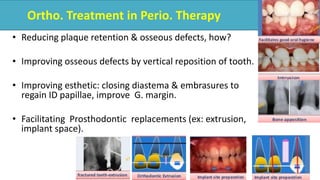
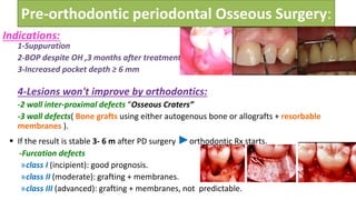
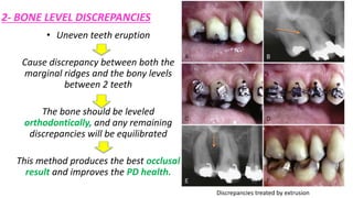
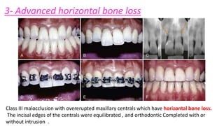
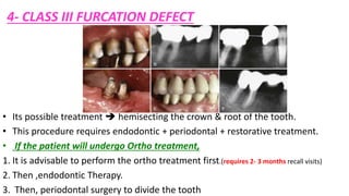
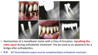
Ortho. Treatment in Perio. Therapy
• Reducing plaque retention & osseous defects, how?
• Improving osseous defects by vertical reposition of tooth.
• Improving esthetic: closing diastema & embrasures to
regain ID papillae, improve G. margin.
• Facilitating Prosthodontic replacements (ex: extrusion,
implant space).
Pre-orthodontic periodontal therapy:
Non-surgical treatment
– Remove etiological factors(SRP)
– Individualized home-care program(OHI)
– Re-evaluated after 3 months,
– if stable► start orthodontic Rx after another 3 months (total 6
months)- full effect SRP not obtained till 4-6 months.
– Patient is re-evaluated every 2-3 months by periodontist through
orthodontic treatment.
Pre-orthodontic periodontal Osseous Surgery:
1-Suppuration
2-BOP despite OH ,3 months after treatment
3-Increased pocket depth ≥ 6 mm
4-Lesions won't improve by orthodontics:
-2 wall inter-proximal defects “Osseous Craters”
-3 wall defects( Bone grafts using either autogenous bone or allografts + resorbable
membranes ).
 If the result is stable 3- 6 m after PD surgery ►orthodontic Rx starts.
-Furcation defects
»class I (incipient): good prognosis.
»class II (moderate): grafting + membranes.
»class III (advanced): grafting + membranes, not predictable.
Indications:
Treatment of crater before Ortho. Treatment Treatment of 3 wall defect before ortho. Treatment
6mm
3mm
Orthodontic Treatment of Osseous Defects
• Are 1 or 2 wall osseous Defects (often are found around mesially tipped or supererupted teeth)
1- Hemiseptal defects
In the case of the tipped tooth, uprighting and
eruption of the tooth levels the bony defect.
In case of supererupted tooth, intrusion and leveling
of the adjacent CEJ help to level the osseous defect.
2- BONE LEVEL DISCREPANCIES
• Uneven teeth eruption
Cause discrepancy between both the
marginal ridges and the bony levels
between 2 teeth
The bone should be leveled
orthodontically, and any remaining
discrepancies will be equilibrated
This method produces the best occlusal
result and improves the PD health.
Discrepancies treated by extrusion
3- Advanced horizontal bone loss
Class III malocclusion with overerupted maxillary centrals which have horizontal bone loss.
The incisal edges of the centrals were equilibrated , and orthodontic Completed with or
without intrusion .
4- CLASS III FURCATION DEFECT
• Its possible treatment  hemisecting the crown & root of the tooth.
• This procedure requires endodontic + periodontal + restorative treatment.
• If the patient will undergo Ortho treatment,
1. It is advisable to perform the ortho treatment first.(requires 2- 3 months recall visits)
2. Then ,endodontic Therapy.
3. Then, periodontal surgery to divide the tooth
• Hemisection of a mandibular molar with a Class III furcation →pushing the
roots apart during orthodontic treatment →to be used as an abutment for a
bridge after orthodontics.
• N.B RCT & periodontal surgery must be completed before orthodontic treatment.
5- IMPLANTS
• The implant must remain embedded in bone for 4 -6 months after placement
before it can be loaded as an orthodontic anchor.
• If the implant won’t be an anchor…… it may be placed after the orthodontic Rx.
6- ROOT PROXIMITY
• This approach generally improves the PD health of this area:
1.This opens the embrasure beneath the tooth contact(correct
gingival defects)
2.Provides additional bone support.
3.Increase OH accessibility.
Forced eruption(extruded 4mm) by orthodontic treatment then
crown lengthening surgery was done
Pre-operative
• The overextension of prepration of
the crown margin will cause
invasion of the BW persistent
inflammation of the gingiva.
• Ex: Erupt the fracture root margin
coronally to be properly restored.
BUT if the fracture extends too
apically…….extraction.
7- FRACTURED TEETH & FORCED
ERUPTION
The root: crown ratio should be about 1 : 1.
• If less too little root remaining in the bone for stability.
1- Root length
2. Root shape /form:
Broad & non-tapering>> thin & tapered.
• A thin tapered root provides a narrower cervical region
compromise the esthetics of the final restoration.
• The root canal should not be > 1/3 of the overall internal width
to provide adequate strength for the final restoration.
• If the root canal is wide thin preparation  early fracture.
• 3 -Level of the fracture: If the fractured crown 2 -3 mm apical to Bone level, it is
difficult to attach to the root to erupt it.
• 4-Relative importance of the tooth :
Example: If the patient is 70 years and both adjacent teeth have prosthetic crowns,
so construct a fixed bridge is better.
However, if the patient is 15 years of age and the adjacent teeth are unrestored,
forced eruption is much better.
• 5-Esthetics: high lip line and displays 2 - 3 mm of gingiva when smiling, and any
type of restoration in this area will be more obvious.
• 6-Endodontic/periodontal prognosis:
• If the tooth has a significant periodontal defect and can’t retain the root.
• If the tooth root has a vertical fracture, the poor prognosis…….extraction
The root may be erupted rapidly or slowly:
• Rapidly  the bone will be left behind temporarily, and a circumferential
fiberotomy is performed to prevent bone from following the erupted root.
• Slowly  the G & bone follows the tooth, this may requires crown
lengthening(w or wout bone reduction).
• As the root erupts, the G. moves coronally with the tooth. So, the clinical
crown becomes shorter after extrusion …..Gingival SURGERY
Orthodontic treatment for Gingival Discrepancies(Uneven G.
margin)
4 factors contribute to IDEAL GINGIVAL FORM:
1. The G. margin of the 2 centrals  at the same level.
2. The G. margin of the centrals  more apically to the laterals &
at the same level of the canines.
3. The labial G. margins mimic the CEJs.
4. A papilla should exist between each tooth. The height of the
papilla is halfway between the incisal edge & the G. margin of
each ant. tooth.
(So, the papilla occupies ½ interproximal contact, and the adjacent
teeth form the ½ of the contact) The papilla/contact ratio is 1 : 1.
Surgical correction of G. margin
discrepancies.
Orthodontic movement to
reposition the G. margin.
Significant Abrasion and Over eruption
• Abraded teeth are impossible for crown preparation due to the lack of crown length.
• Two options are available:
1. Extensive crown lengthening (Apically displaced flap+ removing sufficient bone)
(contraindicated in short, tapered roots because it will affect the root: crown & open G. embrasures
of anterior teeth).
2. Orthodontically intrude the teeth and move the G. margin apically.
• it is necessary to hold these teeth for at least 6 months in the intruded position to make the
principal fibers of the periodontium accommodate to the new intruded position.
Ortho. intrusion of severely abraded and overerupted teeth >> crown lengthening.
Open Gingival Embrasures
• This open space is usually caused by
:
1. Tooth shape
2. Root angulation
3. Periodontal bone loss
 Closing open contact.
 Restore OR reshape open embrasure “Interproximal
stripping and close space”.
 Correct the root angulation “Converge root angulations”
 Periodontal surgery to augment interdental papilla.
Treatment of black triangle
TOOTH SHAPE ROOT ANGULATION
IATROGENIC EFFECTS ASSOCIATED WITH ORTHO. TX.
N.B Root resorption can be repaired by cementum deposition.
Response of periodontium to ortho. Tooth movement
23
zones of compression & tension
Strong forces (>>> capillary blood pressure)
Crushed ► ischemia ►hyalinized ►delayed
tooth movement.
Light forces (<<<capillary blood pressure)
tooth movement continuous with simultaneous
bone resorption + formation.
24
Presence Of Periodontitis Or Any Active Periodontal dis.
Will Contraindicate Orthodontic Rx
Thank you

Perio ortho relationship

  • 2.
    Ortho. Treatment inPerio. Therapy • Reducing plaque retention & osseous defects, how? • Improving osseous defects by vertical reposition of tooth. • Improving esthetic: closing diastema & embrasures to regain ID papillae, improve G. margin. • Facilitating Prosthodontic replacements (ex: extrusion, implant space).
  • 3.
    Pre-orthodontic periodontal therapy: Non-surgicaltreatment – Remove etiological factors(SRP) – Individualized home-care program(OHI) – Re-evaluated after 3 months, – if stable► start orthodontic Rx after another 3 months (total 6 months)- full effect SRP not obtained till 4-6 months. – Patient is re-evaluated every 2-3 months by periodontist through orthodontic treatment.
  • 4.
    Pre-orthodontic periodontal OsseousSurgery: 1-Suppuration 2-BOP despite OH ,3 months after treatment 3-Increased pocket depth ≥ 6 mm 4-Lesions won't improve by orthodontics: -2 wall inter-proximal defects “Osseous Craters” -3 wall defects( Bone grafts using either autogenous bone or allografts + resorbable membranes ).  If the result is stable 3- 6 m after PD surgery ►orthodontic Rx starts. -Furcation defects »class I (incipient): good prognosis. »class II (moderate): grafting + membranes. »class III (advanced): grafting + membranes, not predictable. Indications:
  • 5.
    Treatment of craterbefore Ortho. Treatment Treatment of 3 wall defect before ortho. Treatment 6mm 3mm
  • 6.
    Orthodontic Treatment ofOsseous Defects • Are 1 or 2 wall osseous Defects (often are found around mesially tipped or supererupted teeth) 1- Hemiseptal defects In the case of the tipped tooth, uprighting and eruption of the tooth levels the bony defect. In case of supererupted tooth, intrusion and leveling of the adjacent CEJ help to level the osseous defect.
  • 7.
    2- BONE LEVELDISCREPANCIES • Uneven teeth eruption Cause discrepancy between both the marginal ridges and the bony levels between 2 teeth The bone should be leveled orthodontically, and any remaining discrepancies will be equilibrated This method produces the best occlusal result and improves the PD health. Discrepancies treated by extrusion
  • 8.
    3- Advanced horizontalbone loss Class III malocclusion with overerupted maxillary centrals which have horizontal bone loss. The incisal edges of the centrals were equilibrated , and orthodontic Completed with or without intrusion .
  • 9.
    4- CLASS IIIFURCATION DEFECT • Its possible treatment  hemisecting the crown & root of the tooth. • This procedure requires endodontic + periodontal + restorative treatment. • If the patient will undergo Ortho treatment, 1. It is advisable to perform the ortho treatment first.(requires 2- 3 months recall visits) 2. Then ,endodontic Therapy. 3. Then, periodontal surgery to divide the tooth
  • 10.
    • Hemisection ofa mandibular molar with a Class III furcation →pushing the roots apart during orthodontic treatment →to be used as an abutment for a bridge after orthodontics. • N.B RCT & periodontal surgery must be completed before orthodontic treatment.
  • 11.
    5- IMPLANTS • Theimplant must remain embedded in bone for 4 -6 months after placement before it can be loaded as an orthodontic anchor. • If the implant won’t be an anchor…… it may be placed after the orthodontic Rx.
  • 12.
    6- ROOT PROXIMITY •This approach generally improves the PD health of this area: 1.This opens the embrasure beneath the tooth contact(correct gingival defects) 2.Provides additional bone support. 3.Increase OH accessibility.
  • 13.
    Forced eruption(extruded 4mm)by orthodontic treatment then crown lengthening surgery was done Pre-operative • The overextension of prepration of the crown margin will cause invasion of the BW persistent inflammation of the gingiva. • Ex: Erupt the fracture root margin coronally to be properly restored. BUT if the fracture extends too apically…….extraction. 7- FRACTURED TEETH & FORCED ERUPTION
  • 14.
    The root: crownratio should be about 1 : 1. • If less too little root remaining in the bone for stability. 1- Root length 2. Root shape /form: Broad & non-tapering>> thin & tapered. • A thin tapered root provides a narrower cervical region compromise the esthetics of the final restoration. • The root canal should not be > 1/3 of the overall internal width to provide adequate strength for the final restoration. • If the root canal is wide thin preparation  early fracture.
  • 15.
    • 3 -Levelof the fracture: If the fractured crown 2 -3 mm apical to Bone level, it is difficult to attach to the root to erupt it. • 4-Relative importance of the tooth : Example: If the patient is 70 years and both adjacent teeth have prosthetic crowns, so construct a fixed bridge is better. However, if the patient is 15 years of age and the adjacent teeth are unrestored, forced eruption is much better. • 5-Esthetics: high lip line and displays 2 - 3 mm of gingiva when smiling, and any type of restoration in this area will be more obvious. • 6-Endodontic/periodontal prognosis: • If the tooth has a significant periodontal defect and can’t retain the root. • If the tooth root has a vertical fracture, the poor prognosis…….extraction
  • 16.
    The root maybe erupted rapidly or slowly: • Rapidly  the bone will be left behind temporarily, and a circumferential fiberotomy is performed to prevent bone from following the erupted root. • Slowly  the G & bone follows the tooth, this may requires crown lengthening(w or wout bone reduction). • As the root erupts, the G. moves coronally with the tooth. So, the clinical crown becomes shorter after extrusion …..Gingival SURGERY
  • 17.
    Orthodontic treatment forGingival Discrepancies(Uneven G. margin) 4 factors contribute to IDEAL GINGIVAL FORM: 1. The G. margin of the 2 centrals  at the same level. 2. The G. margin of the centrals  more apically to the laterals & at the same level of the canines. 3. The labial G. margins mimic the CEJs. 4. A papilla should exist between each tooth. The height of the papilla is halfway between the incisal edge & the G. margin of each ant. tooth. (So, the papilla occupies ½ interproximal contact, and the adjacent teeth form the ½ of the contact) The papilla/contact ratio is 1 : 1.
  • 18.
    Surgical correction ofG. margin discrepancies. Orthodontic movement to reposition the G. margin.
  • 19.
    Significant Abrasion andOver eruption • Abraded teeth are impossible for crown preparation due to the lack of crown length. • Two options are available: 1. Extensive crown lengthening (Apically displaced flap+ removing sufficient bone) (contraindicated in short, tapered roots because it will affect the root: crown & open G. embrasures of anterior teeth). 2. Orthodontically intrude the teeth and move the G. margin apically. • it is necessary to hold these teeth for at least 6 months in the intruded position to make the principal fibers of the periodontium accommodate to the new intruded position. Ortho. intrusion of severely abraded and overerupted teeth >> crown lengthening.
  • 20.
    Open Gingival Embrasures •This open space is usually caused by : 1. Tooth shape 2. Root angulation 3. Periodontal bone loss  Closing open contact.  Restore OR reshape open embrasure “Interproximal stripping and close space”.  Correct the root angulation “Converge root angulations”  Periodontal surgery to augment interdental papilla. Treatment of black triangle
  • 21.
    TOOTH SHAPE ROOTANGULATION
  • 22.
    IATROGENIC EFFECTS ASSOCIATEDWITH ORTHO. TX. N.B Root resorption can be repaired by cementum deposition.
  • 23.
    Response of periodontiumto ortho. Tooth movement 23 zones of compression & tension Strong forces (>>> capillary blood pressure) Crushed ► ischemia ►hyalinized ►delayed tooth movement. Light forces (<<<capillary blood pressure) tooth movement continuous with simultaneous bone resorption + formation.
  • 24.
    24 Presence Of PeriodontitisOr Any Active Periodontal dis. Will Contraindicate Orthodontic Rx
  • 25.

Editor's Notes

  • #3 Treatment of malocclusion and malaligmnet of teeth and trauma from occlusion ………….. Easily OHI and reduce bone defects………….reduce plaque retention Improving G by treatment of diestma and ID recsssion and treatment of open embrassures Vertical repostioning can improve certain types of bone defects
  • #4 Patient is re-evaluated every 2-3 months by periodontist through ortho.treatment ►as chronic periodontitis have short exacerbations and long remissions (may be years)
  • #5 The extent of the osseous surgery depends on the type of defect (e.g., crater, hemiseptal defect, three-wall defect, or furcation lesion). The prudent clinician knows which defects can be improved with orthodontic treatment and which defects require preorthodontic, periodontal, or surgical intervention. GTR
  • #6 This patient had a 6-mm probing defect distal to the maxillary 6 (A). When this area was flapped (B), a cratering defect was apparent. Osseous surgery was used to alter the bony architecture on the buccal and lingual surfaces to eliminate the defect (C and D). After 6 weeks, the probing pocket defect had been reduced to 3 mm and orthodontic appliances were placed on the teeth (E). By eliminating the crater before orthodontic therapy, the patient could maintain the area during and after orthodontic treatment (F). This patient had a significant periodontal pocket (A) distal to the mandibular right first molar. Periapical radiograph (B) confirmed the osseous defect. A flap was elevated (C), revealing a deep, three-wall osseous defect. Freeze-dried bone (D) was placed in the defect. Six months after the bone graft, orthodontic treatment was initiated (E). The final periapical radiograph shows that the preorthodontic bone graft helped regenerate bone and eliminate the defect distal to the molar (F).
  • #7 This patient was missing the mandibular 5 , and the first molar had tipped mesially (A). Pretreatment periapical radiograph (B) revealed a significant hemiseptal osseous defect on the mesial side of the molar. To eliminate the defect, the molar was erupted, and the occlusal surface was equilibrated (C). The eruption was stopped when the bone defect was leveled (D). The posttreatment intraoral photograph (E) and periapical radiograph (F) show that the periodontal health had been improved by correcting the hemiseptal defect orthodontically.
  • #8 During orthodontic treatment, when teeth are being extruded to level hemiseptal defects, the patient should be monitored regularly. Initially, the hemiseptal defect has a greater sulcular depth and is more difficult for the patient to clean. As the defect is ameliorated through tooth extrusion, interproximal cleaning becomes easier. The patient should be recalled every 2 to 3 months during the leveling process to control inflammation in the interproximal region.
  • #9 Before orthodontic treatment, this patient had a significant class III malocclusion (A). The maxillary central incisors had overerupted (B) relative to the occlusal plane. Pretreatment periapical radiograph (C) showed that significant horizontal bone loss had occurred. To avoid creating a vertical periodontal defect by intruding the central incisors, the brackets were placed to maintain the bone height (D). The incisal edges of the centrals were equilibrated (E), and the orthodontic treatment was completed without intruding the incisors (F).
  • #10 Furcation involvement needs special consideration and the worse to handle or maintain and may be become worse by ortho If a patient with a Class III furcation defect will be undergoing orthodontic treatment, a possible method for treating the furcation is to eliminate it by hemisecting the crown and root of the tooth This procedure requires endodontic, periodontal, and restorative treatment. Pic…..furcation grade iii in abutment lower 7 Ortho is done then endo then perio then resto
  • #11 Hemi section of a mandibular molar with a Class III furcation……pushing the roots apart during orthodontic treatment……to be used as an abutment for a bridge after orthodontics, N.B Endodontic therapy, and periodontal surgery must be completed before the start of orthodontic treatment About 7 or 8 mm may be created between the roots of the hemisected molar This process eliminates the original ,furcation problem and allows the patient to clean the area with greater efficiency
  • #12 It must be placed precisely so that it not only provides an anchor for tooth movement
  • #13 Crown recontouring is needed for occlusal adjustment because the roots are moving apart. So ,the crowns may develop an unusual occlusal contact with the opposing arch.
  • #15 Broad and non-tapering rather than thin and taperedtapered root provides a narrower cervical region after the tooth has been erupted 4 mm. This could compromise the esthetic appearance of the final restoration. If the root canal is wide…..the distance between the external root surface and root canal filling will be narrow. In these patients, the walls of the crown preparation are thin, which could result in early fracture of the restored root.
  • #16 5-Esthetics: If the patient has a high lip line and displays 2 to 3 mm of gingiva when smiling, any type of restoration in this area will be more obvious. Keeping the patient's own tooth would be much more esthetic than any type of implant or prosthetic replacement.
  • #17 If all these factors are favorable, forced eruption of the fractured root is indicated. The orthodontic mechanics necessary to erupt the tooth can vary from elastic traction to orthodontic banding and bracketing. If a large portion of the tooth is still present, orthodontic bracketing is necessary. If the entire crown has fractured, leaving only the root, elastic traction from a bonded bar may be possible. The root may be erupted rapidly or slowly. If the movement is performed rapidly, the alveolar bone will be left behind temporarily, and a circumferential fiberotomy may be performed to prevent bone from following the erupted root. However, if the root is erupted slowly, the bone follows the tooth. In this situation, the erupted root requires crown lengthening to expose the correct amount of tooth to create the proper ferrule, resistance form, and retention for the final restoration. After the tooth root has been erupted, it must be stabilized to prevent it from intruding back into the alveolus. The reason for reintrusion is the orientation of the principal fibers of the periodontium. During forced eruption, the periodontal fibers become oriented obliquely and stretched as the root moves coronally. These fibers eventually reorient themselves after about 6 months. Before this occurs, the root can reintrude significantly. Therefore, if this type of treatment is performed, an adequate period of stabilization is necessary to avoid significant relapse and reintrusion of the root. As the root erupts, the gingiva moves coronally with the tooth. As a result, the clinical crown length becomes shorter after extrusion (see Figure 50-10). In addition, the gingival margin may be positioned more incisally than the adjacent teeth. In these patients, gingival surgery is necessary to create ideal gingival margin heights. The type of surgery varies, depending on whether bone removal is necessary. If bone has followed the root during eruption, a flap is elevated, and the appropriate amount of bone is removed to match the bone height of the adjacent teeth. If the bone level is flat between adjacent teeth, a simple excisional gingivectomy corrects the gingival margin discrepancy. After gingival surgery, an open gingival embrasure may exist between the erupted root and adjacent teeth (see Figure 50-10). The space occurs because the narrower root portion of the erupted tooth has been moved into the oral cavity. This space may be closed in two ways: (1) overcontouring of the replacement restoration and (2) reshaping of the crown of the tooth and movement of the root to close the space. The second method often helps improve the overall shape of the final crown on the restored tooth.
  • #20 patients have destructive dental habits, such as a protrusive bruxing habit, that can result in significant wear of the maxillary and mandibularincisors and compensatory overeruption of these teeth The restoration of these abraded teeth is often impossible because of the lack of crown length to achieve adequate retention and resistance form for the crown preparations. Two options are available. One option is extensive crown lengthening by elevating a flap, removing sufficient bone, and apically positioning the flap to expose adequate tooth length for crown preparation. However, this type of procedure is contraindicated in the patient with short, tapered roots because it could adversely affect the final root-to-crown ratio and potentially open gingival embrasures(black triangle formation) between the anterior teeth. The other option for improving The restorability of these short abraded teeth is to intrude the teeth orthodontically and move the gingival margins apically (see Figure 50-13). It is possible to intrude up to four maxillary incisors by using the posterior teeth as anchorage during the intrusion process. When abraded teeth are significantly intruded, it is necessary to hold these teeth for at least 6 months in the intruded position with orthodontic brackets or archwires (or both), or some type of bonded retainer. The principal fibers of the periodontium must accommodate to the new intruded position, a process that could take a minimum of 6 months in most adult patients. Orthodontic intrusion of severely abraded and overerupted teeth is usually a distinct advantage over periodontal crown lengthening, unless the patient has extremely long and broad roots or has had extensive horizontal periodontal bone loss.
  • #21 The interproximal contact between the maxillary central incisors consists of two parts: the tooth contact and the papilla. The papilla/contact ratio is 1 : 1. Half the space is occupied by papilla, and half is formed by the tooth contact. If the patient has an open embrasure, the first aspect that must be evaluated is whether the problem is caused by the papilla or the tooth contact. If the papilla is the problem, the cause is usually a lack of bone support because of an underlying periodontal problem. In some situations, a deficient papilla can be improved with orthodontic treatment. By closing open contacts, the interproximal gingiva can be squeezed and moved incisally. This type of movement may help create a more esthetic papilla between two teeth despite alveolar bone loss. Another possibility is to erupt adjacent teeth when the interproximal bone level is positioned apically. Most open embrasures between the central incisors are caused by problems with tooth contact. The first step in the diagnosis of this problem is to evaluate a periapical radiograph of the central incisors. If the root angulation is divergent, the brackets should be repositioned so that the root position can be corrected (Figure 50-14). In these patients the incisal edges may be uneven and require restoration with either composite or porcelain restorations. If the periapical radiograph shows that the roots are in their correct relationship, the open gingival embrasure is caused by a triangular tooth shape (Figure 50-15). If the shape of the tooth is the problem, two solutions are possible: (1) restore the open gingival embrasure or (2) reshape the tooth by flattening the incisal contact and closing the space (see Figure 50-15). This second option results in lengthening of the contact until it meets the papilla. In addition, if the embrasure space is large, closing the space squeezes the papilla between the central incisors. This helps create a 1:1 ratio between the contact and papilla and restores uniformity to the heights between the midline and adjacent papillae.
  • #22  triangle shaped centrals are the cause of the open embrasures …….The mesial surfaces of the centrals were contoured then diastema was closed by ortho …tooth contact moved gingivally and the papillae moved incisally
  • #23 Effect of bands on periodontuim……………..gingivitis and root resorption and bone loss
  • #24 Bone resorption in pressure zone Bone deposition in tension zone On Pressure zone……Intial period of movement 6-8 days to compress PDL ……cause cur of BV(become avascular)…..hyalinization (free cell zone)….then periodontal regeneratiion eliminate hylaization Tooth movement result from controlled force Response of perio. depends on magnitude & duration of these force