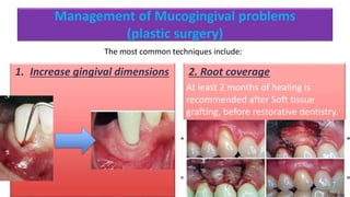
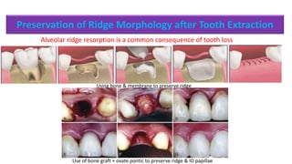
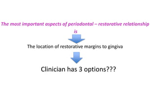
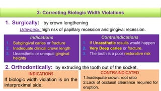
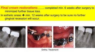
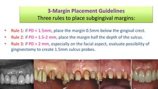
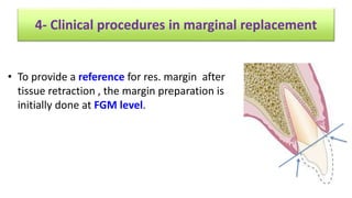
Preparation of the Periodontium
for Restorative Dentistry
Dr. DIANA ABO EL OLA
To achieve the long-term comfort & good functioning predictable treatment.
Active PD infection must be treated before restorative, esthetic, and implant
dentistry
SO, healthy periodontuim ……………… successful comprehensive dentistry
1- To ensure stable GM before tooth preparation avoid shrinkage
& bleeding.
2- To provide adequate tooth length for retention & accessibility.
3- After the resolution of infl., teeth may repositioned or changes
may occur in the ST….. may interfere with prosthetic procedures.
4- The Quality ,quantity & topography of the periodontuim
provides structural defense factors in maintaining health.
SEQUENCE OF TREATMENT
The preparation of the periodontium for restorative dentistry can be
divided into 2 phases:
(2) Pre-prosthetic periodontal
surgeries
-Management of Mucogingival
problems.
-Preservation of ridge morphology
after tooth extraction.
-Crown-lengthening procedures.
-Alveolar ridge reconstruction.
(1)Control of PD infl. with or without
surgery
Emergency Rx
Extraction of hopeless teeth
OH instructions
Scaling and root planing
Re-evaluation
Periodontal surgery
Adjunctive orthodontic therapy
Management of Mucogingival problems
(plastic surgery)
1. Increase gingival dimensions 2. Root coverage
At least 2 months of healing is
recommended after Soft tissue
grafting, before restorative dentistry.
The most common techniques include:
Preservation of Ridge Morphology after Tooth Extraction
Alveolar ridge resorption is a common consequence of tooth loss
Using bone & membrane to preserve ridge
Use of bone graft + ovate pontic to preserve ridge & ID papillae
Crown lengthening procedures
It can be done with or without osseous reduction( apical position flap )
Alveolar Ridge Reconstruction
Indications:
• to provide adequate anatomic dimensions for esthetic pontic or implants.
For Small defects may be treated with ST ridge augmentation
For larger defects and in those sites receiving dental implants, hard tissue
modalities are used
RESTORATIVE- PERIO
INTERRELATIONSHIPS
The most important aspects of periodontal – restorative relationship
is
The location of restorative margins to gingiva
Clinician has 3 options???
Clinician has 3 options
Supragingival margins
(less contact to GM.)
Equigingival margin
(even with the GM.)
Subgingival margin
(risk of violation of biological
width)
• Equigingival margin →not desirable →retain more plaque→ Gingival
inflammation.
FROM A PERIODONTAL VIEW POINT
Supra gingival and
equigingival are well
tolerated
Greatest biological
risk occurs in
subgingival margin
• Subgingival margin should be placed not more than 0.5mm into
the sulcus.
(1) to create resistance and retention for restorative prepration (inadequate
clinical crown)
(2) to make significant contour for caries removal or tooth defects or fracture
(3) to mask the tooth/restoration interface(for esthetic purpose)
BUT
BW violation
Sulcus =0.69mm
JE=0.97mm
CT=1.07mm
Total distance from GM to bone =3mm
SO
(flat G. scallops, thick cortical plates & inc.
thickness of KG.)……… little apical migration and intrabony pocket
formation are observed.
(highly scalloped & ??)……….more susceptible to G.
recession than the thick periodontium.
• When the margin is placed too far below the gingival crest, 2 different responses can
be observed:
Biological considerations :
• 1-Evaluation of BW
• 2-Correction of BW Violations
• 3-Margin Placement Guidelines
• 4-Managing Interproximal Embrasures
• 5-Correcting Open G. Embrasures Restoratively
• 6-Managing G. Embrasure Form for Patients with G. Recession
• 7-Pontic Design
1-Biological width evaluation
Radiographically
Can identify interproximal violation of
biological width only, Y?
Using periodontal probe
Pushing probe through attachment
tissue from sulcus to bone
If distance <2mmviolation confirmed If distance <3mmviolation confirmed
2- Correcting Biologic Width Violations
1. Surgically: by crown lengthening
Drawback: high risk of papillary recession and gingival recession.
Indications
1. Subgingival caries or fracture.
2. Inadequate clinical crown length
3. Unaesthetic or unequal gingival
heights.
Contraindications
1. If Unaesthetic results would happen.
2. Very Deep caries or fracture.
3. The tooth is a poor restorative risk.
2. Orthodontically: by extruding the tooth out of the socket,
INDICATIONS
If biologic width violation is on the
interproximal side.
CONTRAINDICATED
1.Inadequate crown: root ratio
2.Lack of occlusal clearance required for
eruption.
Gingivectomy
(Soft tissue surgery)
Or
Osseous surgery
Or
Orthodontic treatment
Indicated in case of pseudopockets - gummy smile-
probing depth >3mm
Needs adequate Keratinized gingiva.
Scalloped marginal incision +crestal bone reduction at
least 2mm away from restoration margin+ apically
displaced flap (ADF)/UDF.
Disadv: risk of ID recession (black triangle)
1)Slow ortho. Extrusion + apically positioned flap
Or
2)Rapid ortho. Extrusion (preventing gingiva& bone to
follow the tooth)
Indicated in case of narrow zone of attached gingiva
Crownlengthening
• Gingivectomy
ADF with osseous surgery
Final crown restorations…….. completed min. 6 weeks after surgery to
minimized further tissue loss.
In esthetic areas  min. 12 weeks after surgery to be sure no further
gingival recession will occur.
Ortho. Treatment
• In the case of caries or tooth fracture
The surgery should provide at least 4 mm
from the apical extent of the caries or
fracture to the bone crest.
3-Margin Placement Guidelines
Three rules to place subgingival margins:
• Rule 1: if PD < 1.5mm, place the margin 0.5mm below the gingival crest.
• Rule 2: if PD = 1.5-2 mm, place the margin half the depth of the sulcus.
• Rule 3: if PD > 2 mm, especially on the facial aspect, evaluate possibility of
gingivectomy to create 1.5mm sulcus probes.
• To provide a reference for res. margin after
tissue retraction , the margin preparation is
initially done at FGM level.
4- Clinical procedures in marginal replacement
5- Managing interproximal embrasures
Papillary height related to
1.Level of the bone
2.Biologic width
3.Embrasure form
• Too Wide Embrasure Flattened & Blunt Papilla
• Too Narrow Embrasure Inflamed Papilla (papilla may grow out)
• Ideal Embrasure Healthy & pointed Papilla(sulcus 3mm)
Tapered teeth+ wide embrasure Narrow embrasure Ideal embrasure
• GM about 3 mm above the facial bone
• Tip of the papilla about 4.5 -5 mm above
interproximal bone.
Same biologic width for both (2mm)
•Interprox. area Sulcus is deeper
than in the facial surface.
Distance from inter proximal contact to bone level ≤ 5mm papilla always filled 6mm (56%) 7mm(37%)
Interproximal tooth contact
6- Correcting Open Gingival Embrasures by
Restoration
• The papilla is inadequate in height because of
(1) Bone loss
(2) Too high interproximal contact coronally(long embrasure)
• If the papilla is apical to the adjacent papillae; the interproximal
bone levels should be evaluated .
• If the bone is apical to the adj. bone levels bone loss is the cause.
at the same level  the open embrasure is the cause.
1- Diverging roots
2- Tapered Tooth shape
It can be corrected by moving the contact point to the tip of the papilla by
restoration OR margins prepared 1-1.5mm below papillae
7-Managing open Embrasure Form for Patients with
Gingival Recession
1. Carry the interproximal contacts apically  to eliminate the open embrasures
2. Multiple-unit restorations tissue-colored ceramics porcelain papillae
directly on the restoration.
The interproximal contacts should be moved apically to
minimize large food traps but still leave enough space for
interdental brush for hygiene.
In esthetic areas:
In the post. areas
Correcting the open embrasures by periodontal surgery
preoperative: loss of ID papillae + class IV CT graft from tuberosity +bone from
tuberosity
Coronally positioned flap
Bone graft is fixed by Ti screw &
covered by ST graft
8-Pontic design
SANITARY RIDGE LAP MODIFIED RIDGE OVATE
👍🏻
Thank you

Perio resorative inter-relationship.

  • 1.
    Preparation of thePeriodontium for Restorative Dentistry Dr. DIANA ABO EL OLA
  • 2.
    To achieve thelong-term comfort & good functioning predictable treatment. Active PD infection must be treated before restorative, esthetic, and implant dentistry SO, healthy periodontuim ……………… successful comprehensive dentistry
  • 3.
    1- To ensurestable GM before tooth preparation avoid shrinkage & bleeding. 2- To provide adequate tooth length for retention & accessibility. 3- After the resolution of infl., teeth may repositioned or changes may occur in the ST….. may interfere with prosthetic procedures. 4- The Quality ,quantity & topography of the periodontuim provides structural defense factors in maintaining health.
  • 4.
    SEQUENCE OF TREATMENT Thepreparation of the periodontium for restorative dentistry can be divided into 2 phases: (2) Pre-prosthetic periodontal surgeries -Management of Mucogingival problems. -Preservation of ridge morphology after tooth extraction. -Crown-lengthening procedures. -Alveolar ridge reconstruction. (1)Control of PD infl. with or without surgery Emergency Rx Extraction of hopeless teeth OH instructions Scaling and root planing Re-evaluation Periodontal surgery Adjunctive orthodontic therapy
  • 5.
    Management of Mucogingivalproblems (plastic surgery) 1. Increase gingival dimensions 2. Root coverage At least 2 months of healing is recommended after Soft tissue grafting, before restorative dentistry. The most common techniques include:
  • 6.
    Preservation of RidgeMorphology after Tooth Extraction Alveolar ridge resorption is a common consequence of tooth loss Using bone & membrane to preserve ridge Use of bone graft + ovate pontic to preserve ridge & ID papillae
  • 7.
    Crown lengthening procedures Itcan be done with or without osseous reduction( apical position flap )
  • 8.
    Alveolar Ridge Reconstruction Indications: •to provide adequate anatomic dimensions for esthetic pontic or implants. For Small defects may be treated with ST ridge augmentation For larger defects and in those sites receiving dental implants, hard tissue modalities are used
  • 9.
  • 10.
    The most importantaspects of periodontal – restorative relationship is The location of restorative margins to gingiva Clinician has 3 options???
  • 11.
    Clinician has 3options Supragingival margins (less contact to GM.) Equigingival margin (even with the GM.) Subgingival margin (risk of violation of biological width)
  • 12.
    • Equigingival margin→not desirable →retain more plaque→ Gingival inflammation. FROM A PERIODONTAL VIEW POINT Supra gingival and equigingival are well tolerated Greatest biological risk occurs in subgingival margin
  • 13.
    • Subgingival marginshould be placed not more than 0.5mm into the sulcus. (1) to create resistance and retention for restorative prepration (inadequate clinical crown) (2) to make significant contour for caries removal or tooth defects or fracture (3) to mask the tooth/restoration interface(for esthetic purpose) BUT BW violation
  • 14.
  • 15.
    (flat G. scallops,thick cortical plates & inc. thickness of KG.)……… little apical migration and intrabony pocket formation are observed. (highly scalloped & ??)……….more susceptible to G. recession than the thick periodontium. • When the margin is placed too far below the gingival crest, 2 different responses can be observed:
  • 16.
    Biological considerations : •1-Evaluation of BW • 2-Correction of BW Violations • 3-Margin Placement Guidelines • 4-Managing Interproximal Embrasures • 5-Correcting Open G. Embrasures Restoratively • 6-Managing G. Embrasure Form for Patients with G. Recession • 7-Pontic Design
  • 17.
    1-Biological width evaluation Radiographically Canidentify interproximal violation of biological width only, Y? Using periodontal probe Pushing probe through attachment tissue from sulcus to bone If distance <2mmviolation confirmed If distance <3mmviolation confirmed
  • 18.
    2- Correcting BiologicWidth Violations 1. Surgically: by crown lengthening Drawback: high risk of papillary recession and gingival recession. Indications 1. Subgingival caries or fracture. 2. Inadequate clinical crown length 3. Unaesthetic or unequal gingival heights. Contraindications 1. If Unaesthetic results would happen. 2. Very Deep caries or fracture. 3. The tooth is a poor restorative risk. 2. Orthodontically: by extruding the tooth out of the socket, INDICATIONS If biologic width violation is on the interproximal side. CONTRAINDICATED 1.Inadequate crown: root ratio 2.Lack of occlusal clearance required for eruption.
  • 19.
    Gingivectomy (Soft tissue surgery) Or Osseoussurgery Or Orthodontic treatment Indicated in case of pseudopockets - gummy smile- probing depth >3mm Needs adequate Keratinized gingiva. Scalloped marginal incision +crestal bone reduction at least 2mm away from restoration margin+ apically displaced flap (ADF)/UDF. Disadv: risk of ID recession (black triangle) 1)Slow ortho. Extrusion + apically positioned flap Or 2)Rapid ortho. Extrusion (preventing gingiva& bone to follow the tooth) Indicated in case of narrow zone of attached gingiva Crownlengthening
  • 20.
  • 21.
    Final crown restorations……..completed min. 6 weeks after surgery to minimized further tissue loss. In esthetic areas  min. 12 weeks after surgery to be sure no further gingival recession will occur. Ortho. Treatment
  • 22.
    • In thecase of caries or tooth fracture The surgery should provide at least 4 mm from the apical extent of the caries or fracture to the bone crest.
  • 23.
    3-Margin Placement Guidelines Threerules to place subgingival margins: • Rule 1: if PD < 1.5mm, place the margin 0.5mm below the gingival crest. • Rule 2: if PD = 1.5-2 mm, place the margin half the depth of the sulcus. • Rule 3: if PD > 2 mm, especially on the facial aspect, evaluate possibility of gingivectomy to create 1.5mm sulcus probes.
  • 24.
    • To providea reference for res. margin after tissue retraction , the margin preparation is initially done at FGM level. 4- Clinical procedures in marginal replacement
  • 25.
    5- Managing interproximalembrasures Papillary height related to 1.Level of the bone 2.Biologic width 3.Embrasure form • Too Wide Embrasure Flattened & Blunt Papilla • Too Narrow Embrasure Inflamed Papilla (papilla may grow out) • Ideal Embrasure Healthy & pointed Papilla(sulcus 3mm) Tapered teeth+ wide embrasure Narrow embrasure Ideal embrasure
  • 26.
    • GM about3 mm above the facial bone • Tip of the papilla about 4.5 -5 mm above interproximal bone. Same biologic width for both (2mm) •Interprox. area Sulcus is deeper than in the facial surface. Distance from inter proximal contact to bone level ≤ 5mm papilla always filled 6mm (56%) 7mm(37%) Interproximal tooth contact
  • 27.
    6- Correcting OpenGingival Embrasures by Restoration • The papilla is inadequate in height because of (1) Bone loss (2) Too high interproximal contact coronally(long embrasure) • If the papilla is apical to the adjacent papillae; the interproximal bone levels should be evaluated . • If the bone is apical to the adj. bone levels bone loss is the cause. at the same level  the open embrasure is the cause.
  • 28.
    1- Diverging roots 2-Tapered Tooth shape It can be corrected by moving the contact point to the tip of the papilla by restoration OR margins prepared 1-1.5mm below papillae
  • 29.
    7-Managing open EmbrasureForm for Patients with Gingival Recession 1. Carry the interproximal contacts apically  to eliminate the open embrasures 2. Multiple-unit restorations tissue-colored ceramics porcelain papillae directly on the restoration. The interproximal contacts should be moved apically to minimize large food traps but still leave enough space for interdental brush for hygiene. In esthetic areas: In the post. areas
  • 30.
    Correcting the openembrasures by periodontal surgery preoperative: loss of ID papillae + class IV CT graft from tuberosity +bone from tuberosity Coronally positioned flap Bone graft is fixed by Ti screw & covered by ST graft
  • 31.
    8-Pontic design SANITARY RIDGELAP MODIFIED RIDGE OVATE 👍🏻
  • 32.

Editor's Notes

  • #5 Inflammation had healed after periodontal treatment
  • #6 Plastic surgery: Gingival graft was done to increase KG and deepen the vestibule for partial fixed denture CT graft under double papillae flap for root coverage
  • #7 Preservation of ridge is so important for implant or pontic placement Using Bone graft (deprotinized bovine bone +calcium sulfate)+ membrane D: provisional ovate pontic extending 2mm into socket and supporting the tissues E& F: after 8 weeks G: after permanent restoration
  • #8 To increase retention of restoration, more esthetic results ,preserve BW(=2mm) It can be done with or without osseous reduction The picture shows : apically repositioned flap with bone recontouring
  • #9 A:alv. Ridge defect dt loss of Lt central incisor B: inscion is done C: pouch for receiving graft D: ST graft from the palate E:ST graft into pouch F: removable ovate pontic in light contact with graft site G: swelling form more natural appearance Treatment of defect for implant replacement Full thickness flap +bone graft (deprotinzed boven bone +autogenous bone graft )& Ti reinforced un resorbable memebrane+sutures Bone formation and implant replacement F:
  • #12 Equigingival margin( at crest of G. margin) is not desired as it is the most retained plaque and not esthetic……..causing gingival inflammation According to the concept of “extension for prevention”; margins of restoration have to be placed on self cleansing regions of teeth Restoration with SUPRA GINGIVAL MARGIN gave the most favorable gingival response(placed on non esthetical areas- least impact with G>- preparation and manipulation and impression are easier) Restoration with Subgingival margins(below G. – not much accessible like the other margins-greatest biological risk-may violates the G. attachment apparatus) not as accessible as supragingival or equigingival margins for finishing procedures If the margin is placed too far below the gingival tissue crest, it will violate the gingival attachment apparatus: Gingival inflammation and loss of attachment with pocket formation have been observed, Surface roughness of the restoration and tooth-restoration interface favors plaque retention From a periodontal viewpoint, both supragingival and equigingival margins are well tolerated
  • #13 Restoration with Subgingival margins (below G. – not much accessible like the other margins-greatest biological risk-may violates the G. attachment apparatus) not as accessible as supragingival or equigingival margins for finishing procedures If the margin is placed too far below the gingival tissue crest, it will violate the gingival attachment apparatus: Gingival inflammation and loss of attachment with pocket formation have been observed, Surface roughness of the restoration and tooth-restoration interface favors plaque retention
  • #15 Violation on the biologic width may result in: Gingival inflammation CAL Bone loss
  • #16 Highly scalloped, thin G…… more prone to recession than a flat thick fibrotic periodontuim.
  • #18 X- ray cant identify BW facially or lingually because tooth superimposition
  • #19 Orthodontic treatment: Indications? Contraindications? Contraindicated when……….
  • #20  Apically displaced flap : Indication : narrow zone of attached G, crown lengthening –root caries- fracture Contraindicated: not used in single tooth in esthetic zone Orthodontic treatment: Slow ortho. Extrusion ………eruption of tooth slowly bringing the alv. Bone and g. tissue with it Rapid ortho. Extrusion …..prevent bone and G. to follow the tooth. Stabilization for at least 12 weeks( 3 months)
  • #21 ADF with bone reduction 1-Reverse bevel incision 2-Mucoperiosteal flap elevation 3-Sometimes we may need vertical incision 4-Include one tooth in each side (for proper contour) Note :if we have narrow gingival width we do intrasulcular incision 5-Osseous reduction with rotary hand piece (end cutting bur) then manually with chisel in a scalloped manner, Surgical crown lengthening may include the removal of soft tissue or both soft tissue and alveolar bone. Reduction of soft tissue alone is indicated if there is adequate attached gingiva and more than 3 mm of tissue coronal to the bone crest (Figure 71-10). This may be accomplished by either gingivectomy or flap technique (see Chapters 63–66). Inadequate attached gingiva and less than 3 mm of soft tissue require a flap procedure and bone recontouring (Figure 71-11). In the case of caries or tooth fracture, to ensure margin placement on sound tooth structure and retention form, the surgery should provide at least 4 mm from the apical extent of the caries or fracture to the bone crest (Figure 71-12). Inadequate attached gingiva and less than 3 mm of soft tissue require an Apically positioned flap with/without Bone Recontouring
  • #23 In the case of caries or tooth fracture, to ensure margin placement on sound tooth structure and retention form, the surgery should provide at least 4 mm from the apical extent of the caries or fracture to the bone crest
  • #24 This case: is 78 y old woman with unsatisfied results of restoration placed 6 months earlier PD=3mm There were 2 options: gingivectomy- place original margin half of the sulcus Gingivectomy was done and margin were placed 0.5 mm below tissues After 6 months restoration was done
  • #26 Pic 1 show: excessive large embrasure dt tapered shape of teeth…………so blunted flattened papillae is the result -Ideal interproximal embrasure should house the gingival papillae without impinging on it Proper proximal contact is essential to prevent food impaction The ideal contact should be 2-3 mm coronal to the attachment ,which coincide with the depth of the average interproximal sulcus
  • #27 When the gingival level of the interproximal tooth contacts measured 5 mm or less to the alveolar bone, the papilla always filled the space. When the contact was 6 mm from bone, only 56% of the papillae could fill the space. Finally, when the contact was 7 mm from bone, only 37% of the papillae could fill the spaces. -The gingival margin is 3mm above facial or lingual bone while papillae is 4.5-5mm above proximal bone……so the papillae will have 1-1.5 mm deeper sulcus.
  • #29 A: open embrasure dt tapered form of the teeth B: the dentist add material to restoration subgingivally to close embrasure by moving contact to tip of papillae……….cause overhanging……unable to do flossing………no OHI C: the correct method: margins r prepared 1-1.5 mm below tip of papillae without damaging the attachment apparatus
  • #30 In post. Areas…..where the inter-root widths are significantly greater, it is often impossible to carry the proximal contacts to contact the tissue without creating large overhangs In these situations the contact should be moved apically to minimize any large food traps while still leaving an embrasure of a convenient size to be accessible for OHI.
  • #31 A: preoperative: loss of interdental papillae + facial gingival recession class iv B: CT graft from tuberosity +bone from tuberosity C: bone graft is fixed on interproximal area by Ti screw J: coronally positioned flap K: after healing 3 months Final: after restoration placement
  • #32 A: sanitary(3mm from ridge) B: ridge lap pontic (saddle like)(very difficult for OHI) C: modified ridge pontic(facial concave) D: ovate pontic(more esthetic-the ideal) A+D…..convex……easily for cleaning B+C…..concave……more difficult for cleaning