Welcome…
Perio Restorative Inter Relationship
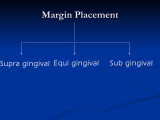
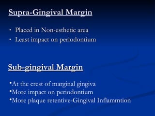
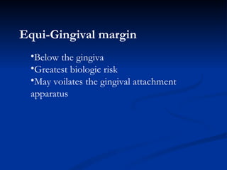
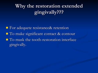
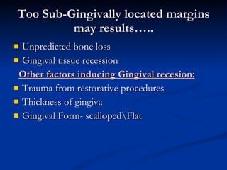
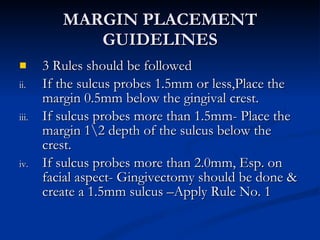
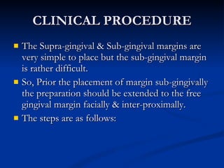
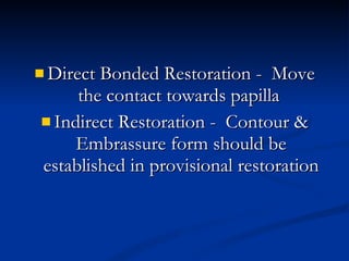
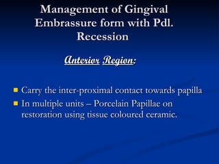
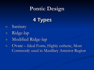
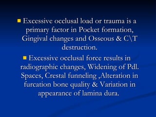
The relationship between periodontal health & the restoration of teeth is intimate & inseparable. For long term survival of restoration the perio dontium must remain healthy or vice-versa
BIOLOGIC CONSIDERATIONS ESTHETIC CONSIDERATIONS  OCCLUSAL CONSIDERATIONS SPECIAL CONSIDERATIONS
BIOLOGIC CONSIDERATIONS
BIOLOGIC CONSIDERATIONS Margin Placement & Biologic Width: Biologic Width:  The C\T attachment occupies 1.07mm of space above the crest of alveolar bone &that the junctional epithelium below the base of the gingival sulcus ocupies another 0.97mm of space above the C\T attachment.
Evaluation of Biologic-Width By Radiographs By using a Periodontal probe
Correction of Biologic-Width Voilation SURGICALLY:   By removing bone away from proximity to the restorative margins. Drawback:   High risk of inter papillary recession. 2.  ORTHODONTICALLY:  By extruding the tooth out of the socket.
Two Methods By applying low extrusive forces By rapid orthodontic extrusion
Margin Placement Supra gingival Sub gingival Equi gingival
Supra-Gingival Margin Placed in Non-esthetic area Least impact on periodontium Sub-gingival Margin At the crest of marginal gingiva More impact on periodontium More plaque retentive-Gingival Inflammtion
Equi-Gingival margin   Below the gingiva Greatest biologic risk May voilates the gingival attachment apparatus
Why the restoration extended gingivally??? For adequete resistance& retention To make significant contact & contour To mask the tooth-restoration interface gingivally.
Too Sub-Gingivally located margins may results….. Unpredicted bone loss Gingival tissue recession Other factors inducing Gingival recesion: Trauma from restorative procedures Thickness of gingiva Gingival Form- scalloped\Flat
MARGIN PLACEMENT GUIDELINES 3 Rules should be followed If the sulcus probes 1.5mm or less,Place the margin 0.5mm below the gingival crest. If sulcus probes more than 1.5mm- Place the margin 1\2 depth of the sulcus below the crest. If sulcus probes more than 2.0mm, Esp. on facial aspect- Gingivectomy should be done & create a 1.5mm sulcus –Apply Rule No. 1
CLINICAL PROCEDURE The Supra-gingival & Sub-gingival margins are very simple to place but the sub-gingival margin is rather difficult. So, Prior the placement of margin sub-gingivally the preparation should be extended to the free gingival margin facially & inter-proximally. The steps are as follows:
TISSUE RETRATION For protection from abrasion  For proper access By Gingival Retraction Cords
For Rule 1 Margin 2 Cords are used 1 st   cord is placed 0.5mm below the prepared margin . 2 nd  Cord is displaces the first cord apically & sits b\w the margin & tissue. This process : Protects the tissues Creates the correct axial reduction  Establishes a desired sub-gingival level margin.
For Rule 2 Margin 2 Large diameter Cords are used. Thick cord is placed for impression. Electro - surgery is necessary to remove  overhanging restoration.
PROVISIONAL RESTORATION 3 Critical areas which must be appropriate to maintain the health & position of the Gingival tissues: Marginal Fit Contour Surface finish
Poorly adapted margins, Over contoured or under contoured restoration  &  Rough or Porous surface can cause: Inflammation Gingival Recession  Overgrowth
MARGIN FIT Open margins can provide shelter for micro-organisms and may be responsible for inflammatory response.
CROWN CONTOUR Ideal contour provides access for hygiene & has fullness to create desired gingival form and good esthetics.
SUB-GINGIVAL DEBRIS Leaving debris below the tissue during the restorative procedure can cause adverse Pdl. Changes.  Causes: Retraction cords Impression material Provisional material  Cements  -  Temporary \ permanent
HYPERSENSITIVITY TO DENTAL MATERIALS Non-Precious alloys shows hyper sensitive  inflammatory response.
ESTHETIC CONSIDERATIONS
Inter-proximal Embrasure Form Any change in shape or form of embrassure Change in height & form of the papilla Food impaction,Accumulation of micro-organisms & Plaque accumulation
Too Wide Embrasure – Flattened & Blunt Papilla Too Narrow Embrasure – Inflammed Papilla Ideal Embrasure – Healthy & pointed Papilla
Restorative Correction of Open Gingival Embrasure 2 Causes: In adequete papillary height due to bone loss Highly located inter-proximal contact Corrected by Restorative Procedure
Direct Bonded Restoration -  Move the contact towards papilla  Indirect Restoration -  Contour & Embrassure form should be established in provisional restoration
Management of Gingival Embrassure form with Pdl. Recession  Anterior   Region : Carry the inter-proximal contact towards papilla In multiple units – Porcelain Papillae on restoration using tissue coloured ceramic.
Posterior Region: Contacts should be moved far enough apically.
Pontic Design 4 Types  Sanitary Ridge-lap Modified Ridge-lap Ovate  – Ideal Form, Highly esthetic, Most Commonly used in Maxillary Anterior Region
OCCLUSAL CONSIDERATIONS
Excessive occlusal load or trauma is a primary factor in Pocket formation, Gingival changes and Osseous & C\T destruction. Excessive occlusal force results in radiographic changes, Widening of Pdl. Spaces, Crestal funneling ,Alteration in furcation bone quality & Variation in appearance of lamina dura.
SPECIAL CONSIDERATIONS
Restoration of Root – Resected Tooth One piece cast or core is indicated Development of appropriate contour for hygienic access Avoid any excess convexities
Splinting Apllied   to : Bonded External Appliances Intra Coronal Appliances Indirect Cast Restorations
Any inflammation of the Pdl,. Supporting tissues must be controlled before making a decision for splinting Bcoz Inflammation may cause mobility in presence of normal occlusal forces & Pdl. Support.
Anterior Aesthetic Surgery More Imp. In Anterior Region Gingivectomy, Apically displaced flaps with osseous recontouring & Use of Orthodontics in positioning the gingiva apically or coronally by Extruding or Intruding the teeth. Computer Imaging for Visual preview A Stone cast of patient`s own teeth may be used – Composite or Acrylic veneer is constructed on the cast extending gingivally in a correct position.
Pdl. Treatment before Restorative Management Sequence of Treatment: Extraction of “Hopeless” teeth – Construction of Temp. partial denture Periodontal Therapy is performed 2 month after Pdl. Therapy – Relocate the margins – Construct the Final Restoration
Phase 1 Therapy Controlling active dental diseases Pdl. Surgical Procedures: Crown lengthening surgery  –  To prevent exposure of crown margin & Root surface after placement. Ridge – augmentation  :  For correcting Ridge discrepancies – 2 Techniques The Roll Technique The C\T Tissue Tunnel Technique
Phase 2 Therapy
Thank You…

Perio restorative inter relationship

  • 1.
  • 2.
  • 3.
    The relationship betweenperiodontal health & the restoration of teeth is intimate & inseparable. For long term survival of restoration the perio dontium must remain healthy or vice-versa
  • 4.
    BIOLOGIC CONSIDERATIONS ESTHETICCONSIDERATIONS OCCLUSAL CONSIDERATIONS SPECIAL CONSIDERATIONS
  • 5.
  • 6.
    BIOLOGIC CONSIDERATIONS MarginPlacement & Biologic Width: Biologic Width: The C\T attachment occupies 1.07mm of space above the crest of alveolar bone &that the junctional epithelium below the base of the gingival sulcus ocupies another 0.97mm of space above the C\T attachment.
  • 7.
    Evaluation of Biologic-WidthBy Radiographs By using a Periodontal probe
  • 8.
    Correction of Biologic-WidthVoilation SURGICALLY: By removing bone away from proximity to the restorative margins. Drawback: High risk of inter papillary recession. 2. ORTHODONTICALLY: By extruding the tooth out of the socket.
  • 9.
    Two Methods Byapplying low extrusive forces By rapid orthodontic extrusion
  • 10.
    Margin Placement Supragingival Sub gingival Equi gingival
  • 11.
    Supra-Gingival Margin Placedin Non-esthetic area Least impact on periodontium Sub-gingival Margin At the crest of marginal gingiva More impact on periodontium More plaque retentive-Gingival Inflammtion
  • 12.
    Equi-Gingival margin Below the gingiva Greatest biologic risk May voilates the gingival attachment apparatus
  • 13.
    Why the restorationextended gingivally??? For adequete resistance& retention To make significant contact & contour To mask the tooth-restoration interface gingivally.
  • 14.
    Too Sub-Gingivally locatedmargins may results….. Unpredicted bone loss Gingival tissue recession Other factors inducing Gingival recesion: Trauma from restorative procedures Thickness of gingiva Gingival Form- scalloped\Flat
  • 15.
    MARGIN PLACEMENT GUIDELINES3 Rules should be followed If the sulcus probes 1.5mm or less,Place the margin 0.5mm below the gingival crest. If sulcus probes more than 1.5mm- Place the margin 1\2 depth of the sulcus below the crest. If sulcus probes more than 2.0mm, Esp. on facial aspect- Gingivectomy should be done & create a 1.5mm sulcus –Apply Rule No. 1
  • 16.
    CLINICAL PROCEDURE TheSupra-gingival & Sub-gingival margins are very simple to place but the sub-gingival margin is rather difficult. So, Prior the placement of margin sub-gingivally the preparation should be extended to the free gingival margin facially & inter-proximally. The steps are as follows:
  • 17.
    TISSUE RETRATION Forprotection from abrasion For proper access By Gingival Retraction Cords
  • 18.
    For Rule 1Margin 2 Cords are used 1 st cord is placed 0.5mm below the prepared margin . 2 nd Cord is displaces the first cord apically & sits b\w the margin & tissue. This process : Protects the tissues Creates the correct axial reduction Establishes a desired sub-gingival level margin.
  • 19.
    For Rule 2Margin 2 Large diameter Cords are used. Thick cord is placed for impression. Electro - surgery is necessary to remove overhanging restoration.
  • 20.
    PROVISIONAL RESTORATION 3Critical areas which must be appropriate to maintain the health & position of the Gingival tissues: Marginal Fit Contour Surface finish
  • 21.
    Poorly adapted margins,Over contoured or under contoured restoration & Rough or Porous surface can cause: Inflammation Gingival Recession Overgrowth
  • 22.
    MARGIN FIT Openmargins can provide shelter for micro-organisms and may be responsible for inflammatory response.
  • 23.
    CROWN CONTOUR Idealcontour provides access for hygiene & has fullness to create desired gingival form and good esthetics.
  • 24.
    SUB-GINGIVAL DEBRIS Leavingdebris below the tissue during the restorative procedure can cause adverse Pdl. Changes. Causes: Retraction cords Impression material Provisional material Cements - Temporary \ permanent
  • 25.
    HYPERSENSITIVITY TO DENTALMATERIALS Non-Precious alloys shows hyper sensitive inflammatory response.
  • 26.
  • 27.
    Inter-proximal Embrasure FormAny change in shape or form of embrassure Change in height & form of the papilla Food impaction,Accumulation of micro-organisms & Plaque accumulation
  • 28.
    Too Wide Embrasure– Flattened & Blunt Papilla Too Narrow Embrasure – Inflammed Papilla Ideal Embrasure – Healthy & pointed Papilla
  • 29.
    Restorative Correction ofOpen Gingival Embrasure 2 Causes: In adequete papillary height due to bone loss Highly located inter-proximal contact Corrected by Restorative Procedure
  • 30.
    Direct Bonded Restoration- Move the contact towards papilla Indirect Restoration - Contour & Embrassure form should be established in provisional restoration
  • 31.
    Management of GingivalEmbrassure form with Pdl. Recession Anterior Region : Carry the inter-proximal contact towards papilla In multiple units – Porcelain Papillae on restoration using tissue coloured ceramic.
  • 32.
    Posterior Region: Contactsshould be moved far enough apically.
  • 33.
    Pontic Design 4Types Sanitary Ridge-lap Modified Ridge-lap Ovate – Ideal Form, Highly esthetic, Most Commonly used in Maxillary Anterior Region
  • 34.
  • 35.
    Excessive occlusal loador trauma is a primary factor in Pocket formation, Gingival changes and Osseous & C\T destruction. Excessive occlusal force results in radiographic changes, Widening of Pdl. Spaces, Crestal funneling ,Alteration in furcation bone quality & Variation in appearance of lamina dura.
  • 36.
  • 37.
    Restoration of Root– Resected Tooth One piece cast or core is indicated Development of appropriate contour for hygienic access Avoid any excess convexities
  • 38.
    Splinting Apllied to : Bonded External Appliances Intra Coronal Appliances Indirect Cast Restorations
  • 39.
    Any inflammation ofthe Pdl,. Supporting tissues must be controlled before making a decision for splinting Bcoz Inflammation may cause mobility in presence of normal occlusal forces & Pdl. Support.
  • 40.
    Anterior Aesthetic SurgeryMore Imp. In Anterior Region Gingivectomy, Apically displaced flaps with osseous recontouring & Use of Orthodontics in positioning the gingiva apically or coronally by Extruding or Intruding the teeth. Computer Imaging for Visual preview A Stone cast of patient`s own teeth may be used – Composite or Acrylic veneer is constructed on the cast extending gingivally in a correct position.
  • 41.
    Pdl. Treatment beforeRestorative Management Sequence of Treatment: Extraction of “Hopeless” teeth – Construction of Temp. partial denture Periodontal Therapy is performed 2 month after Pdl. Therapy – Relocate the margins – Construct the Final Restoration
  • 42.
    Phase 1 TherapyControlling active dental diseases Pdl. Surgical Procedures: Crown lengthening surgery – To prevent exposure of crown margin & Root surface after placement. Ridge – augmentation : For correcting Ridge discrepancies – 2 Techniques The Roll Technique The C\T Tissue Tunnel Technique
  • 43.
  • 44.