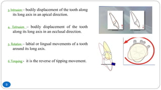
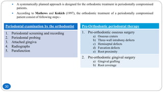
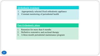
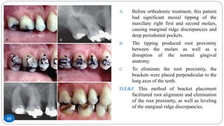
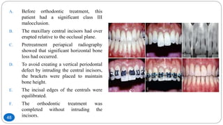
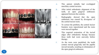
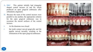
There are many benefits to integrating orthodontics and periodontics in the management of adult patients with underlying periodontal defects. The key to treating these patients is communication and proper diagnosis before orthodontic therapy. Not all periodontal problems are treated in the same way. It should be remembered that overall success of orthodontic treatment depends on the combined effort and close monitoring of the case, by an orthodontist and a periodontist.
Vip Call Girls Anna Salai Chennai 👉 8250192130 ❣️💯 Top Class Girls Available
Perio-Ortho Interdisciplinary approch
1. Raina J.P Khanam
Dept. Of Periodontics
2nd year PG
Perio-Ortho
Interdisciplinary approch
1
2. Contents
Introduction
Benefits of orthodontic therapy
Types of tooth movement
Molecular biology in orthodontic tooth movement
Cellular biology in orthodontic tooth movement
Extracellular remodelling during orthodontic tooth movement
Events after application of orthodontic forces
Sequence of orthodontic treatment in periodontally compromised patients
Periodontal examination by the orthodontist
Pre-orthodontic osseous surgery
Orthodontic treatment of osseous defects
Orthodontic treatment of gingival discrepancies
Advances in surgeries
Clinical situation where orthodontic treatment should be avoided
Plaque controlling aids
Conclusion
2
3. Introduction
Many adults who seek routine restorative dentistry have problems with tooth
malposition that compromise their ability to clean and maintain their dentitions. If these
individuals also are susceptible to periodontal disease, tooth malposition may be an
exacerbating factor that could cause premature loss of specific teeth.
Orthodontic appliances have become smaller, less noticeable, and easier to maintain
during orthodontic therapy. Many adults are taking advantage of the opportunity to
have their teeth aligned to improve the esthetics of their smiles. If these individuals also
have underlying gingival or osseous periodontal defects, these defects often can be
improved during orthodontic therapy if the orthodontist is aware of the situation and
designs the appropriate tooth movement.3
5. The following six factors should be considered:
Aligning crowded or malposed maxillary or mandibular anterior teeth permits the
patient, better access to clean all surfaces of their teeth adequately. This
could be a tremendous advantage for patients who are susceptible to
periodontal bone loss or do not have the dexterity to maintain their oral
hygiene.
Vertical orthodontic tooth repositioning can improve certain types of osseous
defects in periodontal patients.
Orthodontic treatment can improve the esthetic relationship of the maxillary
gingival margin levels before restorative dentistry.
5
6. Orthodontic therapy also benefits the patient with a severe fracture of a maxillary
anterior tooth that requires forced eruption to permit adequate restoration of
the root.
Orthodontic treatment allows open gingival embrasures to be corrected to regain
lost papilla. If these open gingival embrasures are located in the maxillary
anterior region, they can be unesthetic.
Orthodontic treatment could improve adjacent tooth position before implant
placement or tooth replacement. This is especially true for the patient who
has been missing teeth for several years and has drifting and tipping of the
adjacent dentition.
6
8. 1. TIPPING
Controlled tipping – it occurs when a tooth tips about a centre of rotation of its apex.
There is a lingual movement of the crown with minimal movement of root in labial
direction.
Uncontrolled tipping – that occurs about a centre of rotation, with crown moving in one
direction and root in opposite direction.
2. Bodily movement - all the points on the tooth will move an equal distance in the same
direction signifying bodily displacement or translation.
8
9. 3. Intrusion – bodily displacement of the tooth along
its long axis in an apical direction.
4. Extrusion – bodily displacement of the tooth
along its long axis in an occlusal direction.
5. Rotation – labial or lingual movements of a tooth
around its long axis.
6. Torquing - it is the reverse of tipping movement.
9
11. Orthodontic forces result in areas of pressure and tension around teeth
which initiate the local inflammatory reaction.
This reaction is mediated by various mediators which act on their target
cells.
The most important of these mediators are:-
1. Cytokines
2. Growth factors
3. Transcription factors
11
12. 1. Cytokines
Cytokines are important signaling molecules responsible for various biological actions.
They act on nearby cells in an autocrine or paracrine fashion to facilitate cell to cell
communication.
The cytokines involved in bone metabolism include:-
1) IL-1
2) TNF-
3) IL-2
4) IL-3
5) IL-6
6) IL-8
7) IFN
8) Osteoclast differentiation factor (ODF)
IL-1 and TNF- have shown to be potent stimulators of bone resorption.
12
13. IL-1 acts as strong
chemoattractant for
leukocytes and
stimulating
fibroblasts,
endothelial cells,
osteoclasts and
osteoblasts.
TNF- can directly
stimulate the
differentiation of
osteoclast
progenitors to
osteoclasts in the
presences of
macrophage colony
stimulating factor
(M-CSF)
It has been
demonstrating that
during orthodontic
treatment IFN- can
cause bone
resorption by
apoptosis of effector
T-cells.
13
14. 2. Growth factors
Growth factors play a vital role during bone remodelling such as:-
1. Fibroblast growth factor (FGF)
2. Epidermal growth factor (EGF)
3. Platelet derived growth factor (PDGF)
4. Transforming growth factor- (TGF-)
5. Insulin growth factor (IGF)
6. Bone morphogenetic proteins (BMP)
7. Connective tissue growth factor (CTGF)
14
15. Transforming
growth factors
beta (TGF-)
family includes
TGF-1, activins,
inhibins and BMP
which play a vital
role bone
metabolism.
The function of
fibroblast growth
factor (FGF) and
insulin growth
factor (IGF) is to
act on fibroblasts,
endothelial cells,
myoblasts,
chondrocytes and
osteoblasts.
Platelet derived
growth factor (PDGF)
is secreted by platelets
which migrate from
the blood vessels to
the extra vascular
space because of
inflammation resulting
from orthodontic
forces.
Connective tissue
growth factor.
CTGF protein is
associated with
the extracellular
matrix
remodelling
during anabolic
bone remodelling.
15
16. 3. Transcription factors
The non-collagenous bone matrix proteins include:-
1. Osteopointin
2. Bone sialoprotien
3. Osteocalcin
4. Osteonectin
These proteins play an important role in initial mineralization of bone.
16
18. 1. Osteoblasts
The present data suggest that osteoblasts lining the bony socket are directly
responsive to strain orthodontic forces through the proprioceptive receptor system.
This osteoblast response to a great extent depends on Integrins, the cell membrane
proteins.
Integrins translate these mechanical strain signals which in turn stimulate gene
dependent synthesis of ligands which allow intracellular communication.
These changes initiate undermining bone resorption resulting in orthodontic tooth
movement.
18
19. 2. Osteocytes
The present data suggest that osteocytes are very proprioceptive.
It has been shown that mechanical strain within physiological limits causes
increased expressions of glucose-6-phosphate dehydrogenase, 3H-urinidine, c-fos
and IGF in the osteocytes measured within 6hr after application of intermittent
loading at physiological strain magnitude.
19
20. 3. Osteoclasts
They resorb the bone matrix by dissolving the bone minerals.
Active osteoclasts can be identified by their higher content of tartrate-resistant acid
phosphatase (TRAP).
Due to increased vascularity, there is increased capillary blood supply and increased
no. of cells including osteoclasts and fibroblasts.
Under light forces, bone resorption takes place just below the area of pressure on the
PDL side (frontal resorption).
Under heavy forces, because of excessive compression of PDL, its necrosis results
and the area become devoid of cells. The area becomes hyalinised (undermining
resorption).
20
22. PDL plays a very important role during orthodontic tooth movement.
It is primarily made up of Type I and Type III collagen fibers with predominantly
Type I fibers.
Elasticity to PDL is provided by the principal and oxytalan fibers.
The extracellular matrix of PDL is made up of components like:-
1.Glycoproteins
2.Proteoglycans
3.Fibromodulin
4.Fibronectin
These molecules facilitate the cellular migration and proliferation.
22
23. When orthodontic forces are applied on teeth, these causes biophysical and biochemical changes in
the extracellular matrix of PDL and also effect constituent cells of the periodontium and dental
pulp.
Due to orthodontic forces, areas of pressure and tension are created in the PDL which cause fluid
displacement in the extracellular matrix of PDL.
This fluid displacement causes physiological activation of PDL fibroblasts, osteocytes, osteoblasts.
23
24. The nociceptive stimulus causes PDL fibers associated neurons to release neuropeptides such as
Substance P, Calcitonin gene-related peptide.
Under the influence of these peptides as well as prostaglandins and other humoral factors capillary
dilatation in PDL occurs, resulting in the release of immune components from the capillaries.
The migration of these cells from capillaries to ECM is facilitated by vascular endothelial growth
factor, secreted by endothelial cells and osteoblasts.
24
25. Phosphorylation of cellular proteins including extracellular signal-regulated kinases (ERK) occurs
because of changes in cytoskeleton, which trigger signal transduction via
integrins/fibronectin/kinase pathway.
The MMP are involved in connective tissue remodelling by their action of degradation of matrix
components.
These matrix degrading enzymes increase in pressure areas around the tooth subjected to
orthodontic forces.
25
26. Particularly MMP-1, MMP-8, MMP-9, MMP-3, MMP-13 are increased in these sites.
Along with this, PgE2 and COX 2 mRNA are also increased at the compression sites during
remodelling.
Recent evidence suggest important roles of other molecules such as Sox-9, parathyroid hormone-
related protein and Indian Hedgehog protein in orthodontic tooth movement.
26
28. There are 3 histological events:-
The first event is related to alteration of blood vasculature. After the application of orthodontic forces the
blood supply is altered both in areas of tension as well as pressure areas. This causes decreased oxygen
level at the compressed area and probably increased oxygen level at tension areas.
The second event is the generation of an electrical signal. Due to bending of bone and deformation of the
crystallization structure, a piezoelectric signal or more appropriately referred to as a bioelectric potential in
the form of a small voltage of current is generated.
Thirdly, the physical distortion imposed by peripheral forces on paradental tissues such as nerve fibers
and terminals results in the release of various neurotransmitters such as Substance P, Vasointestinal
polypeptide and Calcitonin gene-related peptide.
28
30. A systematically planned approach is designed for the orthodontic treatment in periodontally compromised
patients.
According to Mathews and Kokich (1997), the orthodontic treatment of a periodontally compromised
patient consist of following steps:-
Periodontal examination by the orthodontist
1. Periodontal screening and recording
2. Periodontal probing
3. Attached gingiva
4. Radiographs
5. Parafunction
Pre-Orthodontic periodontal therapy
1. Pre-orthodontic osseous surgery
a) Osseous craters
b) Three-wall intrabony defects
c) Hemiseptal defects
d) Furcation defects
e) Root proximity
2. Pre-orthodontic gingival surgery
a) Gingival grafting
b) Root coverage
30
31. Orthodontic treatment
1. Appropriately selected fixed orthodontic appliance
2. Constant monitoring of periodontal health
Post-Orthodontic phase
1. Retention for more than 6 months
2. Definitive restorative and occlusal therapy
3. A three-month periodontal maintenance program
31
33. An orthodontist must incorporate compulsory periodontal examination during the initial
consultation with the patient.
Periodontal screening and recording (PSR) is a simple, quick and effective method of recording the
periodontal status of the patient with minimum documentation.
In this examination, a score is given to each area which helps the examiner to decide what kind of
periodontal treatment is required for a particular patient.
The width of attached gingiva is examined and areas with less than 2mm of attached gingiva
should be evaluated by a periodontist.
Along with this, patients with thin gingival biotype and thin alveolar bone should also be evaluated
by the periodontist.
33
34. Radiographic examination is required for the evaluation of the generalized bone topography.
IOPA, bitewing or panoramic may be advised as required.
For evaluation of periodontal osseous lesions, panoramic radiographs are not as suitable as
periapical or bitewing radiographs.
The most common areas that are missed in a panoramic radiograph includes interproximal craters
between upper molars, infrabony defects on the mesial of the upper first bicuspid and defects
around the lower incisors.
The crestal bone is more clearly visible in bitewing radiographs.
Patients who are bruxers or clenchers must be identified during the initial consultation because it
has been shown that these parafunctional habits can cause extensive osseous breakdown during
orthodontic therapy.
These patients may need a night guard while undergoing active orthodontic treatment.
34
36. The extent of the osseous surgery depends on the type of defect such as:-
1. Crater
2. Hemiseptal defect
3. Three-wall defect
4. Furcation lesion
The prudent clinician knows which defects can be improved with
orthodontic treatment and which defects require pre-orthodontic,
periodontal, surgical intervention.
36
37. 1. Osseous Craters
An osseous crater is an interproximal, two-wall defect that does not improve
with orthodontic treatment.
Some shallow craters (4 to 5mm pocket) may be maintainable non-surgically
during orthodontic treatment.
However, if surgical correction is necessary, this type of osseous lesion can
easily be eliminated by reshaping the defect and reducing the pocket depth.
37
38. A. This patient had a 6-mm probing defect
distal to the maxillary right first molar.
B. When this area was flapped, a cratering
defect was apparent.
C&D. Osseous surgery was performed to alter
the bony architecture of the buccal and
lingual surfaces to eliminate the defect.
E. After 6 weeks, the probing pocket defect
had been reduced to 3 mm and
orthodontic appliances were placed on
the teeth.
F. Because the crater was eliminated before
orthodontic therapy, the patient could
maintain the area during and after
orthodontic treatment.
38
39. 2. Three-wall intrabony defect
Three-wall defects are amenable to pocket reduction with regenerative
periodontal therapy.
Bone grafts using either autogenous bone from the surgical site or allograft
along with the use of resorbable membranes have been successful in filling
three-wall defects.
If the result of periodontal therapy is stable 3 to 6 months after periodontal
surgery, orthodontic treatment may be initiated.
39
40. A. This patient had a significant
periodontal pocket distal to the
mandibular right first molar.
B. Periapical radiograph confirmed the
osseous defect.
C. A flap was elevated, revealing a deep,
three-wall osseous defect.
D. Freeze-dried bone graft was placed in
the defect.
E. Six months after the bone graft,
orthodontic treatment was initiated.
F. The final periapical radiograph shows
that the pre-orthodontic bone graft
helped regenerate bone and eliminate
the defect distal to the molar.
40
42. 1. Hemiseptal defects
Hemiseptal defects are one-or two-wall osseous defects that often are
found around mesially tipped teeth or teeth that have super-erupted.
Usually, these defects can be eliminated with the appropriate orthodontic
treatment.
In the case of the tipped tooth, uprighting and eruption of the tooth,
levels the bony defect.
If the tooth is super-erupted, intrusion and leveling of the adjacent
cemento-enamel junctions can help level the osseous defect.
42
43. A. This patient was missing the mandibular
left second premolar and the first molar
had tipped mesially.
B. Pretreatment periapical radiograph
revealed a significant hemiseptal osseous
defect on the mesial side of the molar.
C. To eliminate the defect, the molar was
erupted, and the occlusal surface was
equilibrated.
D. The eruption was stopped after the bone
defect was leveled.
E. The post-treatment intraoral photograph.
F. Periapical radiograph show that the
periodontal health was improved by
orthodontic correction of the hemiseptal
defect.
43
44. A. This patient showed over eruption of
the maxillary right first molar and a
marginal ridge defect between the
second premolar and first molar.
B. The pretreatment periapical radiograph
showed that the interproximal bone
was flat.
C&D. To avoid creating a hemiseptal defect,
the occlusal surface of the first molar
was equilibrated.
E&F. The malocclusion was corrected
orthodontically.
44
45. In these techniques it may require endodontic therapy and restoration of the
tooth because of the required amount of reduction of the length of the
crown.
This approach is acceptable if the treatment results in a more favorable
bone contour between the teeth.
Some patients have a discrepancy between both the marginal ridges and the
bony levels between two teeth. However, these discrepancies may not be of
equal magnitude; orthodontic leveling of the bone may still leave a
discrepancy in the marginal ridges.
The patient should be recalled every 2 to 3 months during the leveling
process to control inflammation in the interproximal region.45
46. A. Before orthodontic treatment, this patient
had significant mesial tipping of the
maxillary right first and second molars,
causing marginal ridge discrepancies and
deep periodontal pockets.
B. The tipping produced root proximity
between the molars as well as a
disruption of the normal gingival
anatomy.
C. To eliminate the root proximity, the
brackets were placed perpendicular to the
long axes of the teeth.
D,E&F. This method of bracket placement
facilitated root alignment and elimination
of the root proximity, as well as leveling
of the marginal ridge discrepancies.
46
47. 2. Advancedhorizontal bone loss
After orthodontic treatment has been planned, one of the most important factors that
determine the outcome of orthodontic therapy is the location of the bands and
brackets on the teeth.
In a periodontally healthy individual, the position of the brackets is usually
determined by the anatomy of the crowns of the teeth.
Anterior brackets should be positioned relative to the incisal edges. Posterior bands
or brackets are positioned relative to the marginal ridges.
If the incisal edges and marginal ridges are at the correct level, the cemento-enamel
junction (CEJ) will also be at the same level. This relationship creates a flat, bony
contour between the teeth.
However, if a patient has underlying periodontal problems and significant alveolar
bone loss around certain teeth, using the anatomy of the crown to determine bracket
placement is not appropriate.47
48. A. Before orthodontic treatment, this
patient had a significant class III
malocclusion.
B. The maxillary central incisors had over
erupted relative to the occlusal plane.
C. Pretreatment periapical radiography
showed that significant horizontal bone
loss had occurred.
D. To avoid creating a vertical periodontal
defect by intruding the central incisors,
the brackets were placed to maintain
bone height.
E. The incisal edges of the centrals were
equilibrated.
F. The orthodontic treatment was
completed without intruding the
incisors.48
49. 3. Furcation defects
These lesions require special attention in the patient undergoing orthodontic treatment.
Furcation lesions require special consideration because they are the most difficult
lesions to maintain and can worsen during orthodontic therapy.
These patients need to be maintained on a 2 to 3 month recall schedule.
Class I defects have good prognosis and can be treated by osseous surgical correction.
Class II defects can be treated with grafting and regenerative therapy with barrier
membranes.
49
50. If a patient with a class III furcation defect will be undergoing orthodontic
treatment, a possible method for treating the furcation is to eliminate it by
hemisecting the crown and root of the tooth. However, this procedure requires
endodontic, periodontal and restorative treatment. If the patient will be
undergoing orthodontic treatment, it is advisable to perform the orthodontic
treatment first. This is especially true if the roots of the teeth will not be
moved apart. In these patients the molar to be hemisected remains intact
during orthodontics. This patient would require 2 or 3 month recall visits to
ensure that the furcation defect does not lose bone during orthodontic
treatment.
50
51. A&B. This patient had a class III furcation
defect before orthodontic treatment.
C. Orthodontic treatment was performed and
the furcation defect was maintained by the
periodontist with 2-month recalls until
after orthodontic treatment.
D. After appliance removal, the tooth was
hemisected.
E. And the roots were restored and splinted
together.
F. The final periapical radiograph shows that
the furcation defect was eliminated by
hemisection and restoration of the two
root fragments.
51
52. Hemisected molar is used as an abutment for a bridge after orthodontics,
moving the roots apart orthodontically permits a favorable restoration and
splinting across the adjacent edentulous spaces. In these patients,
hemisection, endodontic therapy, and periodontal surgery must be completed
before the start of orthodontic treatment. After completion of these
procedures, bands or brackets can be placed on the root fragments and coil
springs are used to separate the roots. The amount of separation is determined
by the size of the adjacent edentulous spaces and the occlusion in the
opposing arch. About 7 or 8 mm may be created between the roots of the
hemisected molar. This process eliminates the original furcation problem and
allows the patient to clean the area with greater efficiency.
52
53. A&B. Before orthodontic treatment, this
patient had a class III furcation defect in
the mandibular left second molar.
C. Because the patient had an edentulous
space mesial to the molar, the tooth was
hemisected.
D. The root fragments were separated
orthodontically.
E&F. After orthodontic treatment, the root
fragments were used as abutments to
stabilize a multiunit posterior bridge.
53
54. In some molars with class III furcation defects, the tooth may have short
roots, advanced bone loss, fused roots, or other problems that prevent
hemisection and crowning of the remaining roots. In these patients,
extracting the root with a furcation defect and placing an implant may be
more advisable. In some patients the implant can be used as an anchor to
facilitate pre-restorative orthodontic treatment. The implant must remain
embedded in bone for 4 to 6 months after placement before it can be loaded
as an orthodontic anchor. It must be placed precisely so that it not only
provides an anchor for tooth movement, but also may be used an eventual
abutment for a crown or fixed bridge.
54
55. A. This patient was missing several teeth
in the mandibular left posterior
quadrant.
B. The mandibular left third molar had a
class III furcation defect and short
roots.
C. The third molar was extracted, and two
implants were placed in the
mandibular left posterior quadrant.
D. The implants were used as anchors to
facilitate orthodontic treatment.
E&F. And help reestablish the left posterior
occlusion.
55
56. 4. Root proximity
When roots of posterior teeth are close together, the ability to
maintain periodontal health and accessibility for restoration of
adjacent teeth may be compromised.
However, for the patient undergoing orthodontic therapy, the
roots can be moved apart, and bone will form between the
adjacent roots.
Radiographs are needed to monitor the progress of orthodontic
root separation. Generally, 2 to 3 mm of root separation
provides an adequate bone and embrasure space to improve
periodontal health.
56
57. 5. Fractured teethand forced eruption
Occasionally, children and adolescents may fall and injure their anterior teeth.
If the injuries are minor and result in small fractures of enamel, these can be restored with light-
cured composite or porcelain veneers.
In some patients, however, the fracture may extend beneath the level of the gingival margin and
terminate at the level of the alveolar ridge, so in that case, restoration of the fractured crown is
impossible because the tooth preparation would extend to the level of the bone. This over-extension
of the crown margin could result in an invasion of the biologic width of the tooth and cause
persistent inflammation of the marginal gingiva.
It may be beneficial in such cases to erupt the fractured root out of the bone and move the fracture
margin coronally so that it can be properly restored. However, if the fracture extends too far
apically, it may be better to extract the tooth and replace it with an implant or bridge.57
58. A. This patient had a severe fracture of the
maxillary right central incisor that extended
apical to the level of the alveolar crest on the
lingual side.
B. Radiographic image.
C. To restore the tooth adequately, the fractured
root was extruded 4mm.
D. As the tooth erupted, the gingival margin
followed the tooth.
E&F. Gingival surgery was required to lengthen the
crown of the central incisor, so that the final
restoration had sufficient support for
resistance and retention as well as the
appropriate gingival margin relationship with
the adjacent central incisor.
58
59. The following six criteria are used to determine whether the tooth should be forcibly
erupted or extracted:
1. Root length- If a tooth fracture extends to the level of the bone, it must be erupted 4 mm. The
first 2.5mm moves the fracture margin far enough away from the bone to prevent a biologic width
problem. The other 1.5 mm provides the proper amount of support for adequate resistance form of
the crown preparation. The root/crown ratio should be about 1:1. If the root/crown ratio is less
than this amount, there may be too little root remaining in the bone for stability. In that situation, it
may be prudent to extract the root and place a bridge or implant.
2. Root form- The shape of the root should be broad and non-tapering rather than thin and tapered.
A thin, tapered root provides a narrower cervical region after the tooth has been erupted 4mm. This
could compromise the esthetic appearance of the final restoration.
59
60. 4. Relative importance of the tooth- If the patient is 70 years of age and both adjacent teeth have
prosthetic crowns, it would be more prudent to construct a fixed bridge. However, if the patient is
15 years of age and the adjacent teeth are un-restored, forced eruption would be much more
conservative and appropriate.
3. Level of the fracture- If the entire crown is fractured 2 to 3 mm apical to the level of the
alveolar bone, it is difficult to treat.
60
61. 6. Endodontic/periodontal prognosis- If the tooth has a significant periodontal defect, it may not
be possible to retain the root. In addition, if the tooth root has a vertical fracture, the prognosis
would be poor, and extraction of the tooth would be the proper course of therapy.
5. Esthetics- If the patient has a high lip line and displays 2 to 3 mm of gingiva when smiling, any
type of restoration in this area will be more obvious. Keeping the patient’s own tooth would be
much more esthetic than any type of implant or prosthetic replacement.
61
62. After the tooth root has been erupted, it must be stabilized to prevent it from intruding
back into the alveolus. The reason for reintrusion is the orientation of the principal
fibers of the periodontium. During forced eruption, the periodontal fibers become
oriented obliquely and stretched as the root moves coronally. These fibers eventually
reorient themselves after about 6 months.
As the root erupts, the gingiva moves coronally with the tooth. As a result, the clinical
crown length becomes shorter after extrusion. A gingival surgery is necessary to create
ideal gingival margin heights.
If bone has followed the root during eruption, a flap is elevated, and the appropriate
amount of bone is removed to match the bone height of the adjacent teeth.
62
63. 6. Hopeless teethmaintainedfor orthodontic anchorage
Patients with advanced periodontal disease may have specific teeth diagnosed as hopeless, which
would be extracted before orthodontic therapy.
However, these teeth can be useful for orthodontic anchorage if the periodontal inflammation can
be controlled.
In moderate to advanced cases, flaps are reflected for debridement of the roots to control
inflammation around the hopeless tooth during the orthodontic process. The important factor is to
maintain the health of the bone around the adjacent teeth.
After orthodontic treatment, there is a 6-month period of stabilization before reevaluating the
periodontal status.
In most cases, however, the hopeless tooth requires extraction, especially if other restorations are
planned in the segment.
63
64. A. This patient had an impacted mandibular
right second molar.
B. The mandibular right first molar was
periodontally hopeless because of an
advanced class III furcation defect.
C. The impacted second molar was
extracted, but the first molar was
maintained as an anchor to help upright
the third molar orthodontically.
D. After orthodontic up righting of the third
molar, the first molar was extracted and a
bridge was placed to restore the
edentulous space.
64
66. 1. Uneven gingival margins
The relationship of the gingival margins of the six maxillary anterior teeth plays an important
role in the esthetic appearance of the crowns.
The following four factors contribute to ideal gingival form:
1. The gingival margins of the two central incisors should be at the same level.
2. The gingival margins of the central incisors should be positioned more apically than the lateral incisors
and at the same level as the canines.
3. The contour of the labial gingival margins should mimic the CEJs of the teeth.
4. A papilla should exist between each tooth, and the height of the tip of the papilla is usually halfway
between the incisal edge and the labial gingiva.
66
67. A. This patient had a protrusive bruxing habit that
resulted in abrasion and over eruption of the
maxillary right central incisor.
B. The objective was to level the gingival margins
during orthodontic therapy.
C. Although gingival surgery was a possibility, the
labial sulcular depth of the maxillary right central
incisor was only 1 mm, and the CEJ was located
at the bottom of the sulcus.
D. Therefore, the best solution involved positioning
the orthodontic brackets to facilitate intrusion of
the right central incisor.
E. This permitted the restorative dentist to restore
the portion of the tooth that the patient had
abraded.
F. Correct gingival margin levels and crown lengths
at the end of treatment.
67
68. To make the correct decision, it is necessary to evaluate four criteria.
First, the relationship between the gingival margin of the maxillary central incisors and
the patient’s lip line should be assessed when the patient smiles.
Second, is to evaluate the labial Sulcular depth over the two central incisors.
Third, is to evaluate the relationship between the shortest central incisor and the adjacent
lateral incisors.
Fourth, is to determine whether the incisal edges have been abraded. This is best
accomplished by evaluating the teeth from an incisal perspective. If one incisal edge is
thicker labio-lingually than the adjacent tooth, this may indicate that it has been abraded
and the tooth has over erupted.
68
69. 2. Significant abrasion andOver-eruption
Occasionally, patients have destructive dental habits, such as a protrusive bruxing habit, that can
result in significant wear of the maxillary and mandibular incisors and compensatory over
eruption of these teeth .
The restoration of these abraded teeth is often impossible because of the lack of crown length to
achieve adequate retention and resistance form for the crown preparations.
Two options are available. One option is extensive crown lengthening by elevating a flap,
removing sufficient bone, and apically positioning the flap to expose adequate tooth length for
crown preparation.
The other option for improving the restorability of these short abraded teeth is to intrude the teeth
orthodontically and move the gingival margins apically.
When abraded teeth are significantly intruded, it is necessary to hold these teeth for at least 6
months in the intruded position with orthodontic brackets or archwires (or both), or some type of
bonded retainer.69
70. A. This patient had a protrusive bruxing habit that
caused severe abrasion of the maxillary anterior
teeth, resulting in the loss of over half of the
crown length of the incisors.
B. Maxillary cast.
C. Two possible options existed for gaining crown
length to restore the incisors. One option was an
apically positioned flap with osseous
recontouring, which would expose the roots of
the teeth.
D. The less destructive option was to intrude the
four incisors orthodontically, level the gingival
margins.
E. And allow the dentist to restore the abraded
incisal edges.
F. The orthodontic option was clearly successful
and desirable in this patient.
70
71. 3. Open gingival embrasures
The presence of a papilla between the maxillary central incisors is a key esthetic factor in any
individual.
Occasionally, adults have open gingival embrasures or lack gingival papillae between their central
incisors.
These unesthetic areas are often difficult to resolve with periodontal therapy. However,
orthodontic treatment can correct many of these open gingival embrasures.
This open space is usually caused by
(1) tooth shape
(2) root angulation
(3) periodontal bone loss
71
72. A. This patient initially had overlapped
maxillary central incisors.
B. After initial orthodontic alignment of the
teeth, an open gingival embrasure
appeared between the centrals.
C. Radiography showed that the open
embrasure was caused by divergence of
the central incisor roots.
D. To correct the problem, the central incisor
brackets were repositioned and the roots
were moved together.
E. This required restoration of the incisal
edges after orthodontic therapy because
these teeth had worn unevenly before
therapy.
F. As the roots were paralleled, the tooth
contact moved gingivally and the papilla
moved incisally, resulting in elimination of
the open gingival embrasure.
72
73. A, B&C. This patient initially had triangular
shaped central incisors (A and B), which
produced an open gingival embrasure after
orthodontic alignment.
D. Because the roots of the central incisors were
parallel to one another, the appropriate solution
for the open gingival embrasure was to
recontour the mesial surfaces of the central
incisors.
E. As the diastema was closed.
F. the tooth contact moved gingivally and the
papilla moved incisally, resulting in the
elimination of the open gingival embrasure.
73
75. PERIODONTALLY ACCELERATED OSTEOGENIC
ORTHODONTICS
Periodontal accelerated osteogenic orthodontics (PAOO) is a clinical procedure that combines
selective alveolar corticotomy, particulate bone grafting and the application of orthodontic forces.
Kole et al (1959) -first to describe modern day PAOO.
Rapid tooth movement was by eliminating the resistance of cortical bone by circumscribing
corticotomy cuts around the tooth.
75
76. Wilko et al (2001) suggested that Rapid tooth movement was due to demineralization/
remineralization process consistent with the initial phase of regional acceleratory phenomenon
(RAP).
REGIONALACCELERATORY PHENOMENON-
Introduced by Frost in 1983.
REGIONAL- Demineralization occurs at both the cut sites as well as adjacent bone.
ACCELERATORY- Exaggerated bone response in cuts that extends to the bone marrow.
It is a local response to noxious stimulus which describes a process by which tissue forms faster
than normal regeneration process. RAP begins within few days of injury, peaks at 1-2 months and
lasts for 4 months in bone.
As long as tooth movement continues RAP is prolonged.
When RAP dissipates, osteopenia disappears and radiographic image of normal spongiosa
appears.
When orthodontic tooth movement is completed it favors an environment for alveolar
remineralization.
76
77. CONCEPTS
The technique given by Wilko and Ferguson came to be known as PAOO.
This technique is a combination of-
Bone activation.(selective alveolar decortication, ostectomies and bone thinning)
Alveolar augmentation using bone grafts.
Orthodontic treatment.
TREATMENT PROCEDURE
Orthodontic brackets are placed and light wire engaged a week before surgery.
Subsequent orthodontic adjustments made at 2 week interval.
Treatment plan for teeth that will undergo bone activation is prepared by the combined team.
Procedure to be performed under i.v. or oral sedation as it may take 3-4 hrs to complete.
A full thickness flap is raised.
77
78. Decortication which Can be done with high speed hand piece, implant drill or piezo knife.
Vertical grooves are placed in the inter-radicular area, b/w the root prominence in the alveolar
bone. This groove extends from a point 2-3 mm below the crest and to a point 2 mm beyond the
apices of the roots.
If the thickness of alveolar bone is estimated to be 1 to 2mm , perforations are omitted to avoid
damage to roots.
Care should be taken to ensure <1.5 mm of bone remains over the tooth.
78
79. Grafting procedure - Bone graft is wetted with clindamycin phosphate/ bacteriostatic agent.
Wetting also permits its easy placement.
Decalcified freeze dried bone allograft (DFDBA) or combination of DFDBA or FDBA can be
used.
Suturing done with non resorbable suture material and placed for a minimum of 2 weeks.
Antibiotics and painkillers are administered at the clinician preference.
Patient may experience oedema and ecchymosis which are self limiting.
Pt recalled every week for gentle prophylaxis every week for 1st month and then monthly
thereafter.
79
81. 1. Uncontrolled infection and inflammation
2. Lack of retention for stabilization of teeth in their new position.
3. Inadequate space into which teeth can not be moved.
4. Movement of teeth against occlusal opposition or into occlusal trauma.
5. Movement of teeth in conditions where periodontal health, function or esthetics
will not improve.
6. Movement of teeth against inadequate anchorage.
7. Movement of teeth into unfavourable environment.
8. Lack of patient motivation and cooperation.
9. Tooth movement in patients with systemic problems that cannot be treated or are
difficult to control.
81
83. Toothbrush
1. Manual toothbrush – made up of nylon. (V-trim)
2. Electronic toothbrush – having head of circulatory and back and forth vibratory
motion.
3. End-tuft brush – having head with 7 tufts of tightly packed soft nylon bristles. They
reach deep into smaller areas.
4. Sulcabrushes – are designed to clean the teeth along the gingival margin.
83
84. Interdental aids
1. Super floss – kind of dental floss which is supplied in pre-cut
segments.
2. Oral irrigation devices – used for sulcus irrigation using latex
free rubber tip.
84
85. Charter’s brushing technique
Introduced by charter in 1848. It is a very useful technique for patients having orthodontic appliances
and in patients who recently underwent periodontal surgery. In this brushing technique, the brush is
placed at an angle of 45 to the long axis of the teeth in an opposite direction as recommended in
bass technique, ie; the bristles are directed coronally. After adaptation of the brush in place, the
bristles point away from the gingiva but towards the interproximal surfaces of the teeth and short
back and forth vibratory strokes are given. The bristles of the brush are then pressed on the
occlusal surfaces of the teeth and using a slight rotary motion, pits and fissure are cleaned.
Disadvantage of this technique is that does not efficiently clean the interdental areas due to angulation
of the bristles in a coronal direction.
85
86. 86
The 3 Best Mouthwashes For Braces
Colgate Phos-Flur Ortho Defense Anti-cavity Fluoride Rinse
The Best For Fighting Cavities. ACT Anti-cavity Fluoride Rinse
The Best On A Budget. Listerine Total Care Anti-cavity
87. Conclusion
There are many benefits to integrating orthodontics and periodontics in the
management of adult patients with underlying periodontal defects. The key
to treating these patients is communication and proper diagnosis before
orthodontic therapy. Not all periodontal problems are treated in the same
way. It should be remembered that overall success of orthodontic treatment
depends on the combined effort and close monitoring of the case, by an
orthodontist and a periodontist.
87