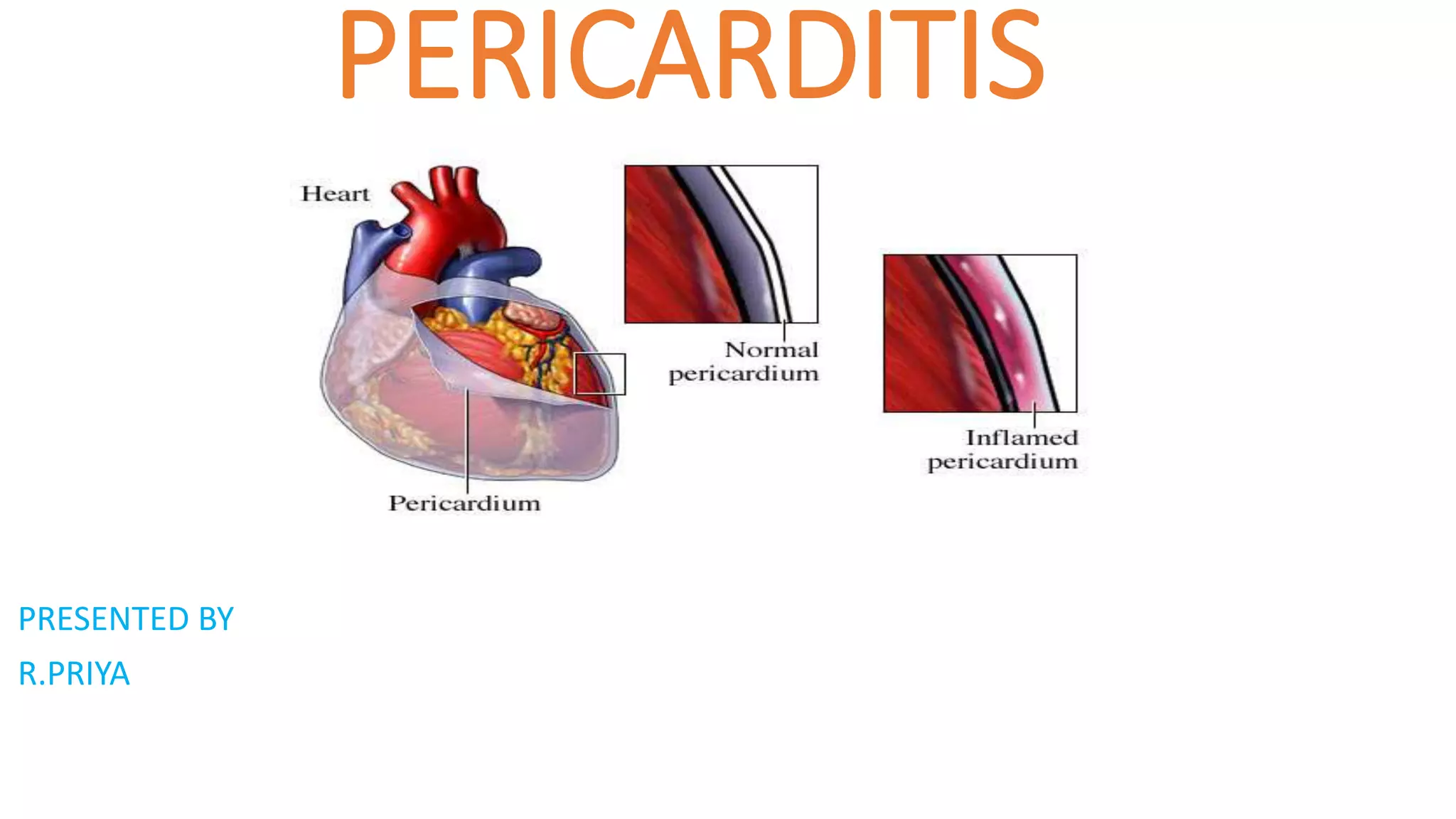
Pericarditis is an inflammation of the pericardium that commonly affects men aged 20-50. It has several causes including viral or bacterial infections, certain cancers, autoimmune diseases, or physical trauma. Symptoms include chest pain that increases with deep breathing or lying flat. Diagnosis involves electrocardiograms, echocardiograms, and blood tests. Treatment focuses on reducing inflammation with medications like aspirin, ibuprofen, or colchicine. Surgery may be required in severe cases to drain fluid or remove the pericardium.
Cardiomyopathy, or heart muscle disease, is a type of progressive heart disease in which the heart is abnormally enlarged, thickened, and/or stiffened. As a result, the heart muscle's ability to pump blood is less efficient, often causing heart failure and the backup of blood into the lungs or rest of the body. The disease can also cause abnormal heart rhythms.
CARDIAC TAMPONADE ( Cardiac emergency) • Cardiac Tamponade is a life threatening complication caused by excessive accumulation of fluid in the pericardium. Or • Compression of all cardiac chambers due to excessive accumulation of pericardial fluid leading to compromised cardiac out put.
Acute Rheumatic Fever and Rheumatic Heart Disease, are two common conditions in children between 3-15 years of age following a Group B Streptococcal throat infection. We discuss these two conditions in the slides above, as well as their management.
This presentation is about pericarditis it's
pathophysiology Sign and symptoms classification types diagnostics treatment. This presentation also includes video demonstrations at the end
congenital heart disease is a heart defect, it is caused by the exposure of teracogenic substances during pregnancy ,it may lead to the congenital defect .
ACYANOTIC DISEASE- Non cyanotic heart diseasesNelsonNgulube
ETIOLOGY AND EPIDEMIOLOGY
Congenital heart disease occurs in 8 per 1,000 births. The spectrum of lesions ranges from asymptomatic to fatal. Although most cases of congenital heart disease are multifactorial, some lesions are associated with chromosomal disorders, single gene defects, teratogens, or maternal metabolic disease (see Table139-2).
Congenital heart defects can be divided into three pathophysiological groups (Table 143.1).
1. Left-to-right shunts
2. Right-to-left shunts
3. Obstructive, stenotic lesions
Acyanotic congenital heart disease includes left-to-right shunts resulting in an increase in pulmonary blood flow (patent ductus arteriosus [PDA], ventricular septal defect [VSD], atrial septal defect [ASD]) and obstructive lesions (aortic stenosis, pulmonary stenosis, coarctation of the aorta), which usually have normal pulmonary blood flow.
VENTRICULAR SEPTAL DEFECTEtiology and Epidemiology
The ventricular septum is a complex structure that can be divided
into four components. The largest component is the muscular
septum. The inlet or posterior septum comprises endocardial
cushion tissue. The subarterial or supracristal septum com
prises conotruncal tissue. The membranous septum is below
the aortic valve and is relatively small. VSDs occur when any of these components fail to develop normally (Fig. 143.1). VSD,
the most common congenital heart defect, accounts for 25% of all congenital heart disease. Perimembranous VSD
In heart valve disease, one or more of the valves in your heart doesn't work properly.
Your heart has four valves that keep blood flowing in the correct direction. In some cases, one or more of the valves don't open or close properly. This can cause the blood flow through your heart to your body to be disrupted.
Your heart valve disease treatment depends on the heart valve affected and the type and severity of the valve disease. Sometimes heart valve disease requires surgery to repair or replace the heart valve.Your heart has four valves that keep blood flowing in the correct direction. These valves include the mitral valve, tricuspid valve, pulmonary valve and aortic valve. Each valve has flaps (leaflets or cusps) that open and close once during each heartbeat. Sometimes, the valves don't open or close properly, disrupting the blood flow through your heart to your body.
Heart valve disease may be present at birth (congenital). It can also occur in adults due to many causes and conditions, such as infections and other heart conditions.
Heart valve problems may include:
Regurgitation. In this condition, the valve flaps don't close properly, causing blood to leak backward in your heart. This commonly occurs due to valve flaps bulging back, a condition called prolapse.
Stenosis. In valve stenosis, the valve flaps become thick or stiff, and they may fuse together. This results in a narrowed valve opening and reduced blood flow through the valve.
Atresia. In this condition, the valve isn't formed, and a solid sheet of tissue blocks the blood flow between the heart chambers.Several factors can increase your risk of heart valve disease, including:
Older age
History of certain infections that can affect the heart
History of certain forms of heart disease or heart attack
High blood pressure, high cholesterol, diabetes and other heart disease risk factors
Heart conditions present at birth (congenital heart disease)Heart valve disease can cause many complications, including:
Heart failure
Stroke
Blood clots
Heart rhythm abnormalities
Death
Heart rhythm problems (heart arrhythmias) occur when the electrical impulses that coordinate your heartbeats don't work properly, causing your heart to beat too fast, too slow or irregularly.
Heart arrhythmias (uh-RITH-me-uhs) may feel like a fluttering or racing heart and may be harmless. However, some heart arrhythmias may cause bothersome — sometimes even life-threatening — signs and symptoms.
Heart arrhythmia treatment can often control or eliminate fast, slow or irregular heartbeats. In addition, because troublesome heart arrhythmias are often made worse — or are even caused — by a weak or damaged heart, you may be able to reduce your arrhythmia risk by adopting a heart-healthy lifestyle.Arrhythmias may cause you to feel premature heartbeats, or you may feel that your heart is racing or beating too slowly. Other signs and symptoms may be related to your heart not pumping effectively due to the fast or slow heartbeat. These include shortness of breath, weakness, dizziness, lightheadedness, fainting or near fainting, and chest pain or discomfort. Seek urgent medical care if you suddenly or frequently experience any of these signs and symptoms at a time when you wouldn't expect to feel them.Ventricular fibrillation is one type of arrhythmia that can be deadly. It occurs when the heart beats with rapid, erratic electrical impulses. This causes the lower chambers in your heart (ventricles) to quiver uselessly instead of pumping blood. Without an effective heartbeat, blood pressure plummets, cutting off blood supply to your vital organs.f slow heartbeats (bradycardias) don't have a cause that can be corrected, doctors often treat them with a pacemaker because there aren't any medications that can reliably speed up the heart.
A pacemaker is a small device that's usually implanted near your collarbone. One or more electrode-tipped wires run from the pacemaker through your blood vessels to your inner heart. If your heart rate is too slow or if it stops, the pacemaker sends out electrical impulses that stimulate your heart to beat at a steady rate.
The terms leukopenia and neutropenia are often used interchangeably. However, they refer to slightly different conditions. Leukopenia is an umbrella term that refers to a reducation in any of the white blood cell types.
Neutropenia is a type of leukopenia but refers specifically to a decrease in neutrophils, the most common type of white blood cell. A person’s neutrophil count is an important indicator of their infection risk.
In disseminated intravascular coagulation, abnormal clumps of thickened blood (clots) form inside blood vessels. These abnormal clots use up the blood's clotting factors, which can lead to massive bleeding in other places. Causes include inflammation, infection and cancer.
Symptoms include blood clots and bleeding, possibly from many sites in the body.
The goal is to treat the underlying cause and provide supportive care through intravenous fluids and blood transfusions.
Leukocyte is another name for white blood cell (WBC). These are the cells in your blood that help your body fight infections and some diseases.
When the number of white cells in your blood is higher than normal, it’s called leukocytosis. This usually happens because you’re sick, but sometimes it’s just a sign that your body is stressed.is a condition that affects all types of white blood cells. Other illnesses, such as neutrophilia, lymphocytosis, and granulocytosis, target specific types of white blood cells. Normal white blood cell counts are 4,300-10,800 white blood cells per microliter. Leukocyte or white blood cell levels are considered elevated when they are between 15,000-20,000 per microliter. The increased number of leukocytes can occur abnormally as a result of an infection.An abnormally large number of leukocytes, as observed in acute infections, inflammation, hemorrhage, and other conditions. A white blood cell count of 10,000/mm3 (or more) usually indicates leukocytosis Most examples of leukocytosis represent a disproportionate increase in the number of cells in the neutrophilic series, and the term is frequently used synonymously with the designation neutrophilia. Leukocytosis of 15,000-25,000/mm3 is frequently observed in various pathologic conditions, and values as high as 40,000 are not unusual; occasionally, as in some examples of leukemoid reactions, white blood cell counts may range up to 100,000/mm3.Leukocytosis is usually a response to an infection or inflammation, so it’s not a cause for alarm. However, it can be caused by serious diseases such as leukemia and other cancers, so it’s important that your doctor diagnose the cause of an increased WBC when it’s found. Leukocytosis associated with pregnancy or in response to exercise is normal and nothing to worry about.
Leukemia is a cancer of blood-forming tissues, including bone marrow. Many types exist such as acute lymphoblastic leukemia, acute myeloid leukemia, and chronic lymphocytic leukaemia. Many patients with slow-growing types of leukaemia don't have symptoms. Rapidly growing types of leukaemia may cause symptoms that include fatigue, weight loss, frequent infections, and easy bleeding or bruising.Treatment is highly variable. For slow-growing leukemias, treatment may include monitoring. For aggressive leukemias, treatment includes chemotherapy that's sometimes followed by radiation and stem-cell transplant.
Hemophilia is not one disease but rather one of a group of inherited bleeding disorders that cause abnormal or exaggerated bleeding and poor blood clotting. The term is most commonly used to refer to two specific conditions known as hemophilia A and hemophilia BHemophilia is an inherited genetic condition. This condition isn’t curable, but it can be treated to minimize symptoms and prevent future health complications.
In extremely rare cases, hemophilia can develop after birth. This is called “acquired hemophilia.” This is the case in people whose immune system forms antibodies that attack factors VIII or IX. Hemophilia A is caused by a mutation in the gene for factor VIII, so there is deficiency of this clotting factor. Hemophilia B (also called Christmas disease) results from a deficiency of factor IX due to a mutation in the corresponding gene.
A condition referred to as hemophilia C involves a deficiency of clotting factor XI. This condition is much rarer than hemophilia A and B and typically leads to mild symptoms. It is also not inherited in an X-linked manner and affects persons of both sexes.
Lymphangitis is inflammation of lymphatic channels due to infectious or noninfectious causes. Potential pathogens include bacteria, mycobacteria, viruses, fungi, and parasites. Lymphangitis most commonly develops after cutaneous inoculation of microorganisms into the lymphatic vessels through a skin wound or a distal infection complication.
Swollen lymph nodes usually occur as a result of infection from bacteria or viruses. Rarely, swollen lymph nodes are caused by cancer. Your lymph nodes, also called lymph glands, play a vital role in your body's ability to fight off infections. They function as filters, trapping viruses, bacteria and other causes of illnesses before they can infect other parts of your body. Common areas where you might notice swollen lymph nodes include your neck, under your chin, in your armpits and in your groin.
In some cases, the passage of time .Hard, swollen or tender lymph nodes
Itchy skin, Lump, or mass that can be felt beneath the skin, Rash
Redness, warmth or selling immune system disorders
Lupus — a chronic inflammatory disease that targets your joints, skin, kidneys, blood cells, heart and lungs
Rheumatoid arthritis — a chronic inflammatory disease targeting the tissue that lines your joints (synovium)
Cancers
Lymphoma — cancer that originates in your lymphatic system
Leukemia — cancer of your body's blood-forming tissue, including your bone marrow and lymphatic system
Other cancers that have spread (metastasized) to lymph nodes
Lymphoma is a cancer of the lymphatic system, which is part of the body's germ-fighting network.
The lymphatic system includes the lymph nodes (lymph glands), spleen, thymus gland and bone marrow. Lymphoma can affect all those areas as well as other organs throughout the body.Being older, male, or Caucasian
Having any of the following conditions:
An inherited immune system disorder
An autoimmune disease, Use of immunosuppressant drugs following an organ transplant
High levels of exposure to certain pesticides have been found in some observational studies to slightly increase the risk of NHL in agricultural workers. The risk from low-level and/or periodic exposure to these substances is not certain.
Exposure to radiation THESEare the cause.symptoms. These can include:
night sweats
unintentional weight loss
a high temperature (fever)
a persistent cough or feeling of breathlessness
persistent itching of the skin all over the body, treat meant include like chemotherapy, radiation therapy, bone marrow transplantation, etc
An aneurysm is an enlargement of the artery. it is divided into 3type according to action, more pathology, etc. the treatment of this is commonly surgery some of the procedures also help full for the aneurysm like shutting procedure. the prevention n of this is avoid smoking, exercise...
Raynauds disease is Raynaud's (ray-NOSE) disease that causes some areas of your body — such as your fingers and toes — to feel numb and cold in response to cold temperatures or stress. In Raynaud's disease, smaller arteries that supply blood to your skin become narrow, limiting blood flow to affected areas (vasospasm).This condition causes “attacks” that limit blood supply to fingers and toes, which may get pale, cold and numb. As blood returns, they may start to tingle and hurt. Except in rare cases, it’s typically not serious. There’s no cure, but there are changes you can make to your routine, dress, and diet that can help you manage symptoms. Episodes are typically triggered by cold or emotional stress. The primary treatment is avoiding the cold. Other measures include the discontinuation of nicotine or stimulant use. vaso dilator is effective .statinis effective for this condition
Myocardial infarction is the medical name of a heart attack. A heart attack is a life-threatening condition that occurs when blood flow to the heart muscle is abruptly cut off, causing tissue damage. This is usually the result of a blockage in one or more of the coronary arteries.Symptoms include tightness or pain in the chest, neck, back or arms, as well as fatigue, lightheadedness, abnormal heartbeat and anxiety. Women are more likely to have atypical symptoms than men.
Treatment ranges from lifestyle changes and cardiac rehabilitation to medication, stents, and bypass surgery.
Cellulitis is a bacterial infection of the deep dermis and subcutaneous tissue. It is most commonly caused by S. pyogenes and S. aureus.5 Bacteria may gain access to the dermis via a break in the skin barrier in healthy adults, whereas the hematogenous route is more common in immunocompromised patients.
The affected skin is usually erythematous, swollen, painful, and warm to the touch. Severe cellulitis can be complicated by bullae, pustules, or necrotic tissue. Damage to lymphatic vessels can lead to recurrent episodes of cellulitis.6 In areas of the world endemic for lymphatic filariasis, it is important to rule out this disease in cases of recurrent bouts of lower-extremity cellulitis and lymphangitis.
high blood pressure (hypertension) is a common condition in which the long-term force of the blood against your artery walls is high enough that it may eventually cause health problems, such as heart disease.
Blood pressure is determined both by the amount of blood your heart pumps and the amount of resistance to blood flow in your arteries. The more blood your heart pumps and the narrower your arteries, the higher your blood pressure. A blood pressure reading is given in millimeters of mercury (mm Hg). It has two numbers. The top number (systolic pressure). The first, or upper, number measures the pressure in your arteries when your heartbeats.
Bottom number (diastolic pressure). The second, or lower, number measures the pressure in your arteries between beats.For most adults, there's no identifiable cause of high blood pressure. This type of high blood pressure, called primary (essential) hypertension, tends to develop gradually over many yearsSome people have high blood pressure caused by an underlying condition. This type of high blood pressure, called secondary hypertension, tends to appear suddenly and cause higher blood pressure than does primary hypertension. Various conditions and medications can lead to secondary hypertension, including: Obstructive sleep apnea
Kidney disease
Adrenal gland tumors
Thyroid problems
Certain defects you're born with (congenital) in blood vessels
Certain medications, such as birth control pills, cold remedies, decongestants, over-the-counter pain relievers and some prescription drugs
Illegal drugs, such as cocaine and amphetamines. The risk of high blood pressure increases as you age. Until about age 64, high blood pressure is more common in men. Women are more likely to develop high blood pressure after age 65.
Race. High blood pressure is particularly common among people of African heritage, often developing at an earlier age than it does in whites. Serious complications, such as stroke, heart attack and kidney failure, also are more common in people of African heritage.Weakened and narrowed blood vessels in your kidneys. This can prevent these organs from functioning normally.
Thickened, narrowed, or torn blood vessels in the eyes. This can result in vision loss.
Varicose veins are dilated, often palpable subcutaneous veins with reversed blood flow. They are most commonly found in the legs. Estimates of the prevalence of varicose veins vary. Visible varicose veins in the lower limbs are estimated to affect at least a third of the population. Varicose veins are swollen, twisted veins that you can see just under the skin. They usually occur in the legs, but also can form in other parts of the body. Hemorrhoids are a type of varicose vein.
Your veins have one-way valves that help keep blood flowing toward your heart. If the valves are weak or damaged, blood can back up and pool in your veins. This causes the veins to swell, which can lead to varicose veins.
Varicose veins are very common. You are more at risk if you are older, are female, have obesity, don't exercise, or have a family history of varicose veins. They can also be more common in pregnancy. Visible Blue, Red, or Purple veins in legs. May even bulge
Pain in legs while standing and sitting, Leg cramps, Legs feeling heavy, burning, Radiating pain Numb legs and Bleeding.
Treatment involves compression stockings, exercise, or procedures to close or remove the veins home treatments for varicose veins · 1. Exercise · 2. Compression stockings · 3. Plant extracts · 4. Dietary changes · 5. Eat more flavonoids · 6. Herbal remedies.
Deep vein thrombosis is a blood clotting disorder. causes of this is age above 60 yrs. cancer , obesity, prolonged standing etc. diagnostic evaluation of this doppler study, CT, MRI, etc. medical management of this blood thinner, like aspirin, stockings etc
THROMBOCYTOPENIA is decreased platelet count we call it thrombocytopenias. causes of this are called an infection, cancer condition, some type of the drugs like heparin, etc. signs and symptoms of the is bleeding tendency patiche, purpuraetc/ the management of this is plasma transfusion admin situation of some of the drug immunotherapy is helpful for this condition. surgery splenectomy.
disseminated intravascular coagulation is an abnormal blood clot in the blood vessels called dic. causes of this are any infection, cancer, liver disease, abnormal pregnancy, etc. signs and symptoms of this fever, petechiae, purpura, etc .treatment of this id anticoagulant agent like aspirin, plasma transfusion, etc
Rheumatoid heart disease is a disease. rheumatic fever, rheumatoid heart disease. cause of this is group A hemolytic streptococci infectfection., any autoimmune disease, etc. symptoms of this are fever tiredness, vomiting, chorea, etc treatment of this is in penicillin. surgical manage meant of this valvuloplasty
Under Pressure : Kenneth Kruk's StrategyKenneth Kruk
Kenneth Kruk's story of transforming challenges into opportunities by leading successful medical record transitions and bridging scientific knowledge gaps during COVID-19.
This document is designed as an introductory to medical students,nursing students,midwives or other healthcare trainees to improve their understanding about how health system in Sri Lanka cares children health.
Stem Cell Solutions: Dr. David Greene's Path to Non-Surgical Cardiac CareDr. David Greene Arizona
Explore the groundbreaking work of Dr. David Greene, a pioneer in regenerative medicine, who is revolutionizing the field of cardiology through stem cell therapy in Arizona. This ppt delves into how Dr. Greene's innovative approach is providing non-surgical, effective treatments for heart disease, using the body's own cells to repair heart damage and improve patient outcomes. Learn about the science behind stem cell therapy, its benefits over traditional cardiac surgeries, and the promising future it holds for modern medicine. Join us as we uncover how Dr. Greene's commitment to stem cell research and therapy is setting new standards in healthcare and offering new hope to cardiac patients.
DECODING THE RISKS - ALCOHOL, TOBACCO & DRUGS.pdfDr Rachana Gujar
Introduction: Substance use education is crucial due to its prevalence and societal impact.
Alcohol Use: Immediate and long-term risks include impaired judgment, health issues, and social consequences.
Tobacco Use: Immediate effects include increased heart rate, while long-term risks encompass cancer and heart disease.
Drug Use: Risks vary depending on the drug type, including health and psychological implications.
Prevention Strategies: Education, healthy coping mechanisms, community support, and policies are vital in preventing substance use.
Harm Reduction Strategies: Safe use practices, medication-assisted treatment, and naloxone availability aim to reduce harm.
Seeking Help for Addiction: Recognizing signs, available treatments, support systems, and resources are essential for recovery.
Personal Stories: Real stories of recovery emphasize hope and resilience.
Interactive Q&A: Engage the audience and encourage discussion.
Conclusion: Recap key points and emphasize the importance of awareness, prevention, and seeking help.
Resources: Provide contact information and links for further support.
Navigating Challenges: Mental Health, Legislation, and the Prison System in B...Guillermo Rivera
This conference will delve into the intricate intersections between mental health, legal frameworks, and the prison system in Bolivia. It aims to provide a comprehensive overview of the current challenges faced by mental health professionals working within the legislative and correctional landscapes. Topics of discussion will include the prevalence and impact of mental health issues among the incarcerated population, the effectiveness of existing mental health policies and legislation, and potential reforms to enhance the mental health support system within prisons.
Deep Leg Vein Thrombosis (DVT): Meaning, Causes, Symptoms, Treatment, and Mor...The Lifesciences Magazine
Deep Leg Vein Thrombosis occurs when a blood clot forms in one or more of the deep veins in the legs. These clots can impede blood flow, leading to severe complications.
Chandrima Spa Ajman is one of the leading Massage Center in Ajman, which is open 24 hours exclusively for men. Being one of the most affordable Spa in Ajman, we offer Body to Body massage, Kerala Massage, Malayali Massage, Indian Massage, Pakistani Massage Russian massage, Thai massage, Swedish massage, Hot Stone Massage, Deep Tissue Massage, and many more. Indulge in the ultimate massage experience and book your appointment today. We are confident that you will leave our Massage spa feeling refreshed, rejuvenated, and ready to take on the world.
Visit : https://massagespaajman.com/
Call : 052 987 1315
2. INTRODUCTION
The incidence of acute pericarditis is approximately 27.7 per 100,000
individuals annually. The recurrence of disease is seen in almost 30%
of patients after first episode. The mortality rate of acute pericarditis is
approximately 1.1% in developed countries. Patients of all age groups
may develop acute pericarditis. Although it commonly affects men in 20 to 50
years of age. Pericarditis in developed countries is most commonly due
to malignancy or viral infection. It usually follows respiratory infections, most
commonly echovirus or coxsackie virus. In children, it is most commonly
caused by adenovirus or coxsackie virus. In developing
countries pericarditis is
usually secondary to tuberculosis or HIV infection. Tuberculous pericarditis,
caused by Mycobacterium tuberculosis, is found in approximately 1% of
all autopsied cases of TB and in 1% to 2% of instances of pulmonary TB
3. DEFINITION
Pericarditis is an inflammation of the pericardium.The condition usually clears
up after 3 months, but sometimes attacks can come and go for years.When you
have pericarditis, the membrane around your heart is red and swollen, like the
skin around a cut that becomes inflamed. Sometimes there is extra fluid in the
space between the pericardial layers, which is called pericardial effusion.
Pericarditis can affect anyone, but it is most common in men aged 16 to 65.
4. TYPES
according to the pattern of symptoms and how long symptoms last.
•Acute pericarditis begins suddenly but doesn't last longer than three
weeks. Future episodes can occur. It may be difficult to tell the difference
between acute pericarditis and pain due to a heart attack.
•Recurrent pericarditis occurs about four to six weeks after an episode of
acute pericarditis with no symptoms in between.
•Incessant pericarditis lasts about four to six weeks but less than three
months. The symptoms are continuous.
•Chronic constrictive pericarditis usually develops slowly and lasts
longer than three months.
5. TYPES
Pericarditis Based on Causes
Constrictive pericarditis
When the pericarditis is associated with a thickening or scarring of the
pericardial layers, this starts constricting the heart within the thoracic cavity,
which in turn limits its effective functioning. This condition is known as
constrictive pericarditis.
Pericardial effusion
In a normal person, the pericardial cavity is filled with about 20–50 ml of
fluid. Sometimes, in patients with specific medical conditions (for example,
severe hypothyroidism or kidney failure), or in those who have undergone
invasive cardiac procedures, there may be a gradual accumulation of fluid
within the pericardial cavity.
Such gradual fluid accumulation may often be asymptomatic until the
surrounding structures (lung, stomach, phrenic nerve) start getting
compressed, in which case symptoms related to such compressions (for
example, dyspnea, nausea, fullness of the abdomen) may manifest.
6. TYPES
Cardiac tamponade
Steady pericardial effusion can cause the pericardial fluid volume to increase up
to 80 ml, or even 200 ml. Such high rises in the pericardial fluid volume can lead
to malfunction of the pericardium; therefore, this condition has to be treated as
an emergency.
Viral pericarditis
Viruses that may cause viral pericarditis include coxsackieviruses, influenza
virus, agents of viral enteritis, human immunodeficiency virus (HIV), and
echovirus. Infection in the upper airways is the early symptom upon the onset of
this ailment. This kind of pericarditis is simple and can be handled as an
outpatient procedure.
Purulent pericarditis
This is a rare disease caused by aerobic and anaerobic bacteria. Before the
discovery of antibiotics, pneumonia was believed to be the main cause of this
infection. Recent studies show that procedures such as surgery in the chest
region, hemodialysis, immunosuppression, and chemotherapy are dominant
causes that lead to this type of pericarditis.
7. TYPES
Tuberculous pericarditis
This condition is also seen in a very minor percentage of patients having
pulmonary tuberculosis. Some of the developing countries remain the leading
risk groups of tuberculous pericarditis. Another population at risk is HIV-positive
patients. There is a gradual progression in symptoms of night sweats, dyspnea,
fever, and chill, but any suspected patient needs to be given emergency
treatment in the hospital.
Radiation Pericarditis
This type of pericarditis is caused due to recent mediastinal radiation at any time
from weeks to months after the exposure.
Traumatic pericarditis
Sharp or blunt trauma causes traumatic pericarditis. Invasive cardiac procedures
also may give rise to this type of pericarditis, which includes cardiac diagnostic
catheterization and electrophysiological ablation procedure.
Malignancy
Malignancy pericarditis is mainly caused by metastatic (i.e. spread of cells to new
areas of the body) disease. It is common in metastasized bronchogenic or breast
carcinoma, Hodgkin’s disease and lymphoma, albeit it is rare in primary
mesothelioma and angiosarcoma.
8. CAUSES
The cause of pericarditis is unknown or unproven in many cases. It mostly affects men ages 20 to
50 years.
Pericarditis is often the result of an infection such as:
•Viral infections that cause a chest cold or pneumonia
•Infections with bacteria (less common)
•Some fungal infections (rare)
The condition may be seen with diseases such as:
•Cancer (including leukemia)
•Disorders in which the immune system attacks healthy body tissue by mistake
•HIV infection and AIDS
•Underactive thyroid gland
• Rheumatic fever
• Tuberculosis infections
•Heart surgery
•Heart attack
9. CAUSES
•Tumors
•Autoimmune disease(such as rheumatoid arthritis, lupus, or scleroderma )
•For some people, no cause can be found.
Other causes include:
•Heart attack
•Heart surgery or trauma to the chest, esophagus, or heart
•Certain medicines, such as procainamide, hydralazine, phenytoin, isoniazid can
cyclosporine, hydralazine, warfarin, and heparin, and some drugs used to treat cancer or
suppress the immune system
•Swelling or inflammation of the heart muscle
•Radiation therapy to the chest
•. injury to the chest, such as after a car accident (traumatic pericarditis), other health
problems such as kidney failure (uremic pericarditis), tumors, genetic diseases such as
Familial Mediterranean Fever (FMF), or rarely.
11. SIGNS AND SYMPTOMS
Chest pain is almost always present. The pain:
•May be felt in the neck, shoulder, back, or abdomen
•Often increases with deep breathing and lying flat, and may increase with
coughing and swallowing
•Can feel sharp and stabbing
•Is often relieved by sitting up and leaning or bending forward
You may have fever, chills, or sweating if the condition is caused by an
infection.
Other symptoms may include:
Ankle , feet, and leg swelling
Anxiety
Breathing difficulty when lying down
•Dry cough
•Fatigue
12. DIAGNOSTIC EALUTION
History collection
Physical examination
doctor will listen to your heart. Pericarditis can cause a rubbing or creaking sound, caused by the rubbing of the
inflamed lining of the pericardium.This is called the “pericardial rub” and is best heard when you lean forward,
hold your breath and breathe out. Depending on how bad the inflammation is, your doctor may also hear crackles
in your lungs, which are signs of fluid in the space around the lungs or extra fluid in the pericardium.
Laboratory values can show
increased urea (BUN), or increased blood creatinine in cases of uremic pericarditis. Generally, however,
laboratory values are normal, but if there is a concurrent myocardial infarction (heart attack) or great stress
to the heart, laboratory values may show increased cardiac markers like Troponin (I, T), CK-
MB, Myoglobin, and LDH1 (lactase dehydrogenase isotype 1),increased WBC,esr increased,CRP
Increased
ECG,
Findings which may demonstrate a 12-lead electrocardiogram with diffuse, nn-specific, concave
("saddle-shaped"), ST-segment elevations in all leads except aVR and V1 and PR-segment depression
possible in any lead except aVR; sinus tachycardia, and low-voltage QRS complexes can also be seen if
there is subsymptomatic levels of pericardial effusion. The PR depression is often seen early in the
process as the thin atria are affected more easily than the ventricles by the inflammatory process of the
pericardium.
13. DIAGNOSTIC EVALUATION
•Chest X-ray to see the size of your heart and any fluid in your lungs.
•Electrocardiogram (ECG or EKG) to look for changes in your heart rhythm. In about half of all patients with
pericarditis, the heart rhythm goes through a sequence of four distinct patterns. Some patients do not have any
changes, and if they do, they may be temporary.
•Echocardiogram (echo) to see how well your heart is working and check for fluid or pericardial effusion around the
heart. An echo will show the classic signs of constrictive pericarditis, including a stiff or thick pericardium that
constricts the heart’s normal movement.
•Cardiac MRI to check for extra fluid in the pericardium, pericardial inflammation or thickening, or compression of
the heart. A contrast agent called gadolinium is used during this highly specialized test.
•CT scan to look for calcium in the pericardium, fluid, inflammation, tumors and disease of the areas around the
heart. Iodine dye is used during the test to get more information about the inflammation.This is an important test
for patients who may need surgery for constrictive pericarditis.
•Cardiac catheterization to get information about the filling pressures in the heart.This is used to confirm a
diagnosis of constrictive pericarditis.
•Blood tests can be used to make sure you are not having a heart attack, to see how well your heart is working, test
the fluid in the pericardium and help find the cause of pericarditis. If you have pericarditis, it is common for your
sedimentation rate (ESR)and ultra sensitive C reactive protein levels (markers of inflammation) to be higher than
normal.You may need other tests to check for autoimmune diseases like lupus and rheumatoid arthritis.
14. MANAGEMENT
•Pain relievers. Pericarditis pain can usually be treated with over-the-
counter pain relievers, such as aspirin or ibuprofen (Advil, Motrin IB,
others). Prescription-strength pain relievers also may be used.
•Colchicine (Colcrys, Mitigare). This drug reduces inflammation in the
body. It's used to treat acute pericarditis or if your symptoms tend to come
back. You should not take this drug if you have liver or kidney disease.
Colchicine can also interfere with other drugs. Your doctor will carefully
check your health history before prescribing colchicine.
•Corticosteroids. Corticosteroids are strong medications that fight
inflammation. Your doctor may prescribe a corticosteroid such as
prednisone if your symptoms don't get better with other medications, or if
symptoms keep returning.
15. MANAGEMENT
Surgeries or other procedures
If pericarditis causes fluid buildup around the heart, you may need drainage or
surgery. Treatments include:
•Pericardiocentesis. In this procedure, a doctor uses a sterile needle or a small
tube (catheter) to remove and drain the excess fluid from the pericardial cavity.
You'll receive a numbing medication (local anesthetic) before the procedure. The
doctor uses ultrasound and echocardiogram images to guide the needle and
tube to the correct location in the body. This drain stays in place for several days
while you are in the hospital.
•Pericardiectomy. This surgery removes the entire pericardium. It may be done
if the sac surrounding your heart has become permanently rigid due to
constrictive pericarditis.