TOPIC: Pericarditis
MODULE 1: Cardiovascular and
Respiratory System
Bisma amin
Assistant professor
Radiology and imaging technology
LEARNING OUTCOME
OF THE TOPIC
• Identify the symptoms and clinical
manifestations of Pericarditis, Valvular
diseases, Rheumatic Heart Disease,
Heart Failure, Chronic Bronchitis,
Emphysema & Bronchitis.
Pericardium
 Your pericardium is a fluid-filled sac that
surrounds your heart and the roots of the
major blood vessels that extend from your
heart.
 Your pericardium is located in your chest,
where it surrounds your heart.
 Some functions of pericardium are:
 Cushioning your heart from outside
forces and pressure.
 Holding your heart in place.
 Keeping your heart from expanding
too much
 Protecting your heart from infections.
 Providing lubrication to reduce friction
between your heart and surrounding
tissues.
Introduction
 Pericarditis is swelling and irritation of the
thin, saclike tissue surrounding the heart,
called pericardium.
 Pericarditis often causes sharp chest pain.
 The chest pain occurs when the irritated
layers of the pericardium rub against each
other.
 Pericarditis often is mild. It may go away
without treatment
Symptoms
 Chest pain is the most common symptom of
pericarditis
 Most often, pericarditis pain is felt behind
the breastbone or on the left side of the
chest.
 Cough.
 Fatigue or general feeling of weakness or
being sick.
 Swelling of the legs or feet.
 Low-grade fever.
 Pounding or racing heartbeat, also called
heart palpitations.
 Shortness of breath when lying down.
 Swelling of the belly, also called the
abdomen.
Types
 Acute pericarditis: It begins suddenly but
doesn't last longer than four weeks.
• It may be hard to tell the difference
between acute pericarditis and pain
due to a heart attack.
 Chronic pericarditis: Inflammation of the
pericardium that lasts for three months or
longer
 Constrictive pericarditis: A severe form of
pericarditis in which the inflamed layers of
the pericardium stiffen, develop scar tissue,
thicken and stick together
 Infectious pericarditis: develops as the
result of a viral, bacterial, fungal or parasitic
infection.
Cont. Types
 Idiopathic pericarditis: Pericarditis that
doesn’t have a known cause.
 Traumatic pericarditis: develops as the
result of an injury to the chest, such as after
a car accident.
 Uremic pericarditis: develops as the result
of kidney failure.
 Malignant pericarditis: develops as result
of a cancer growing in your body.
Causes
 A complication of a viral infection, most
often a gastrointestinal virus, causes viral
pericarditis.
 A bacterial infection, including tuberculosis,
causes bacterial pericarditis.
 A fungal infection causes fungal pericarditis.
 An infection from a parasite causes parasitic
pericarditis.
 Some autoimmune diseases, such as lupus,
rheumatoid arthritis and scleroderma, can
cause pericarditis.
Cont. causes
 Injury to the chest, such as after a car
accident, causes traumatic pericarditis.
 Kidney failure causes uremic pericarditis.
 Tumors like lymphoma causes malignant
pericarditis.
 Genetic diseases such as Familial
Mediterranean Fever (FMF).
 Medications that suppress the immune
system (This is rare).
Risk factor
 A heart attack.
 Open heart surgery (postpericardiotomy
syndrome).
 Radiation therapy.
 Percutaneous treatment, such as cardiac
catheterization or radiofrequency ablation
(RFA).
How is pericarditis diagnosed?
 Sharp pain in your chest and back of the
shoulders that feel better when you sit up
and lean forward
 chest pain with breathing
 The rubbing of your pericardium’s inflamed
lining causes a rubbing or creaking sound
called the “pericardial rub." They’ll be able
to hear it best when you lean forward, hold
your breath and breathe out
Diagnose
 Chest X-ray to see the size of your heart and
any fluid in your lungs.
 Electrocardiogram (ECG or EKG) to look for
changes in your heart rhythm. In about half
of all people with pericarditis, providers see
some characteristic changes to a normal
heart rhythm.
 Echocardiogram (echo) to see how well
your heart is working and check for fluid (a
pericardial effusion) around your heart
 Cardiac MRI to check for extra fluid in your
pericardium, pericardial inflammation or
thickening, or compression of your heart.
Cont.
 CT scan to look for calcium in the
pericardium, fluid, inflammation, tumors and
disease of the areas around your heart
 Blood tests: sedimentation rate (ESR) and
ultra-sensitive C reactive protein levels
Echocardiogram
Radiological investigations
 Cardiac MRI
Video explaining pericarditis while reading radiological scans
◼ https://www.youtube.com/watch?app=desktop&v=e0qZ5qh4GMA
Summary
 Introduction: 
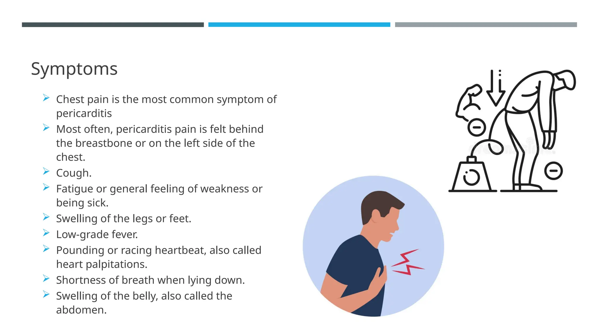
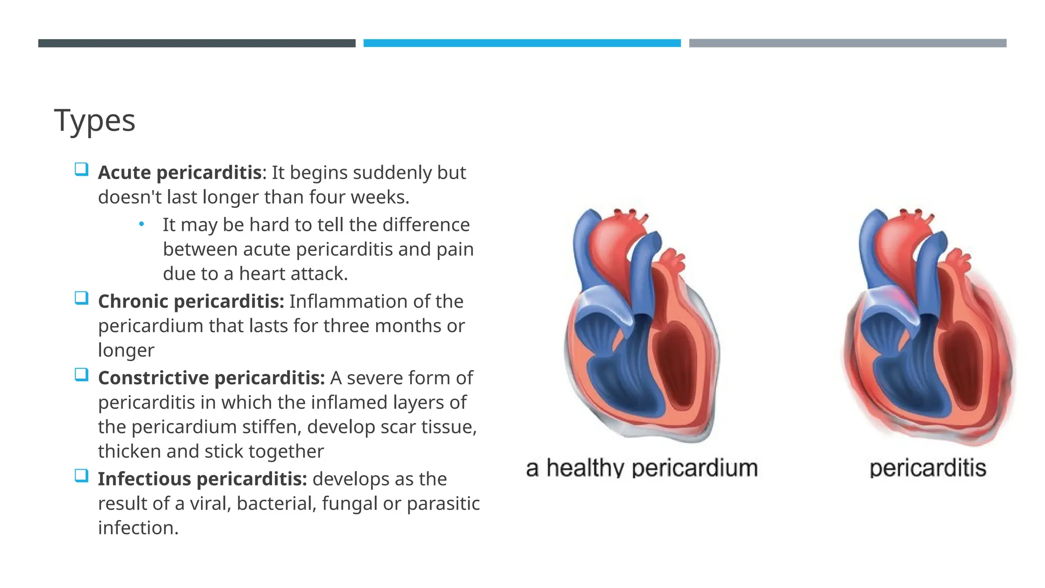
Pericarditis is inflammation of the pericardium, the sac surrounding the heart.
 Symptoms: Chest pain, fever, and shortness of breath are common.
 Types: Acute, chronic, and recurrent pericarditis.
 Diagnosis: Involves ECG, echocardiogram, and blood tests for inflammation markers.
Identify the symptoms and clinical manifestations of Pericarditis

Identify the symptoms and clinical manifestations of Pericarditis

  • 1.
    TOPIC: Pericarditis MODULE 1:Cardiovascular and Respiratory System Bisma amin Assistant professor Radiology and imaging technology
  • 2.
    LEARNING OUTCOME OF THETOPIC • Identify the symptoms and clinical manifestations of Pericarditis, Valvular diseases, Rheumatic Heart Disease, Heart Failure, Chronic Bronchitis, Emphysema & Bronchitis.
  • 3.
    Pericardium  Your pericardiumis a fluid-filled sac that surrounds your heart and the roots of the major blood vessels that extend from your heart.  Your pericardium is located in your chest, where it surrounds your heart.  Some functions of pericardium are:  Cushioning your heart from outside forces and pressure.  Holding your heart in place.  Keeping your heart from expanding too much  Protecting your heart from infections.  Providing lubrication to reduce friction between your heart and surrounding tissues.
  • 4.
    Introduction  Pericarditis isswelling and irritation of the thin, saclike tissue surrounding the heart, called pericardium.  Pericarditis often causes sharp chest pain.  The chest pain occurs when the irritated layers of the pericardium rub against each other.  Pericarditis often is mild. It may go away without treatment
  • 5.
    Symptoms  Chest painis the most common symptom of pericarditis  Most often, pericarditis pain is felt behind the breastbone or on the left side of the chest.  Cough.  Fatigue or general feeling of weakness or being sick.  Swelling of the legs or feet.  Low-grade fever.  Pounding or racing heartbeat, also called heart palpitations.  Shortness of breath when lying down.  Swelling of the belly, also called the abdomen.
  • 6.
    Types  Acute pericarditis:It begins suddenly but doesn't last longer than four weeks. • It may be hard to tell the difference between acute pericarditis and pain due to a heart attack.  Chronic pericarditis: Inflammation of the pericardium that lasts for three months or longer  Constrictive pericarditis: A severe form of pericarditis in which the inflamed layers of the pericardium stiffen, develop scar tissue, thicken and stick together  Infectious pericarditis: develops as the result of a viral, bacterial, fungal or parasitic infection.
  • 7.
    Cont. Types  Idiopathicpericarditis: Pericarditis that doesn’t have a known cause.  Traumatic pericarditis: develops as the result of an injury to the chest, such as after a car accident.  Uremic pericarditis: develops as the result of kidney failure.  Malignant pericarditis: develops as result of a cancer growing in your body.
  • 8.
    Causes  A complicationof a viral infection, most often a gastrointestinal virus, causes viral pericarditis.  A bacterial infection, including tuberculosis, causes bacterial pericarditis.  A fungal infection causes fungal pericarditis.  An infection from a parasite causes parasitic pericarditis.  Some autoimmune diseases, such as lupus, rheumatoid arthritis and scleroderma, can cause pericarditis.
  • 9.
    Cont. causes  Injuryto the chest, such as after a car accident, causes traumatic pericarditis.  Kidney failure causes uremic pericarditis.  Tumors like lymphoma causes malignant pericarditis.  Genetic diseases such as Familial Mediterranean Fever (FMF).  Medications that suppress the immune system (This is rare).
  • 10.
    Risk factor  Aheart attack.  Open heart surgery (postpericardiotomy syndrome).  Radiation therapy.  Percutaneous treatment, such as cardiac catheterization or radiofrequency ablation (RFA).
  • 11.
    How is pericarditisdiagnosed?  Sharp pain in your chest and back of the shoulders that feel better when you sit up and lean forward  chest pain with breathing  The rubbing of your pericardium’s inflamed lining causes a rubbing or creaking sound called the “pericardial rub." They’ll be able to hear it best when you lean forward, hold your breath and breathe out
  • 12.
    Diagnose  Chest X-rayto see the size of your heart and any fluid in your lungs.  Electrocardiogram (ECG or EKG) to look for changes in your heart rhythm. In about half of all people with pericarditis, providers see some characteristic changes to a normal heart rhythm.  Echocardiogram (echo) to see how well your heart is working and check for fluid (a pericardial effusion) around your heart  Cardiac MRI to check for extra fluid in your pericardium, pericardial inflammation or thickening, or compression of your heart.
  • 13.
    Cont.  CT scanto look for calcium in the pericardium, fluid, inflammation, tumors and disease of the areas around your heart  Blood tests: sedimentation rate (ESR) and ultra-sensitive C reactive protein levels Echocardiogram
  • 14.
  • 15.
    Video explaining pericarditiswhile reading radiological scans ◼ https://www.youtube.com/watch?app=desktop&v=e0qZ5qh4GMA
  • 16.
    Summary  Introduction: Pericarditisis inflammation of the pericardium, the sac surrounding the heart.  Symptoms: Chest pain, fever, and shortness of breath are common.  Types: Acute, chronic, and recurrent pericarditis.  Diagnosis: Involves ECG, echocardiogram, and blood tests for inflammation markers.