Pericarditis is inflammation of the pericardium (the fibrous sac surrounding the heart □Pericarditis is inflammation of the pericardium, a sac-like structure with two thin layers of tissue that surround the heart to hold it
SOCIAL AND HISTORICAL CONTEXT - LFTVD.pptxiammrhaywood
More Related Content
Similar to Pericarditis is inflammation of the pericardium (the fibrous sac surrounding the heart □Pericarditis is inflammation of the pericardium, a sac-like structure with two thin layers of tissue that surround the heart to hold it
Similar to Pericarditis is inflammation of the pericardium (the fibrous sac surrounding the heart □Pericarditis is inflammation of the pericardium, a sac-like structure with two thin layers of tissue that surround the heart to hold it (20)
Kisan Call Centre - To harness potential of ICT in Agriculture by answer farm...
Pericarditis is inflammation of the pericardium (the fibrous sac surrounding the heart □Pericarditis is inflammation of the pericardium, a sac-like structure with two thin layers of tissue that surround the heart to hold it
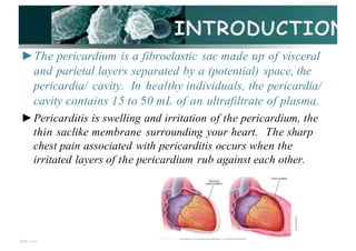
1. ►The pericardium is a fibroelastic sac made up of visceral
and parietal layers separated by a (potential) space, the
pericardia/ cavity. In healthy individuals, the pericardia/
cavity contains 15 to 50 mL of an ultrafiltrate of plasma.
►Pericarditis is swelling and irritation of the pericardium, the
thin saclike membrane surrounding your heart. The sharp
chest pain associated with pericarditis occurs when the
irritated layers of the pericardium rub against each other.
fppt com
· - • < > • - · -
2. □Pericarditis is inflammation of the
pericardium (the fibrous sac
surrounding the heart
□Pericarditis is inflammation of the
pericardium, a sac-like structure with
two thin layers of tissue that
surround the heart to hold it in place
and help it work. lnflammed
pericardium
- - - - - (pericarditis)
Sternu
A D A M
3. ► C. Mycoplasma
► D. Fungal - Histoplasmosis, aspergillosis,
blastomycosis, coccidiodomycosis,
actinomycosis, nocardia, candida
► E. Parasitic - Echinococcus, amebiasis,
toxoplasmosis
► R In{!ctive endocarditis with valve ring
abscess
fppt com
4. 1. Idiopathic
2. Infections
► A. Viral - Coxsackievirus, echovirus, adenovirus,
EBV, CMV, influenza, varicella, rubella, HIV,
hepatitis B, mumps, parvovirus B19,
► B. Bacterial - Staphylococcus, Streptococcus,
pneumococcus, Haemophilus, Neisseria
(gonorrhoeae or meningitidis), Chlamydia (psittaci
or trachomatis), Legionella, tuberculosis,
Salmonella, Lyme disease .
fppt com
5. ■ Pericarditis occurs after pericardectomy
in 5 % - 30% patients.
■ 1 % - 3 % of cases develop after 10 days to 2
months after acute myocardial infarction.
■ In the developed w o r l viruses are
believed to be the cause of about 85% of
cases.
■ In the developing world tuberculosis is a
common cause but it is rare in the
fpptc rn developed world.
6. 4. Cardiac
Myocarditis
Inflammation of
heartmusde
►A. Early infarction
pericarditis
► B. Late postcardiac injury
syndrome (Dressler's
syndrome)
► C. Myocarditis
►D. Dissecting aortic
aneurysm
fppt C m
Abdominal
Aortic Aneurysm
Thoracic
Aortic Aneurysm
7. 4. Cardiac
Myocarditis
Inflammation of
heartmusde
►A. Early infarction
pericarditis
► B. Late postcardiac injury
syndrome (Dressler's
syndrome)
► C. Myocarditis
►D. Dissecting aortic
aneurysm
fppt C m
Abdominal
Aortic Aneurysm
Thoracic
Aortic Aneurysm
8. 3. Neoplasm
►A. Metastatic - Lung or breast cancer,
Hodgkin's disease, leukemia, melanoma
►B. Primary - Rhabdomyosarcoma,
teratoma,fibroma, Lipoma, leiomyoma,
angi•
oma
►C. Paraneoplasm
fppt C m
9. 7. Metabolic
► A. Hypothyroidism - Primarily pericardia[ effusion
► B. Uremia
►C. Ovarian hyperstimulation syndrome
Healthy Thyroid Hypothyroidism
Polycystic
ovary
Hyperstimulated
polycystic ovary
fppt C nl
10. 6.Drugs
►Pericarditis can also develop from a drug
induced lupus syndrome caused by
medications including procainamide,
hydralazine, methyldopa, isoniazid,
mesalazine, and reserpine.
►Doxorubicin: The anthracycline
antineoplastic agents, such as doxorubicin
and cyclophosphamide, have direct cardiac
lpp,c"' toxicity and can cause acute pericarditis
11. 6.Drugs
►Pericarditis can also develop from a drug
induced lupus syndrome caused by
medications including procainamide,
hydralazine, methyldopa, isoniazid,
mesalazine, and reserpine.
►Doxorubicin: The anthracycline
antineoplastic agents, such as doxorubicin
and cyclophosphamide, have direct cardiac
lpp,c"' toxicity and can cause acute pericarditis
12. BASED ON THE CAUSES :-
►Constrictive pericarditis ►Traumatic pericarditis
►Viral pericarditis ►Serous pericarditis
►Purulent pericarditis ►Fiberous pericarditis
b l
.
► d" . ►Hemorrhagic
Tu ercu ous pericar itis peri.card1
·t1
·s
►Radiation Pericarditis ►Adhesive mediastino
pericarditis
fppt C
14. • Trauma
►A. Blunt, Penetrating
►B. Iatrogenic - Catheter and
pacemaker per/orations,
cardiopulmonary resuscitation
• Radiation
fppt C m
15. lpptco
• Purulent or
suppurative
pericarditis :-
It is due to causative
organisms may arise from
direct extension,
hematogenous seeding, or
lymphatic extension, or by
direct introduction during
cardiotomy.
Immunosuppression
facilitates this condition.
16. • Viral ericarditis
Viruses that cause
pericarditis is known
as viral pericarditis
This kind of
pericarditis is simple
and can be handled
as an outpati•
ent
procedure.
fppt
• Tuberculous pericarditis
This condition is also
seen i•
n a very mi•
nor
percentage of patients
having pulmonary
tuberculosis. Some of the
developing countries
remain the leading risk
groups of tuberculous
pericarditis.
17. • Constrictive pericarditis
When the pericarditis is
associated with a
thickening or scarring of
the pericardia/ layers, this
starts constricting the heart
within the thoracic cavity,
which in turn limits its
effective functioning. This
condition is known as
constrictive pericarditis.
fppt C l
N
o
r
m
a
l
h
e
a
r
t P
e
r
i
c
a
r
d
i
a
lcon
s
tric
tio
n
Pericardiun llid®loo
p
e
r
i
c
a
r
d
"
u
m
: I
11.Rl
18. • Fibrous and serofibrinous
pericarditis
• l!.represent the same basic
process and are the most
frequent type of pericarditis.
Common causes include acute
myocardial infarction (Ml),
postinfarction (including
Dressler syndrome), uremia,
radiation and trauma
lppt com
19. • Serous
pericarditis
•
Is usually caused by
noninfectious
inflammation such
as occurs in
rheumatoid arthritis
and systemic lupus
erythematosus .
lppt com
20. • Radiation Pericarditis
This type of pericarditis
mediastinal radiation at
any time from weeks to
months after the
exposure.
• Traumatic
pericarditis
is caused due to recent • Sharp or blunt trauma
causes traumati•
c
pericarditis. Invasive
cardiac procedures also
may give rise to this type
of pericarditis, which
includes cardiac
diagnostic catheterization
and electrophysiological
ablation procedure.
fppt com
21. when microbes are inhaled or ingested, they migrate to
myocardium and cause inflammation
Increased Intra Pericardia/ pressure
'== + - - - - - - - - - , , , =
Compression of the heart
Decreased ventricular filling and emptying
Increase venous
pressure
Decreasea caraiac
output
Decreasea Arterial
pressure
fppt C m
Cardiac Failure
22. • Chronic pericarditis Adhesive
mediastino pericarditis
• Is a reaction that usually follows suppurative
or caseous pericarditis, cardiac surgery, or
irradiation. This condition is rarely caused
by a simple fibrinous exudate. The
pericardia/ potential space is obliterated, and
adhesion of the external surface of the
parietal layer to surrounding structures
fpptc occurs.
23. • Hemorrhagic pericarditis
It involves blood mixed with a fibrinous or
suppurative effusion, and it is most
commonly caused by tuberculosis or direct
neoplastic invasion. This condition can also
occur in severe bacterial infections.
Hemorrhagic pericarditis is common after
cardiac surgery and may cause tamponade.
The clinical significance is similar to
lpplcosuppurativepericarditis
24. •!•In Constrictive Pericarditis:
•!• Pedal edema
•!• Hepatomegaly
•!• Ascites
•!• JVD
•!• Kussmaul's sign
•!• Pericardia/ knock (early
diastolic sound) heard at the
apex
l
ppl
c
o♦
:
♦ Usually - no friction rub
Healthy foot Foot and ankle
witll edema
25. Ewart sign: Ewart's sign is a set of
findings on physical examination in people
with large collections of fluid around their
heart (pericardia/ effusions).Dullness to
percussion ("woody" in quality), egophony,
and bronchial breath sounds may be
appreciated at the inferior angle of the left
scapula when the effusion is large enough to
compress the left lower lobe of the lung•
Beck's triad: failing BP; rising JVP;
fppt com
26. •!•Chest pain beneath the clavicle,
in the neck region worsens with
deep inspiration, relieved with
sitting or leaning forward.It is the
cardinal sign of pericarditis
•!• Mild fever, chills and night
sweats.
•!• Malaise, myalgia
•!• Dyspnea due to constriction or
cardiac tamponade
•!•Palpitation
fppt C m
27. • CT Scan to look for
calcium in the
pericardium, fluid,
inflammation, tumors and
disease of the areas
around the heart. Iodine
dye is used during the test
to get more information
about the inflammation.
• Pericardiocentesis fluid
determine cause; treat
cardiac tamponade
fppt com
16-18 gougeneedle
28. • Echo- for heart wall
movement
shows
• Chest X ra
-v-
a
n enlarged heart and
pericardia/ calcification
• Doppler imaging- to
measure the amount of
blood flow through
your arteries and veins
fpp com
29. • History Collection- regarding the
etiological factors
• Ph•
ysical Examination- check for Ewart's
sign,pedal dema ,hepatomegaly JVD etc..
• CBC- Increased WBC ESR, and CRP
• Cardiac Enz
•
ymes- increased but not as
much as with Ml
• ECG- diffuse St elevation *important to
different from Ml changes (acute
pericarditis}
fppt C m
30. • Pericardia/ effusion.
• Accumulation of fluid in the
pericardia/ sac. may have
symptoms such as:Chest pain
or discomfort, Enlargement
of the veins of the
neck,Fainting,Fast
breathing, Increased heart
rate,Nausea,Pain in the right
upper abdomen,Shortness of
breath,Swelling in the arms
and legs
fppt com
HealthyHeart Heartwitha
Pericardialeffusion:
31. • Can 1ac tampona e
Accumulation of pericardia[ fluid raises intra
pericardial pressure, hence poor ventricular
filling and fall in cardiac output.
The drop in blood pressure can cause blurred
vision, nausea, confusion, and weakness.
I n i t i a l C o n d i t i o n
A n t e n o r w e v
l n l t l a l C o n d i t i o n
A n t enor cur--.away v,ew
R e s u l t i n g C a r d i a c T a m p o n a d e
Anrenor cur-a w a y wew
P.Ork::aN:tium
COV<&f"lnQ the heat"I
C U t o d g e - o·r th& po,rJcard.luni
a u n- o u nt nng t h e - heart
fppt com
- t
n"° p o ric a rd . i a l a a c fllkt
w i t h rtuid c a u s in g s e - ,ere
com rpna.e. 1on ot the h e . a n
32. • Cardiac MRI to check for
extra fluid in the pericardium,
pericardia/ inflammation or
thickening, or compression of
the heart. A contrast agent
called gadolinium is used
during this highly specialized
test.
• Cardiac catheterization
To get information about the
filling pressures in the heart.
This is used to confirm a
diagnosis of constrictive
pericarditis.
fppt com
TIie u f
33. • Aspirin -Aspirin can be given at a dose of 750 to 1000
mg every six to eight hours fallowed by gradual tapering
every week for a treatment period of three to four weeks.
• Corticosteroids Corticosteroids are strong medications
that fight inflammation. Your doctor may prescribe a
corticosteroid such as prednisone if your symptoms don1t
get better with other medications, or if symptoms keep
returning.
• Colchicine anti-inflammatory agent It is recommended as
first-line therapy for acute pericarditis as an adjunct to
aspirin1NSAID therapy. You should not take this drug if
you have liver or kidney disease
fppt C m
34. • ASA or tylenol Acetaminophen decreases fever and pain ,
but does not help inflammation.Adult dosing is 2 regular
strength (325 mg) every 4 hours or 2 extra-strength (500 mg)
every 6 hours. Maximum dose is 4,000 mg per day.
• Aspirin or NSAIDs are recommended as first-line therapy
for acute pericarditis with gastroprotection. Commonly used
NSAID regimens include : lbuprofen - Depending on the
severity of the pericarditis and individual medication
response, a dose of 400 to 800 mg of ibuprofen three times
daily is usually adequate for symptom relief. lbuprofen can
be the preferred NSAID because of its rare side effects,
favorable impact on coronary artery blood flow, and large
fpp C m
35. • Chronic effusive
pericarditis It is an
uncommon pericardia/
syndrome characterized by
concomitant tamponade,
caused by
tense pericardia/ effusion,
and constriction, caused by
thevisceral pericardium. th
e symptoms are chest pain,
lightheadedness, hiccups,
and shortness of breath.
lppt
36. • Pericardiocentesis:- is the aspiration of
fluid f ram the pericardia/ space that
surrounds the heart.
Pericardiocentesis
Subxiphoid Approach
Needle inserted btwn the
xiphoid process and L
costal margin
30°to 45°angle
Aim for L mid-clavicle
Directs needle toward
Anterior wall of R
ventricle
Niuta Joshi MO
Apox of
he rt
fppt com
Subxiphoid
37. • Anticongestive measures such as
diuretics And lnotropics agents
(lnotrtropic agents such as milrinone,
digoxin, dopamine, and dobutamine are
used to increase the force of cardiac
contractions.)
• Anti-anxiety medication (Alprazolam
Diazepam ,Estazolam ,Flurazepam )
• Proton pump inhibitors COmeprazolel
Pantoprazole)
fppt com
38. Indomethacin - Indomethacin (NSAID)can be
administered at a dose of 50 mg three times daily for one to
two weeks fallowed by slow tapering But commonly it is
not rcommended due to its adverse effects
• Penicillin - for Bacterial infection
• ACE Inhibitors - relax the blood vessels in the heart and
help blood flow more easily •
• Beta-blockers are avoided because it decreases the
strength of ventricular contraction (have a negative
inotropic effect)
fppt com
39. Percardiectomy may be
necessary to release both ventricles
from the constrictive and
restrictive inflammation and
scarring Pericardiectomy is
performed through a median
sternotomy, an incision through
the breastbone (sternum) in the
middle front part of the ribs that
allows the surgeon to reach the
heart. The surgeon will remove the
pericardium from the heart, wire
the breastbone and ribs back
together and close the incision
with stitches.
fpp C m
40. • Percutaneous a oon
p e r i c a r d i o t o m y : - is a procedure done to
drain excessfluid in the sac around the heart.
The procedure uses a long thin tube with
a balloon attached. During PBP, a doctor inserts
a needle through the chest wall and into the
tissue around the heart. Once the needle is inside
the pericardium1 the doctor removes it and
replaces it with a long1 thin tube called a
catheter. This tube has an inflatable balloon at
its tip. Repeated inflation of the balloon creates
a small hole or "window11
in the pericardium.
When the hole is large enough1 the doctor
removes the catheter and balloon replaces them
with a new catheter for final draining. This
allows fluid to drain out of the pericardium1
which improves heart function.
fppt C m
41. • Pericardial window a small opening
made in the pericardium1 may be performed to
allow continuous drainage into the chest
cavity.
fppt C m