Pericarditis
Nsg.Instructor
Safoora Qureshi
CON, PIMS
Pericardium:
• Pericardium is the outer most
layer and is made up of two
fibrous serous sacs
Functions:
• Limits cardiac distention
• Restricts excessive movements
• Protect from injury
• Lubricate to prevent friction
• Hold the heart in place
Pericarditis
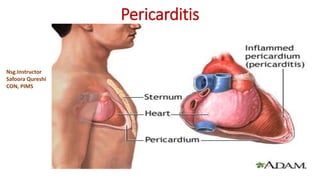
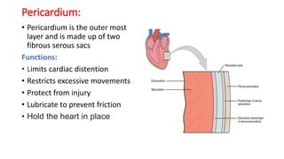
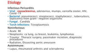
• Pericarditis is inflammation of the pericardium,
• Pericarditis is swelling and irritation of the thin, saclike tissue
surrounding the heart (pericardium).
• Pericarditis often causes sharp chest pain.
• The chest pain occurs when the irritated layers of the
pericardium rub against each other.
Etiology
Infectious Pericarditis
• Viral : coxsackievirus, adenovirus, mumps, varicella zoster, HIV,
Covid-19
• Bacterial: pneumococci, streptococci, staphylococci , tuberculosis,
Septicemia from gram- negative organisms
• Fungal . Candida
• Torch Infections: Toxoplasmosis
Noninfectious:
• Acute MI
• Neoplasms: ca lung, ca breast, leukemia, lymphomas
• Trauma: Thoracic surgery, pacemaker incretion, diagnostic
procedures
• Radiation, Dissecting aortic aneurysm
Autoimmune:
• Lupus, rheumatoid arthritis and scleroderma
Symptoms
Chest pain, is the most common symptom,
• Characterized by Sharp or stabbing pain,
may dull, achy or pressure-like chest pain
• Locate behind the breastbone,
or on the left side of the chest.
• Radiate to the left shoulder and neck
• Get better when sitting up or leaning forward
• Referred to trapezius muscles (shoulder, upper back)
is only the distinction between pericardial pain and angina
Other signs and symptoms
• Fever
• Shortness of breath
• when lying down
• Pericardial friction rub,
• is a scratching, grating, and high-pitched sound one of the
hallmark finding in acute pericarditis
• Cough
• Fatigue
• Palpitation
• Pounding or racing heartbeat)
Types
• Acute pericarditis:
• Begins suddenly but doesn't last longer than three weeks
• Difficult to differentiate between acute pericarditis and MI pain
• Recurrent pericarditis:
• Occurs about four to six weeks after an episode of acute pericarditis
with no symptoms in between.
• Incessant pericarditis:
• Lasts about four to six weeks but less than three months.
• The symptoms are continuous.
• Chronic constrictive pericarditis;
• usually develops slowly and lasts longer than three months.
Complications
• Pericardial effusion
• The fluid buildup can lead to further heart complications.
• Constrictive pericarditis).
• long-term pericarditis develop permanent thickening and scarring of the
heart lining
• The changes prevent the heart from filling and emptying properly.
• Cardiac tamponade)
• Pressure on the heart due to fluid buildup
• Prevents the heart from filling properly.
• Causing a dramatic drop in blood pressure.
• A life-threatening condition requires emergency treatment
Diagnosis
• History and Physical Examination
• Sharp chest pain and shortness of breathing are 2 major clues
• Heart Auscultation:
• Rubbing or creaking sound, called “pericardial rub” is the best
heard when the pt lean forward, hold breath and breathe out.
• Crackles in the lungs, which are signs of pleurisy or pericardial
effusion
• ECG: Normal/specific defused changes over aperid of hours to
days/wks. we
• PR segment depression,
• ST segment elevation and
• T wave flattening
Pericarditis
PR segment depression
ST segment elevation
T flattening
Normal ECG
• Chest X-ray
• to see the size of the heart and any fluid in lungs.
• Echocardiogram (echo) ;
• To find fluid or pericardial effusion around the heart
• Cardiac MRI;
• CT scan;
• to look for calcium in the pericardium, fluid, inflammation, tumors
around the heart
• Cardiac catheterization:
• to confirm a diagnosis of constrictive pericarditis.
• Blood tests
• ESR, CRP,
• To confirm heart attack
• To find the cause of pericarditis.
•
Collaborative Care
Medical treatment:
• Pain Relievers:
• Nonsteroidal anti-inflammatory (NSAIDs) use to treat pain and inflammation
such as aspirin or Ibuprofen
• Antibiotics :
• to reduce inflammation and pain
• Steroids:
• strong anti-inflammatory drug, may used in pericarditis secondary to
autoimmune conditions
• Pericardiocentesis:
• is usually performed for pericardial effusion with cardiac tamponade,
purulent pericarditis and a high suspicious of neoplasm
Nursing management:
• Strict bed rest
• Keep in prop-upped position
• Elevate the bed at 45 degree
• Provide an overbed table for support
• Observe for post anti-inflammatory drugs complications such as upper GI
bleed, gastric upset etc
• Advise to take these drugs with food and milk
• Instruct avoid alcohol beverages
• Take anxiety-reduce measures by explaining performed procedures and
possible cause of pain , treatment and prognosis
• Reassure the client

Paricarditis.pptx

  • 1.
  • 2.
    Pericardium: • Pericardium isthe outer most layer and is made up of two fibrous serous sacs Functions: • Limits cardiac distention • Restricts excessive movements • Protect from injury • Lubricate to prevent friction • Hold the heart in place
  • 3.
    Pericarditis • Pericarditis isinflammation of the pericardium, • Pericarditis is swelling and irritation of the thin, saclike tissue surrounding the heart (pericardium). • Pericarditis often causes sharp chest pain. • The chest pain occurs when the irritated layers of the pericardium rub against each other.
  • 5.
    Etiology Infectious Pericarditis • Viral: coxsackievirus, adenovirus, mumps, varicella zoster, HIV, Covid-19 • Bacterial: pneumococci, streptococci, staphylococci , tuberculosis, Septicemia from gram- negative organisms • Fungal . Candida • Torch Infections: Toxoplasmosis Noninfectious: • Acute MI • Neoplasms: ca lung, ca breast, leukemia, lymphomas • Trauma: Thoracic surgery, pacemaker incretion, diagnostic procedures • Radiation, Dissecting aortic aneurysm Autoimmune: • Lupus, rheumatoid arthritis and scleroderma
  • 6.
    Symptoms Chest pain, isthe most common symptom, • Characterized by Sharp or stabbing pain, may dull, achy or pressure-like chest pain • Locate behind the breastbone, or on the left side of the chest. • Radiate to the left shoulder and neck • Get better when sitting up or leaning forward • Referred to trapezius muscles (shoulder, upper back) is only the distinction between pericardial pain and angina
  • 7.
    Other signs andsymptoms • Fever • Shortness of breath • when lying down • Pericardial friction rub, • is a scratching, grating, and high-pitched sound one of the hallmark finding in acute pericarditis • Cough • Fatigue • Palpitation • Pounding or racing heartbeat)
  • 8.
    Types • Acute pericarditis: •Begins suddenly but doesn't last longer than three weeks • Difficult to differentiate between acute pericarditis and MI pain • Recurrent pericarditis: • Occurs about four to six weeks after an episode of acute pericarditis with no symptoms in between. • Incessant pericarditis: • Lasts about four to six weeks but less than three months. • The symptoms are continuous. • Chronic constrictive pericarditis; • usually develops slowly and lasts longer than three months.
  • 9.
    Complications • Pericardial effusion •The fluid buildup can lead to further heart complications. • Constrictive pericarditis). • long-term pericarditis develop permanent thickening and scarring of the heart lining • The changes prevent the heart from filling and emptying properly. • Cardiac tamponade) • Pressure on the heart due to fluid buildup • Prevents the heart from filling properly. • Causing a dramatic drop in blood pressure. • A life-threatening condition requires emergency treatment
  • 10.
    Diagnosis • History andPhysical Examination • Sharp chest pain and shortness of breathing are 2 major clues • Heart Auscultation: • Rubbing or creaking sound, called “pericardial rub” is the best heard when the pt lean forward, hold breath and breathe out. • Crackles in the lungs, which are signs of pleurisy or pericardial effusion • ECG: Normal/specific defused changes over aperid of hours to days/wks. we • PR segment depression, • ST segment elevation and • T wave flattening
  • 11.
    Pericarditis PR segment depression STsegment elevation T flattening Normal ECG
  • 12.
    • Chest X-ray •to see the size of the heart and any fluid in lungs. • Echocardiogram (echo) ; • To find fluid or pericardial effusion around the heart • Cardiac MRI; • CT scan; • to look for calcium in the pericardium, fluid, inflammation, tumors around the heart • Cardiac catheterization: • to confirm a diagnosis of constrictive pericarditis. • Blood tests • ESR, CRP, • To confirm heart attack • To find the cause of pericarditis. •
  • 13.
    Collaborative Care Medical treatment: •Pain Relievers: • Nonsteroidal anti-inflammatory (NSAIDs) use to treat pain and inflammation such as aspirin or Ibuprofen • Antibiotics : • to reduce inflammation and pain • Steroids: • strong anti-inflammatory drug, may used in pericarditis secondary to autoimmune conditions • Pericardiocentesis: • is usually performed for pericardial effusion with cardiac tamponade, purulent pericarditis and a high suspicious of neoplasm
  • 14.
    Nursing management: • Strictbed rest • Keep in prop-upped position • Elevate the bed at 45 degree • Provide an overbed table for support • Observe for post anti-inflammatory drugs complications such as upper GI bleed, gastric upset etc • Advise to take these drugs with food and milk • Instruct avoid alcohol beverages • Take anxiety-reduce measures by explaining performed procedures and possible cause of pain , treatment and prognosis • Reassure the client