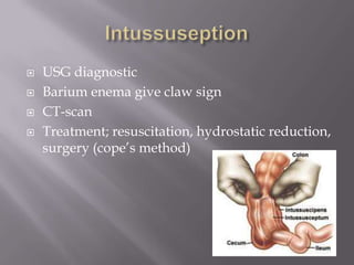
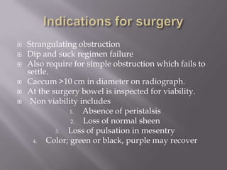
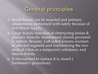
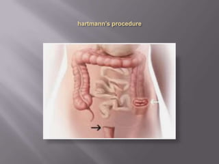
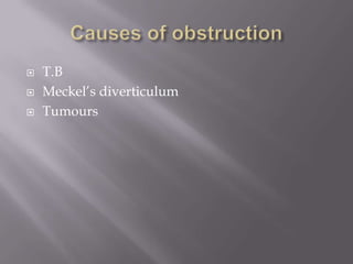
MUSADAQ ASRAR
Shifa College of Medicine
2013-051
 A 95 yr old woman is sent to the ER from
nursing home with complaints of abdominal
pain and distension. It is unclear, but staff at
nursing home thinks it has been about 4 days
since she had a bowel movement.
 The woman has an expressive aphasia and rt.
hemiparesis from a previous stroke.
 Her vitals; pulse 90, B.P 120/80, afebrile.
 Her abdomen is markedly distended with
hyperactive bowel sounds and tympanitic
throughout.
 There is no abdominal tenderness.
 Rectal exam reveals large quantities of soft, brown
stool.
 KUB and upright images reveal marked dilated
colon consistent with obstruction.
Classification.
Can be divided into paralytic and mechanical.
 Mechanical is further classified according to,
1. Speed of onset: acute, chronic, acute on chronic
2. Site: high or low.
3. Nature: simple or strangulated
4. Aetiology.
 In acute obstruction, the onset is rapid and the
symptoms are severe.
 In chronic obstruction, symptoms are insidious
and slowly progressive e.g most case of
carcinoma of large bowel.
 A chronic obstruction may develop acute
symptoms as obstruction becomes complete.
e.g narrow lumen occluded by inspissated
bowel contents.
 Small intestine
 Large intestine
 Simple: when bowel is occluded without
damage to its blood supply.
 Closed loop obstruction
 Strangulated: blood supply of involved
segment is cut off. e.g strangulated hernia,
volvulus, intussusception.
 Gangrene is inevitable if left untreated.
 Causes in the lumen; fecal impaction, gallstone
‘ileus’, food bolus, parasites, intussusception.
 Causes in the wall; congenital atresia, Crohn’s,
tumors, diverticulitis.
 Causes outside the wall; strangulated hernia,
volvulus and obstruction due to adhesions or
bands.
 Neonatal; congenital atresia, stenosis,
imperforate anus, volvulus neonatorum,
hirschsprung’s and meconium ileus.
 Infants; intussusception, hirschsprung’s,
strangulated hernia, and meckel’s
diverticulum.
 Young & middle age; strangulated hernia,
adhesion, bands and Crohn’s disease.
 Elderly; strangulated hernia, carcinoma of
bowel, colonic diverticulitis, impacted faeces.
 Simple occlusion causes Intestine distal to
obstruction to empties rapidly and collapsed.
The bowel above the obstruction becomes
dilated with gas and secretions, increased
peristalsis to over come obstruction causes
which intestinal colic.
 Impaired blood supply due to distention may
cause mucosal ulceration and perforation.
 Integrity of mucosal barrier lost due to
ischemia.
 Secondary peritonitis occurs due to
transudation of organisms in gut.
 Strangulation is followed by gangrene of the
ischemic bowel with perforation.
 Fluid and electrolyte depletion occur due to
copious vomiting and loss into the bowel
lumen.
 Protein loss into the gut and toxemia due to
migration of toxins and intestinal bacteria into
the peritoneal cavity.
 Colicky abdominal pain.
 Distension.
 Absolute constipation.
 Vomiting.
 Small bowel; pariumbilical
 Distal colonic; suprapubic in location
 Postoperative obstruction; colic may be
disguised by general discomfort.
 Usually seen in chronic large bowel
obstruction.
 Volvulus of the sigmoid colon.
 In high intestinal obstruction distension will
not be marked.
 Failure to pass flatus or faeces.
 It is an early feature in large bowel obstruction.
 Late feature in small bowel obstruction.
 Pt. may pass 1 or 2 stools after onset of
obstruction as bowel completely empties below
obstruction.
 Usually occurs in high obstruction, but late in
low bowel obstruction, or may be absent.
 In late stages vomiting becomes faeculent but
not faecal.
 True vomiting of faeces occurs only in
gestrocolic fistula.
 Dehydration
 Elevated pulse
 Temperature usually raised in strangulation.
 Distention and visible peristalsis
 Any hernia or presence of any abdominal scar
suggests adhesions or bands as a cause.
 Mass in intussuseption or carcinoma
 Digital rectal examination.
 Strangulation verses simple must be
distinguished because strangulation ensuing
peritonitis has a high mortality of up to 15%.
 Toxic appearance
 Colicky pain
 Tenderness & abdominal rigidity
 Absent bowel sounds
 Raised white cell count
 Abdominal x rays; erect and supine
 Barium follow through; in cases of small bowel
 barium enema.
 Sigmoidoscopy; chronic obstruction
 Colonoscopy; chronic obstruction
 CT scan
Ladder pattern of dilated loops
haustrations in large bowel obstruction
 IV fluids and nasogastric aspiration.
(dip and suck)
 Nil orally
 2 hourly temperature and pulse
 Abdominal examination 8 hourly.
 Strangulating obstruction
 Dip and suck regimen failure
 Also require for simple obstruction which fails to
settle.
 Caecum >10 cm in diameter on radiograph.
 At the surgery bowel is inspected for viability.
 Non viability includes
1. Absence of peristalsis
2. Loss of normal sheen
3. Loss of pulsation in mesentry
4. Color; green or black, purple may recover
 Small bowel; can be resected and primary
anastomosis performed with safety because of
rich blood supply
 Large bowel; resection of obstructing lesion &
primary ileocolic anastomosis (lesion proximal
to splenic flexure). Left sided lesions; excision
of affected segment and exteriorizing the two
ends of colon as a temporary colostomy and
mucus fistula.
 If not reached to surface it is closed (
hartmann’s procedure)
 T.B
 Meckel’s diverticulum
 Tumours
 Occurs in elderly, constipated patients
 4 time common in men
 X ray shows distended loop of a bowel the
shape of a ‘coffee bean’ arising out of the pelvis
on the left side. Barium enema gives bird beak
appearance.
 Treatment; decompression by sigmoidoscopy.
 Rectal flatus tube placed in situ for 48 h. later
elective resection.
 If decompression is unsuccessful or there are
signs of gangrene or perforation, laparotomy
with resection is under taken, the two ends of
the colon being brought out as a double
barrelled colostomy. Paul- Mikulicz procedure
which is later closed.
 Usually associated with congenital
malformation. Excessively mobile caecum and
ascending colon, defect in rotation, caecum
retain its mesentry.
 AXR; dilated caecum in left upper quadrant
 Treatment; laparotomy, if bowel is viable,
untwisting with caecostomy. Right
hemicolectomy is necessary if the caecum is
infarcted, and to prevent recurrence.
 The mortality rate is high
 Mesenteric embolus; AF, vegetation on valves.
 Mesenteric arterial thrombosis; atheroma,
aortic dissection.
 Mesenteric venous thrombosis; portal
hypertension, may follow splenectomy.
Crohn’s disease, OCPs.
 Vascular occlusion results in infarction of the
affected gut, leads to bleeding.
 Gangrene and perforation occurs
 Classic triad; acute abdominal pain, rectal
bleeding, shock in an elderly who has AF.
 Treatment; early embolectomy and
revascularization before gangrene sets up.
 Lower abdominal colicky pain
 Alternating constipation and diarrhoea
 Abdominal distension
 Sigmoidoscopy to exclude carcinoma
 D.D’s; Ca colon, crohn’s, ischemic colitis
 Complications; large bowel obstruction,
hemorrhage, fistula, perforation and stricture.
 Treatment; resection either by Hartmann’s
procedure followed by subsequent restorative
surgery or a primary anastomosis protected by
a temporary defunctioning loop ileostomy.
 Telescoping of the one segment of bowel into
adjacent segment.
 Ileo-colic
 Ileo-ileal
 Ileo-caecal
 Colo-colic
 Most common in children 5-10month of age
 70-90% idiopathic
 Other association; meckel’s diverticulum,
polyp in children
 Adult intussuseption is associated with peutz-
jegher’s syndrome, lipoma or tumor
 Redcurrent jelly stool.
 Sausage shaped hump in the abdomen and
emptiness in RIF.
 USG diagnostic
 Barium enema give claw sign
 CT-scan
 Treatment; resuscitation, hydrostatic reduction,
surgery (cope’s method)
 Intestinal atresia
 Volvulus neonatorum
 Meconium ileus
 Necrotizing enterocolitis
 Hirschsprung’s disease
 Anorectal atresias
Patient with chronic constipation & abdominal distension

Patient with chronic constipation & abdominal distension

  • 1.
    MUSADAQ ASRAR Shifa Collegeof Medicine 2013-051
  • 2.
     A 95yr old woman is sent to the ER from nursing home with complaints of abdominal pain and distension. It is unclear, but staff at nursing home thinks it has been about 4 days since she had a bowel movement.
  • 3.
     The womanhas an expressive aphasia and rt. hemiparesis from a previous stroke.  Her vitals; pulse 90, B.P 120/80, afebrile.  Her abdomen is markedly distended with hyperactive bowel sounds and tympanitic throughout.  There is no abdominal tenderness.  Rectal exam reveals large quantities of soft, brown stool.  KUB and upright images reveal marked dilated colon consistent with obstruction.
  • 4.
    Classification. Can be dividedinto paralytic and mechanical.  Mechanical is further classified according to, 1. Speed of onset: acute, chronic, acute on chronic 2. Site: high or low. 3. Nature: simple or strangulated 4. Aetiology.
  • 5.
     In acuteobstruction, the onset is rapid and the symptoms are severe.  In chronic obstruction, symptoms are insidious and slowly progressive e.g most case of carcinoma of large bowel.  A chronic obstruction may develop acute symptoms as obstruction becomes complete. e.g narrow lumen occluded by inspissated bowel contents.
  • 6.
     Small intestine Large intestine  Simple: when bowel is occluded without damage to its blood supply.  Closed loop obstruction  Strangulated: blood supply of involved segment is cut off. e.g strangulated hernia, volvulus, intussusception.  Gangrene is inevitable if left untreated.
  • 7.
     Causes inthe lumen; fecal impaction, gallstone ‘ileus’, food bolus, parasites, intussusception.  Causes in the wall; congenital atresia, Crohn’s, tumors, diverticulitis.  Causes outside the wall; strangulated hernia, volvulus and obstruction due to adhesions or bands.
  • 8.
     Neonatal; congenitalatresia, stenosis, imperforate anus, volvulus neonatorum, hirschsprung’s and meconium ileus.  Infants; intussusception, hirschsprung’s, strangulated hernia, and meckel’s diverticulum.  Young & middle age; strangulated hernia, adhesion, bands and Crohn’s disease.  Elderly; strangulated hernia, carcinoma of bowel, colonic diverticulitis, impacted faeces.
  • 9.
     Simple occlusioncauses Intestine distal to obstruction to empties rapidly and collapsed. The bowel above the obstruction becomes dilated with gas and secretions, increased peristalsis to over come obstruction causes which intestinal colic.  Impaired blood supply due to distention may cause mucosal ulceration and perforation.
  • 10.
     Integrity ofmucosal barrier lost due to ischemia.  Secondary peritonitis occurs due to transudation of organisms in gut.  Strangulation is followed by gangrene of the ischemic bowel with perforation.
  • 11.
     Fluid andelectrolyte depletion occur due to copious vomiting and loss into the bowel lumen.  Protein loss into the gut and toxemia due to migration of toxins and intestinal bacteria into the peritoneal cavity.
  • 12.
     Colicky abdominalpain.  Distension.  Absolute constipation.  Vomiting.
  • 13.
     Small bowel;pariumbilical  Distal colonic; suprapubic in location  Postoperative obstruction; colic may be disguised by general discomfort.
  • 14.
     Usually seenin chronic large bowel obstruction.  Volvulus of the sigmoid colon.  In high intestinal obstruction distension will not be marked.
  • 15.
     Failure topass flatus or faeces.  It is an early feature in large bowel obstruction.  Late feature in small bowel obstruction.  Pt. may pass 1 or 2 stools after onset of obstruction as bowel completely empties below obstruction.
  • 16.
     Usually occursin high obstruction, but late in low bowel obstruction, or may be absent.  In late stages vomiting becomes faeculent but not faecal.  True vomiting of faeces occurs only in gestrocolic fistula.
  • 17.
     Dehydration  Elevatedpulse  Temperature usually raised in strangulation.  Distention and visible peristalsis  Any hernia or presence of any abdominal scar suggests adhesions or bands as a cause.  Mass in intussuseption or carcinoma  Digital rectal examination.
  • 18.
     Strangulation versessimple must be distinguished because strangulation ensuing peritonitis has a high mortality of up to 15%.  Toxic appearance  Colicky pain  Tenderness & abdominal rigidity  Absent bowel sounds  Raised white cell count
  • 19.
     Abdominal xrays; erect and supine  Barium follow through; in cases of small bowel  barium enema.  Sigmoidoscopy; chronic obstruction  Colonoscopy; chronic obstruction  CT scan
  • 20.
    Ladder pattern ofdilated loops
  • 21.
    haustrations in largebowel obstruction
  • 22.
     IV fluidsand nasogastric aspiration. (dip and suck)  Nil orally  2 hourly temperature and pulse  Abdominal examination 8 hourly.
  • 23.
     Strangulating obstruction Dip and suck regimen failure  Also require for simple obstruction which fails to settle.  Caecum >10 cm in diameter on radiograph.  At the surgery bowel is inspected for viability.  Non viability includes 1. Absence of peristalsis 2. Loss of normal sheen 3. Loss of pulsation in mesentry 4. Color; green or black, purple may recover
  • 24.
     Small bowel;can be resected and primary anastomosis performed with safety because of rich blood supply  Large bowel; resection of obstructing lesion & primary ileocolic anastomosis (lesion proximal to splenic flexure). Left sided lesions; excision of affected segment and exteriorizing the two ends of colon as a temporary colostomy and mucus fistula.  If not reached to surface it is closed ( hartmann’s procedure)
  • 26.
     T.B  Meckel’sdiverticulum  Tumours
  • 27.
     Occurs inelderly, constipated patients  4 time common in men  X ray shows distended loop of a bowel the shape of a ‘coffee bean’ arising out of the pelvis on the left side. Barium enema gives bird beak appearance.  Treatment; decompression by sigmoidoscopy.  Rectal flatus tube placed in situ for 48 h. later elective resection.
  • 28.
     If decompressionis unsuccessful or there are signs of gangrene or perforation, laparotomy with resection is under taken, the two ends of the colon being brought out as a double barrelled colostomy. Paul- Mikulicz procedure which is later closed.
  • 30.
     Usually associatedwith congenital malformation. Excessively mobile caecum and ascending colon, defect in rotation, caecum retain its mesentry.  AXR; dilated caecum in left upper quadrant  Treatment; laparotomy, if bowel is viable, untwisting with caecostomy. Right hemicolectomy is necessary if the caecum is infarcted, and to prevent recurrence.  The mortality rate is high
  • 31.
     Mesenteric embolus;AF, vegetation on valves.  Mesenteric arterial thrombosis; atheroma, aortic dissection.  Mesenteric venous thrombosis; portal hypertension, may follow splenectomy. Crohn’s disease, OCPs.
  • 32.
     Vascular occlusionresults in infarction of the affected gut, leads to bleeding.  Gangrene and perforation occurs  Classic triad; acute abdominal pain, rectal bleeding, shock in an elderly who has AF.  Treatment; early embolectomy and revascularization before gangrene sets up.
  • 33.
     Lower abdominalcolicky pain  Alternating constipation and diarrhoea  Abdominal distension  Sigmoidoscopy to exclude carcinoma  D.D’s; Ca colon, crohn’s, ischemic colitis  Complications; large bowel obstruction, hemorrhage, fistula, perforation and stricture.
  • 34.
     Treatment; resectioneither by Hartmann’s procedure followed by subsequent restorative surgery or a primary anastomosis protected by a temporary defunctioning loop ileostomy.
  • 35.
     Telescoping ofthe one segment of bowel into adjacent segment.  Ileo-colic  Ileo-ileal  Ileo-caecal  Colo-colic
  • 37.
     Most commonin children 5-10month of age  70-90% idiopathic  Other association; meckel’s diverticulum, polyp in children  Adult intussuseption is associated with peutz- jegher’s syndrome, lipoma or tumor  Redcurrent jelly stool.  Sausage shaped hump in the abdomen and emptiness in RIF.
  • 38.
     USG diagnostic Barium enema give claw sign  CT-scan  Treatment; resuscitation, hydrostatic reduction, surgery (cope’s method)
  • 39.
     Intestinal atresia Volvulus neonatorum  Meconium ileus  Necrotizing enterocolitis  Hirschsprung’s disease  Anorectal atresias