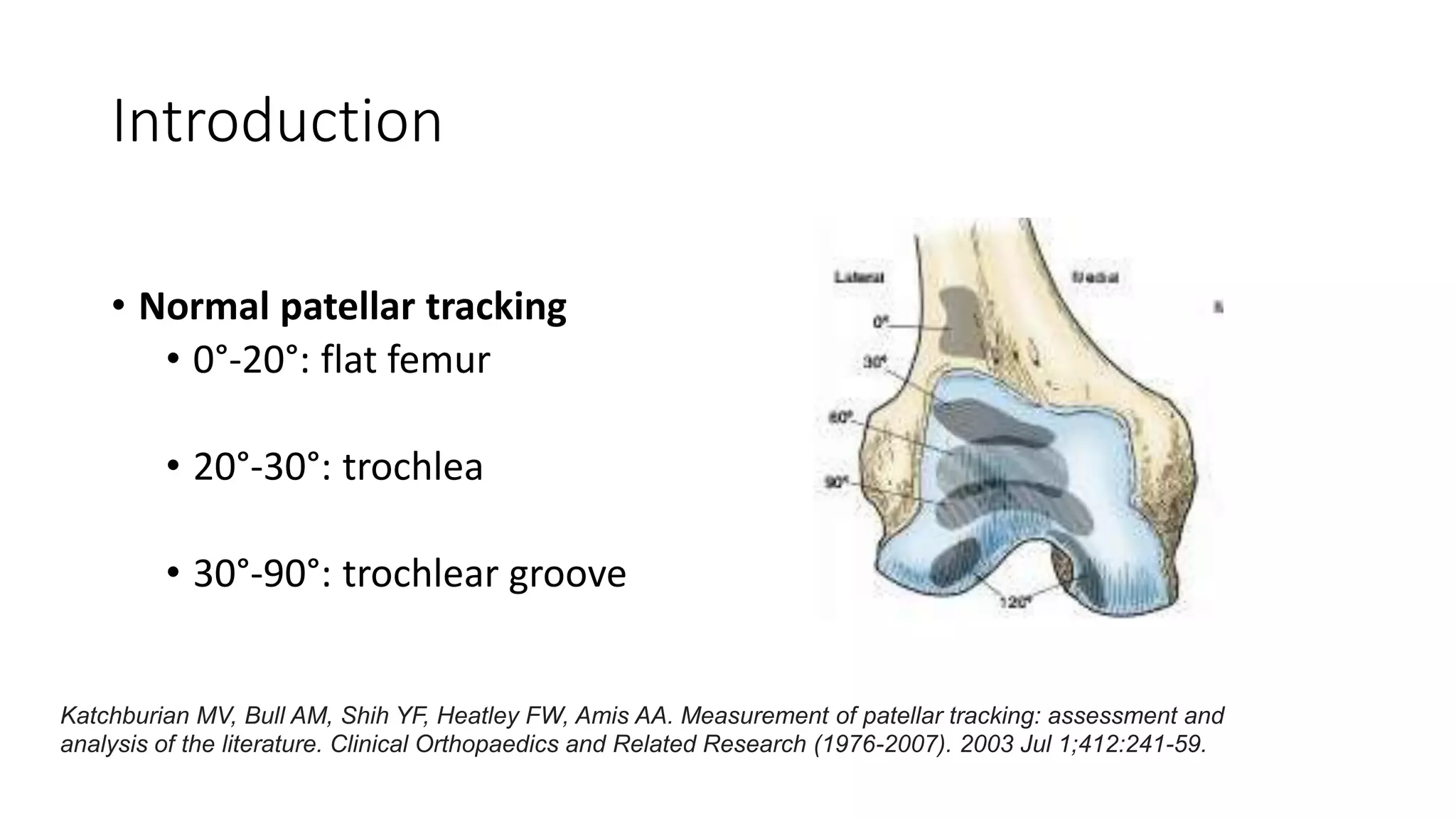
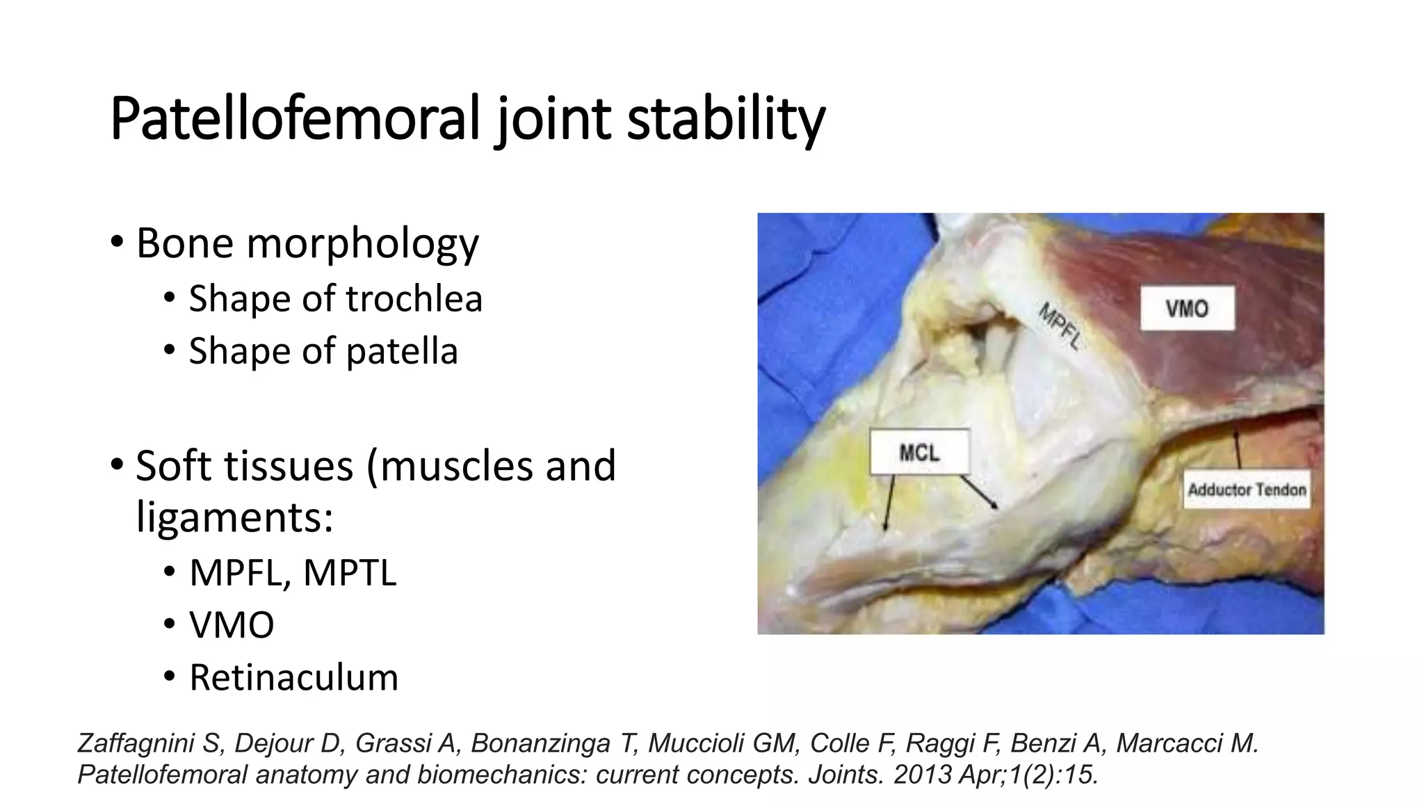
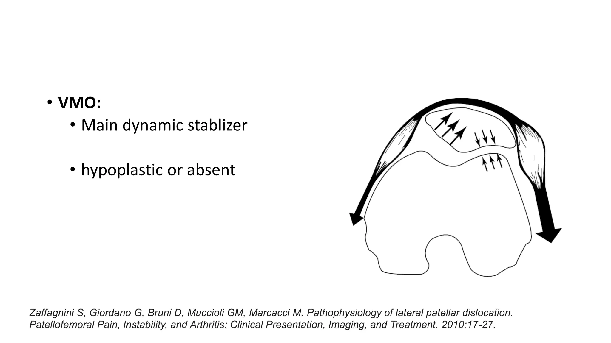
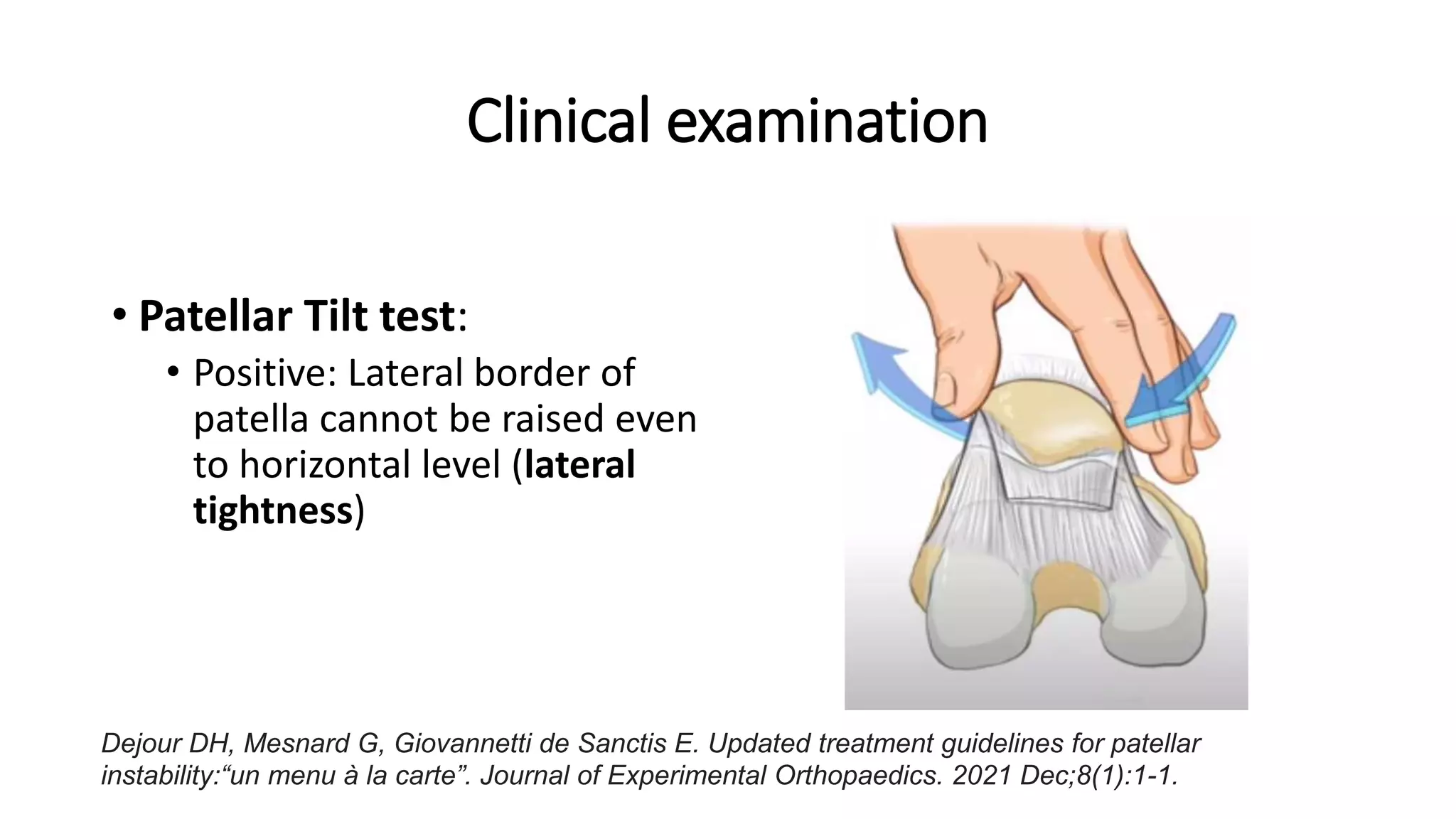
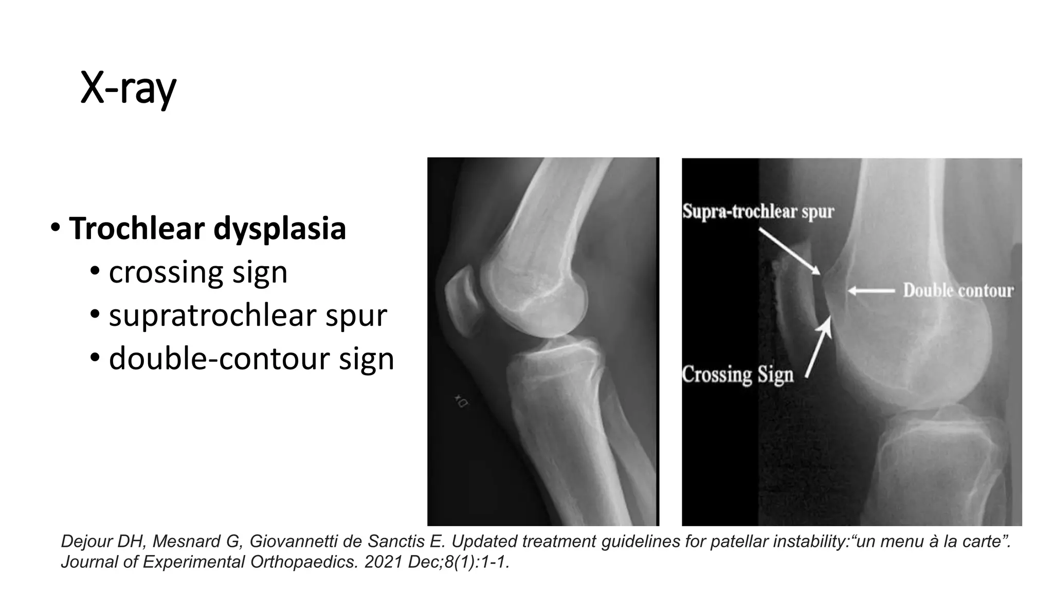
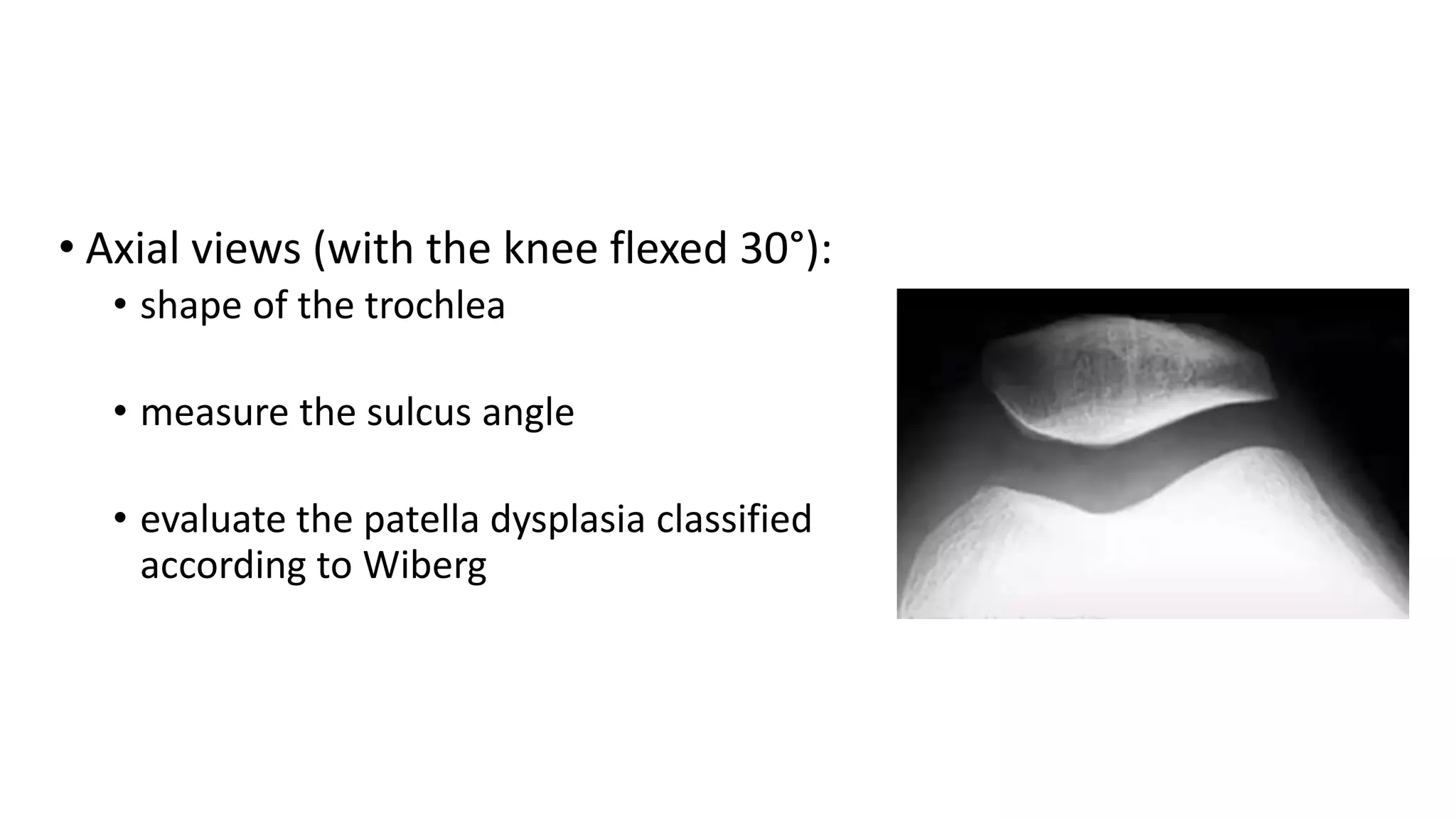
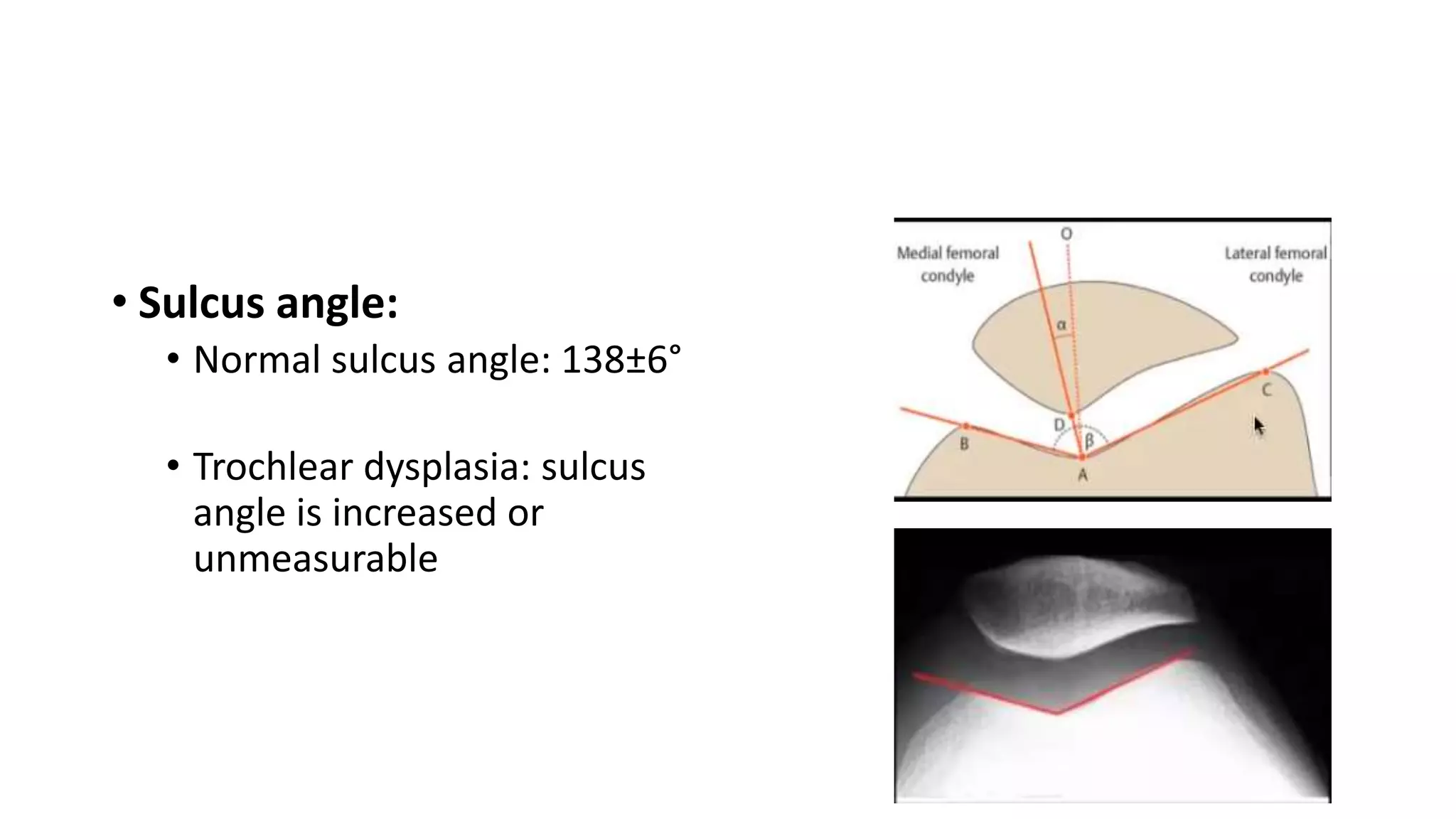
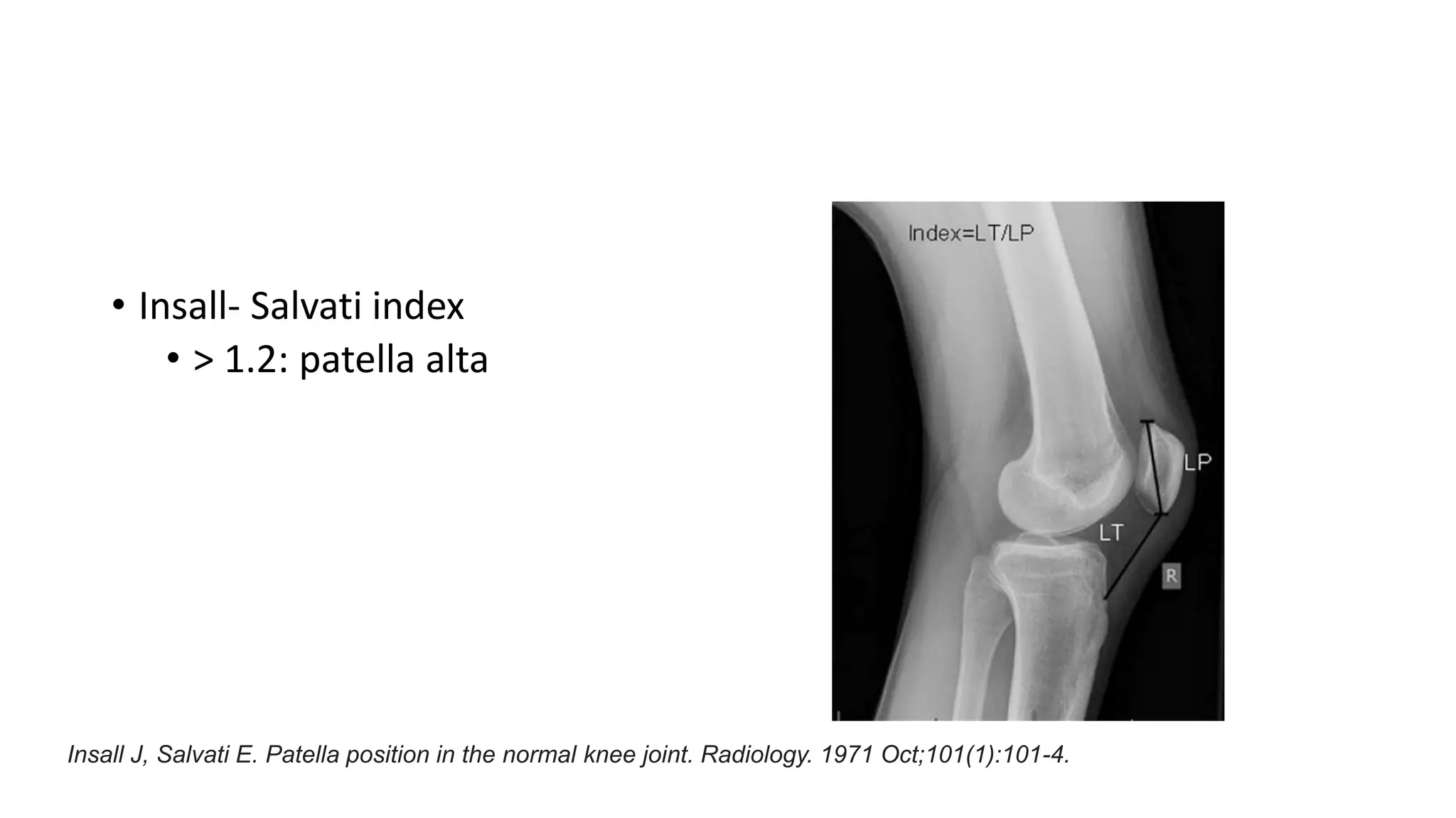
Approach to Patellofemoral
instabilty
Dr Santosh Nepal
Fellow, Arthroscopy and Sports injury
Introduction
• abnormal patellar tracking in relation to the femoral trochlea as the
knee extends/flexes
• multifactorial disease including a spectrum of different conditions
Introduction
• Normal patellar tracking
• 0°-20°: flat femur
• 20°-30°: trochlea
• 30°-90°: trochlear groove
Katchburian MV, Bull AM, Shih YF, Heatley FW, Amis AA. Measurement of patellar tracking: assessment and
analysis of the literature. Clinical Orthopaedics and Related Research (1976-2007). 2003 Jul 1;412:241-59.
Introduction
• Bimechanically a complex joint
numerous capsuloligamentous
structures and muscles that act on
the patella
Zaffagnini S, Dejour D, Grassi A, Bonanzinga T, Muccioli GM, Colle F, Raggi F, Benzi A, Marcacci M. Patellofemoral
anatomy and biomechanics: current concepts. Joints. 2013 Apr;1(2):15.
Patellofemoral joint stability
• Bone morphology
• Shape of trochlea
• Shape of patella
• Soft tissues (muscles and
ligaments:
• MPFL, MPTL
• VMO
• Retinaculum
Zaffagnini S, Dejour D, Grassi A, Bonanzinga T, Muccioli GM, Colle F, Raggi F, Benzi A, Marcacci M.
Patellofemoral anatomy and biomechanics: current concepts. Joints. 2013 Apr;1(2):15.
• MPFL:
• static restraint to lateral translation of
the patella
• Produces 60% of the force directed
medially and thus restrains the lateral
dislocation
• MPFL rupture is the consequence of the
lateral patellar dislocation and never the
cause
Zaffagnini S, Dejour D, Grassi A, Bonanzinga T, Muccioli GM, Colle F, Raggi F, Benzi A, Marcacci M.
Patellofemoral anatomy and biomechanics: current concepts. Joints. 2013 Apr;1(2):15.
• VMO:
• Main dynamic stablizer
• hypoplastic or absent
Zaffagnini S, Giordano G, Bruni D, Muccioli GM, Marcacci M. Pathophysiology of lateral patellar dislocation.
Patellofemoral Pain, Instability, and Arthritis: Clinical Presentation, Imaging, and Treatment. 2010:17-27.
Causes/ predisposing factors
Soft tissue problems
(not able to bring
patella into trochlea)
MPFL tear
Patella alta
Trochlear
dysplasia
Abnormal bony alignment
(not allowing patella to
engage into trochlea)
 Increased femoral anteversion
 External tibia torsion
 Genu valgus
Biomechanics
MPFL tear
Lateral structures overpull
Patella starts from lateral side and
hits the lateral patellar bump
Patella dislocates laterally
Biomechanics
Patella alta
Even in 30° of flexion patella does not
engage in trochlear groove
Patella unstable and chance of
dislocation
dislocation at extension (J-sign)
Biomechanics
Trochlear dysplasia
Flat trochlea/ lateral bump
Patella does not enter the groove and
dislocates
Biomechanics
Valgus knee
Patella initially lying in lateral position
Flexion: patella hits apex of lateral femoral
condyle and dislocates laterally
Increased femoral anteversion
Internal rotation of femur
Lateral femoral condyle first contact with
patella
Dislocates laterally
External tibia torsion
Tibial tuberosity rotated externally (TT-TG
distance > 20mm)
Increased lateral pull on patella
Patella hits lateral femoral condyle and
dislocates laterally
• Dislocation in flexion:
• shorter proximal or distal extensor mechanism
• Trochlear dysplasia
• Dislocation in extension:
• Patella alta
• MPFL insufficiency
Clinical symptoms
• Anterior knee pain
• subjective feeling of unstable knee
Arendt EA, Dejour D. Patella instability: building bridges across the ocean a historic review. Knee Surgery,
Sports Traumatology, Arthroscopy. 2013 Feb;21:279-93.
Clinical examination
• Apprehension test
forced lateral displacement of the
patella produces anxiety and
resistance in patients
Dejour DH, Mesnard G, Giovannetti de Sanctis E. Updated treatment guidelines for patellar instability:“un
menu à la carte”. Journal of Experimental Orthopaedics. 2021 Dec;8(1):1-1.
Clinical examination
• Quadrant test/ patella glide test:
• done in extension and flexion to
evaluate the MPFL competency
• Limited medial passive patellar
glide
• Increased lateral passive patellar
glide
Arendt EA, Dejour D. Patella instability: building bridges across the ocean a historic review. Knee Surgery,
Sports Traumatology, Arthroscopy. 2013 Feb;21:279-93.
Clinical examination
• Patellar Tilt test:
• Positive: Lateral border of
patella cannot be raised even
to horizontal level (lateral
tightness)
Dejour DH, Mesnard G, Giovannetti de Sanctis E. Updated treatment guidelines for patellar
instability:“un menu à la carte”. Journal of Experimental Orthopaedics. 2021 Dec;8(1):1-1.
• J sign and/or abnormal
patellar tracking:
• disengagement of patella from
the trochlea in extension
• patella alta, a short or convex
trochlea
Dejour DH, Mesnard G, Giovannetti de Sanctis E. Updated treatment guidelines for patellar
instability:“un menu à la carte”. Journal of Experimental Orthopaedics. 2021 Dec;8(1):1-1.
Radiographic evaluation
• X-ray
• Lateral
• Axial
• CT scan
• MRI
X-ray
• Trochlear dysplasia
• crossing sign
• supratrochlear spur
• double-contour sign
Dejour DH, Mesnard G, Giovannetti de Sanctis E. Updated treatment guidelines for patellar instability:“un menu à la carte”.
Journal of Experimental Orthopaedics. 2021 Dec;8(1):1-1.
Dejour DH, Mesnard G, Giovannetti de Sanctis E. Updated treatment guidelines for patellar instability:“un menu à la
carte”. Journal of Experimental Orthopaedics. 2021 Dec;8(1):1-1.
• Axial views (with the knee flexed 30°):
• shape of the trochlea
• measure the sulcus angle
• evaluate the patella dysplasia classified
according to Wiberg
• Sulcus angle:
• Normal sulcus angle: 138±6°
• Trochlear dysplasia: sulcus
angle is increased or
unmeasurable
Patella dysplasia (Wiberg classification)
Type I Type II Type III
Fucentese SF, von Roll A, Koch PP, Epari DR, Fuchs B, Schottle PB. The patella morphology in trochlear dysplasia.
The knee. 2006 Mar 1;13(2):145-50.
• Patella alta:
• Caton-Deschamps index (CDI)
(> 1.2: patella alta)
Caton J, Deschamps G, Chambat P, Lerat JL, Dejour H. Patella infera. Apropos of 128 cases. Revue de
chirurgie orthopedique et reparatrice de l'appareil moteur. 1982 Jan 1;68(5):317-25.
• Insall- Salvati index
• > 1.2: patella alta
Insall J, Salvati E. Patella position in the normal knee joint. Radiology. 1971 Oct;101(1):101-4.
• Blackburne- Peel index
• >1 : patella alta
Blackburne JS, Peel TE. A new method of measuring patellar height. The Journal of bone
and joint surgery. British volume. 1977 May;59(2):241-2.
CT scan
• TT-TG (tibial tuberosity-trochlear
groove) distance:
• represents the radiographic
measurement of the lateral quadriceps
vector acting on the patella
Batailler C, Neyret P. Trochlear dysplasia: imaging and treatment options. EFORT open reviews.
2018 May;3(5):240.
MRI
• cartilage status of the patellofemoral
joint
• In traumatic acute dislocation:
osteochondral fragments, MPFL tear
Management
Recurrent patellar dislocation
+MPFLR
Trochlear
dysplasia
Patella alta TT:TG ratio >20
Excessive lateral
tightness
Trochleoplasty TTO
Medialisation
of TT
Lateral release
Dejour DH, Mesnard G, Giovannetti de Sanctis E. Updated treatment guidelines for patellar instability:“un menu
à la carte”. Journal of Experimental Orthopaedics. 2021 Dec;8(1):1-1.
Trochleoplasty
Dejour DH, Mesnard G, Giovannetti de Sanctis
E. Updated treatment guidelines for patellar
instability:“un menu à la carte”. Journal of
Experimental Orthopaedics. 2021 Dec;8(1):1-1.
Tibial tubercle osteotomy (TTO)
• Distalization of TT
• corrects patellar height and restore
patellofemoral stability in patella alta
• Proximalisation of TT
• Corrects the quadriceps tightness
associated with dislocation in flexion
Zaffagnini S, Giordano G, Bruni D, Muccioli GM, Marcacci M. Pathophysiology of lateral patellar dislocation.
Patellofemoral Pain, Instability, and Arthritis: Clinical Presentation, Imaging, and Treatment. 2010:17-27.
• Medialisation of TT
• goal is to change the TT-TG distance, within a range of 10 to 15 mm
• Decreases the lateral pull vector acting on patella
Zaffagnini S, Giordano G, Bruni D, Muccioli GM, Marcacci M. Pathophysiology of lateral patellar dislocation.
Patellofemoral Pain, Instability, and Arthritis: Clinical Presentation, Imaging, and Treatment. 2010:17-27.
Take home messege
• Patellofemoral instability is multifactorial
• Identifying the cause/pathology of patella dislocation is essential to
plan the treatment
• Each pathological abnormalities, either bony or soft tissue, should
be adressed accordingly
THANK YOU

patellofemoral instability.pptx

  • 1.
    Approach to Patellofemoral instabilty DrSantosh Nepal Fellow, Arthroscopy and Sports injury
  • 2.
    Introduction • abnormal patellartracking in relation to the femoral trochlea as the knee extends/flexes • multifactorial disease including a spectrum of different conditions
  • 3.
    Introduction • Normal patellartracking • 0°-20°: flat femur • 20°-30°: trochlea • 30°-90°: trochlear groove Katchburian MV, Bull AM, Shih YF, Heatley FW, Amis AA. Measurement of patellar tracking: assessment and analysis of the literature. Clinical Orthopaedics and Related Research (1976-2007). 2003 Jul 1;412:241-59.
  • 4.
    Introduction • Bimechanically acomplex joint numerous capsuloligamentous structures and muscles that act on the patella Zaffagnini S, Dejour D, Grassi A, Bonanzinga T, Muccioli GM, Colle F, Raggi F, Benzi A, Marcacci M. Patellofemoral anatomy and biomechanics: current concepts. Joints. 2013 Apr;1(2):15.
  • 5.
    Patellofemoral joint stability •Bone morphology • Shape of trochlea • Shape of patella • Soft tissues (muscles and ligaments: • MPFL, MPTL • VMO • Retinaculum Zaffagnini S, Dejour D, Grassi A, Bonanzinga T, Muccioli GM, Colle F, Raggi F, Benzi A, Marcacci M. Patellofemoral anatomy and biomechanics: current concepts. Joints. 2013 Apr;1(2):15.
  • 6.
    • MPFL: • staticrestraint to lateral translation of the patella • Produces 60% of the force directed medially and thus restrains the lateral dislocation • MPFL rupture is the consequence of the lateral patellar dislocation and never the cause Zaffagnini S, Dejour D, Grassi A, Bonanzinga T, Muccioli GM, Colle F, Raggi F, Benzi A, Marcacci M. Patellofemoral anatomy and biomechanics: current concepts. Joints. 2013 Apr;1(2):15.
  • 7.
    • VMO: • Maindynamic stablizer • hypoplastic or absent Zaffagnini S, Giordano G, Bruni D, Muccioli GM, Marcacci M. Pathophysiology of lateral patellar dislocation. Patellofemoral Pain, Instability, and Arthritis: Clinical Presentation, Imaging, and Treatment. 2010:17-27.
  • 8.
    Causes/ predisposing factors Softtissue problems (not able to bring patella into trochlea) MPFL tear Patella alta Trochlear dysplasia Abnormal bony alignment (not allowing patella to engage into trochlea)  Increased femoral anteversion  External tibia torsion  Genu valgus
  • 9.
    Biomechanics MPFL tear Lateral structuresoverpull Patella starts from lateral side and hits the lateral patellar bump Patella dislocates laterally
  • 10.
    Biomechanics Patella alta Even in30° of flexion patella does not engage in trochlear groove Patella unstable and chance of dislocation dislocation at extension (J-sign)
  • 11.
    Biomechanics Trochlear dysplasia Flat trochlea/lateral bump Patella does not enter the groove and dislocates
  • 12.
    Biomechanics Valgus knee Patella initiallylying in lateral position Flexion: patella hits apex of lateral femoral condyle and dislocates laterally
  • 13.
    Increased femoral anteversion Internalrotation of femur Lateral femoral condyle first contact with patella Dislocates laterally
  • 14.
    External tibia torsion Tibialtuberosity rotated externally (TT-TG distance > 20mm) Increased lateral pull on patella Patella hits lateral femoral condyle and dislocates laterally
  • 15.
    • Dislocation inflexion: • shorter proximal or distal extensor mechanism • Trochlear dysplasia • Dislocation in extension: • Patella alta • MPFL insufficiency
  • 16.
    Clinical symptoms • Anteriorknee pain • subjective feeling of unstable knee Arendt EA, Dejour D. Patella instability: building bridges across the ocean a historic review. Knee Surgery, Sports Traumatology, Arthroscopy. 2013 Feb;21:279-93.
  • 17.
    Clinical examination • Apprehensiontest forced lateral displacement of the patella produces anxiety and resistance in patients Dejour DH, Mesnard G, Giovannetti de Sanctis E. Updated treatment guidelines for patellar instability:“un menu à la carte”. Journal of Experimental Orthopaedics. 2021 Dec;8(1):1-1.
  • 18.
    Clinical examination • Quadranttest/ patella glide test: • done in extension and flexion to evaluate the MPFL competency • Limited medial passive patellar glide • Increased lateral passive patellar glide Arendt EA, Dejour D. Patella instability: building bridges across the ocean a historic review. Knee Surgery, Sports Traumatology, Arthroscopy. 2013 Feb;21:279-93.
  • 19.
    Clinical examination • PatellarTilt test: • Positive: Lateral border of patella cannot be raised even to horizontal level (lateral tightness) Dejour DH, Mesnard G, Giovannetti de Sanctis E. Updated treatment guidelines for patellar instability:“un menu à la carte”. Journal of Experimental Orthopaedics. 2021 Dec;8(1):1-1.
  • 20.
    • J signand/or abnormal patellar tracking: • disengagement of patella from the trochlea in extension • patella alta, a short or convex trochlea Dejour DH, Mesnard G, Giovannetti de Sanctis E. Updated treatment guidelines for patellar instability:“un menu à la carte”. Journal of Experimental Orthopaedics. 2021 Dec;8(1):1-1.
  • 21.
    Radiographic evaluation • X-ray •Lateral • Axial • CT scan • MRI
  • 22.
    X-ray • Trochlear dysplasia •crossing sign • supratrochlear spur • double-contour sign Dejour DH, Mesnard G, Giovannetti de Sanctis E. Updated treatment guidelines for patellar instability:“un menu à la carte”. Journal of Experimental Orthopaedics. 2021 Dec;8(1):1-1.
  • 23.
    Dejour DH, MesnardG, Giovannetti de Sanctis E. Updated treatment guidelines for patellar instability:“un menu à la carte”. Journal of Experimental Orthopaedics. 2021 Dec;8(1):1-1.
  • 24.
    • Axial views(with the knee flexed 30°): • shape of the trochlea • measure the sulcus angle • evaluate the patella dysplasia classified according to Wiberg
  • 25.
    • Sulcus angle: •Normal sulcus angle: 138±6° • Trochlear dysplasia: sulcus angle is increased or unmeasurable
  • 26.
    Patella dysplasia (Wibergclassification) Type I Type II Type III Fucentese SF, von Roll A, Koch PP, Epari DR, Fuchs B, Schottle PB. The patella morphology in trochlear dysplasia. The knee. 2006 Mar 1;13(2):145-50.
  • 27.
    • Patella alta: •Caton-Deschamps index (CDI) (> 1.2: patella alta) Caton J, Deschamps G, Chambat P, Lerat JL, Dejour H. Patella infera. Apropos of 128 cases. Revue de chirurgie orthopedique et reparatrice de l'appareil moteur. 1982 Jan 1;68(5):317-25.
  • 28.
    • Insall- Salvatiindex • > 1.2: patella alta Insall J, Salvati E. Patella position in the normal knee joint. Radiology. 1971 Oct;101(1):101-4.
  • 29.
    • Blackburne- Peelindex • >1 : patella alta Blackburne JS, Peel TE. A new method of measuring patellar height. The Journal of bone and joint surgery. British volume. 1977 May;59(2):241-2.
  • 30.
    CT scan • TT-TG(tibial tuberosity-trochlear groove) distance: • represents the radiographic measurement of the lateral quadriceps vector acting on the patella Batailler C, Neyret P. Trochlear dysplasia: imaging and treatment options. EFORT open reviews. 2018 May;3(5):240.
  • 31.
    MRI • cartilage statusof the patellofemoral joint • In traumatic acute dislocation: osteochondral fragments, MPFL tear
  • 32.
    Management Recurrent patellar dislocation +MPFLR Trochlear dysplasia Patellaalta TT:TG ratio >20 Excessive lateral tightness Trochleoplasty TTO Medialisation of TT Lateral release Dejour DH, Mesnard G, Giovannetti de Sanctis E. Updated treatment guidelines for patellar instability:“un menu à la carte”. Journal of Experimental Orthopaedics. 2021 Dec;8(1):1-1.
  • 33.
    Trochleoplasty Dejour DH, MesnardG, Giovannetti de Sanctis E. Updated treatment guidelines for patellar instability:“un menu à la carte”. Journal of Experimental Orthopaedics. 2021 Dec;8(1):1-1.
  • 34.
    Tibial tubercle osteotomy(TTO) • Distalization of TT • corrects patellar height and restore patellofemoral stability in patella alta • Proximalisation of TT • Corrects the quadriceps tightness associated with dislocation in flexion Zaffagnini S, Giordano G, Bruni D, Muccioli GM, Marcacci M. Pathophysiology of lateral patellar dislocation. Patellofemoral Pain, Instability, and Arthritis: Clinical Presentation, Imaging, and Treatment. 2010:17-27.
  • 35.
    • Medialisation ofTT • goal is to change the TT-TG distance, within a range of 10 to 15 mm • Decreases the lateral pull vector acting on patella Zaffagnini S, Giordano G, Bruni D, Muccioli GM, Marcacci M. Pathophysiology of lateral patellar dislocation. Patellofemoral Pain, Instability, and Arthritis: Clinical Presentation, Imaging, and Treatment. 2010:17-27.
  • 36.
    Take home messege •Patellofemoral instability is multifactorial • Identifying the cause/pathology of patella dislocation is essential to plan the treatment • Each pathological abnormalities, either bony or soft tissue, should be adressed accordingly
  • 37.

Editor's Notes

  • #4 Dislocation at 0-20 degree: soft tissue problem Dislocation at > 20 degree: trochear dysplasia
  • #5 abnormalities in one or more of these structures: pathological behavior of the PF joint A mechanical imbalance, often associated with anatomic abnormalities, with laterally overcoming the medially directed forces, may lead to dislocation
  • #6 Imbalance among those would predispose to patellar malalignment and instability.
  • #7 If the MPFL was ruptured the force required to displace the patella laterally was reduced by 50% in the extended knee, decreasing while the knee flexed
  • #8 leading to a biomechanically non efficient action of this muscle
  • #11 An increase in the “free” range of motion, with the patella out of the restraining bony supports, would facilitate lateral dislocation, due to the usual prevalence of the lateral structures with respect to the medial ones Patella Alta, modifying the lever arm between quadriceps and patellar tendons, increases the compression forces in patellofemoral joint leading to cartilage damage
  • #12 At 20°: patella enters trochlea
  • #19 Normal : 1-2 quadrants medially and laterally
  • #21 Maltracking refers to the dynamic malalignment of the patella within the trochlear groove occurring during active or passive range of motion of the knee
  • #31 This value is modified by both femoral/tibial rotation and varus/valgus knee coronal alignment, altering the Q angle, formed by the lines of pull of the quadriceps and patellar tendon intersecting at the center of the patella.
  • #33  Surgical decision making depends on the pathology causing the dislocation
  • #34 Sulcus deepening trochleoplasty has three functions: modifies the trochlear shape with a central groove and oblique medial and lateral facets decreases the patellofemoral joint reaction force by reducing the supratrochlear prominence (spur) reduces the TT-TG value by the groove repositioning (proximal realignment), often without further need of tibial tubercle medialization