SlideShare a Scribd company logo
DR
MOHAMEDIRAQI
ORTHOPEDIC CONSULTANT
RAMP LESION
• Meniscal ramp lesions were studied by Hamberg and
colleagues,in the 1980s but at that time were described
only as “injuries of the posterior aspect of the medial
meniscus.”
• Later, Strobel, introduced the term ramp lesion and
characterized the injury as a longitudinal tear, 2.5 cm in
length, located at the posterior meniscocapsular junction
of medial meniscus.
• In 1991, Morgan ,described a surgical technique for
arthroscopic repair of ramp lesions using a suture hook
through a posteromedial portal.
History
Fig. 1 Arthroscopic trans-notch visualization of the posteromedial
compartment of the left knee (* = posteromedial tibial plateau). Retraction
of the ramp lesion using an arthroscopy probe placed through the
posteromedial portal reveals the close anatomic relationship between the
semimembranosus tendon and the meniscocapsular region.
Anatomy
• The anterior and posterior roots anchor the
meniscus to the tibial plateau, and the
body of the meniscus is attached to the
adjacent joint capsule and to the tibia by
the meniscotibial ligaments.
• The reduced mobility of the medial
meniscus makes it susceptible to injuries,
especially in deep flexion and with
rotational trauma when the pressure is
increased in the posterior horn of the
medial meniscus.
• Capsular arm of the semimembranosus
tendon insertion translates the posterior horn
of the meniscus posteriorly during
knee flexion.
Why Ramp Lesion Is Important
hidden lesion
• A consequence of an underestimation of their incidence
lead to a high rate of missed diagnoses.
• The recent interest in these injuries heralds an increasing
recognition of their importance and an emerging concept
of their association with posteromedial knee instability.
• The expression “hidden lesion,” also has been recently
used to describe this injury, and the term refers to the
difficulty in identifying ramp lesions from standard
anterior arthroscopic portals,
• and also with preoperative MRI, which has low
sensititvity.
• Meniscal ramp lesions are a “HOT TOPIC”
because of increasing recognition that they have
important biomechanical consequences and also
that they occur much more frequently than was
previously understood.
• A systematic exploration of the posteromedial
compartment of the knee via a trans-notch
approach is needed to confirm the presence of a
meniscal ramp lesion.
• If left untreated, meniscal ramp lesions may
contribute to residual antero-posterior instability
& rotation laxity in the anterior cruciate
ligament–reconstructed knee and may also
result in failure of meniscal repair.
Mechanisms
• isolated ACL reconstruction fails to restore normal joint kinematics
and results in residual laxity in the presence of a ramp lesion.
• Ramp lesions are reported to occur frequently
(9.3%–24.0%) in ACL-deficient knees, including in
children and adolescents,
• Thus, a significantly higher prevalence of ramp
tears was reported in patients with chronic ACL
tears compared with patients with acute ACL tears.
• Papageorgiou et al reported that the forces on the
medial meniscus were increased by 200% in
response to anterior tibial loads with a transected
ACL. Likewise, deficiency of the medial meniscus
has been associated with ACLR failure
Incidence
Risk factors for the occurrence of ramp lesions
in ACL-deficient knees
• include male gender, patients younger than 30 years
• revision ACL reconstruction
• chronic injuries
• preoperative side-to-side anteroposterior laxity
difference of 6 mm or more
• the presence of concomitant lateral meniscal tears
• The presence of any of these factors should raise the
index of suspicion for the existence of a ramp lesion.
Objective diagnosis
• there are no specific physical examination tests
for ramp lesions
• Recently, MRI have a broad range of sensitivities
of both 1.5T and 3T
• The most specific sign in the MRI evaluation
of ramp lesions is the hyperintense signal that can
be observed between the meniscus and the
capsule.
• the presence of PMTP edema demonstrated
significantly greater odds for ramp lesions
compared with meniscal body tears.
Meniscus Extrusion
Meniscus Root Tear
Interesting Fact
• All over the world, in orthopedic meetings, the meniscal
root lesion is more extensively debated than the ramp
lesion. This is despite the fact that ramp lesions are a lot
more common (24% in ACLR in our experience).
• This greater focus on root tears is perhaps because of
the well-recognized loss of hoop force distribution and
profound biomechanical consequences of root tears.
However, it is increasingly recognized that ramp lesions
also have important biomechanical consequences
including persistent anteroposterior (AP) and rotational
laxity and an association with higher ACL graft failure
rates.
• Arthroscopy is considered gold standard for
diagnosis of ramp lesions.
• 40 % percent of ramp lesions are not
identified through standard anterior portal
visualization and inspection of the posterior
compartment via a trans-notch view, and
posteromedial probing is required to
identify them.
Classification
• Type 1: Meniscocapsular lesions. These lesions are very peripherally
located in the synovial sheath. Mobility at probing is very low.
•
• Type 2: Partial superior lesions. These lesions are stable and can be
diagnosed only by a trans-notch approach. Mobility at probing is low.
• Type 3: Partial inferior or hidden lesions. The lesions are typically
subtle or not immediately visible even with trans-notch visualization
but can be strongly suggested by significant mobility on probing and
also by identification of abnormal tissue quality on needling.
• Type 4: A complete tear of the red-red zone. Mobility at probing is
very high.
• Type 5: A double tear involving the meniscocapsular junction and a
second more anterior tear of the posterior horn.
Fig. 3
Ramp lesion classification as proposed by Thaunat and
colleagues.
(A) Illustration demonstrating the posterior meniscus-
capsular region and the areas of meniscus
vascularization.
(B) Type 1: Meniscocapsular lesion, located in the synovial
sheath. Mobility at probing is very low.
(C) Type 2: Upper partial lesion. Mobility at probing is low.
These lesions are stable and can be diagnosed only by a trans-notch
approach
(D) Type 3: Lower lesion (“hidden lesion”). significant
mobility on probing . not immediately visible even with trans-notch
visualization
(E) Type 4: Complete injury in the red-red area. Mobility at
probing is very high.
(F) Type 5: Double tear. involving the meniscocapsular junction
and a second more anterior tear of the posterior horn
Treatment options
• isolated tears that are small (less than 10 mm) and stable may
amenable to conservative treatment. abrasion and trephination of
stable lesions (without repair).
• posteromedial portal suture hook device.
• all-inside suture technique
Fig. 8 Image of a curved hook device,
with the tip curvature pointing to the left
side (ideal for suturing meniscal ramp
lesion on a right knee).
• The mobility of the meniscus at probing may lead the surgeon to suspect the presence
of a posterior tear even if it is not visible by standard anterior viewing, because ramp
lesions classified as types III, IV, and V may be highly mobile when pulled.
• To facilitate the passage through this space, a valgus force is applied ,first in extension
and then in flexion. If passage remains difficult, the use of a blunt trocar may be
helpful. Tibial internal rotation may improve visualization of the tear by causing
posterior tibial plateau subluxation and a posterior translation of the medial segment.
Technical Tip
systematic arthroscopic exploration of the
knee joint
• Step 1: Standard arthroscopic exploration
• Step 2: Exploration of posteromedial compartment and probing the
meniscocapsular junction with a needle
• Step 3: Creation of a posteromedial portal
• Step 4: Meniscal repair procedure
Fig. 5 Arthroscopic view with a 30° scope of a right knee through the anterolateral portal.
(A) Probe is inserted through the anteromedial portal in the space between the posterior
cruciate ligament (PCL) and the medial femoral condyle (MFC).
(B) Passage of the arthroscope through the space between the PCL and the MFC after a valgus
force is applied.
(C) Image obtained via trans-notch visualization of the posteromedial region of the knee,
observing the posteromedial capsule and the meniscal ramp lesion. TS, tibial spine.
Two Postero-medial PortalTecnique
Fig. 6 External image of the knee demonstrating how transillumination with the arthroscope
positioned in the trans-notch view &
The knee is positioned at 90° of flexion to minimize the risk of injury to neurovascular structures and
a needle is introduced above the hamstring tendons, 1 cm posterior to the medial tibiofemoral joint
line, pointing toward the lesion
Fig. 7 Arthroscopic image through trans-notch vision showing 2 steps for the creation of
the posteromedial portal.
(A)Insertion of the needle to verify the correct positioning of the portal and
(B) creation of the posteromedial portal with a no. 11 scalpel.
MFC, medial femoral condyle.
Fig. 9 Detailing of a meniscal ramp suture procedure.
(A)Revitalization of the edges of the lesion using a shaver.
(B) Suture device inserted through the posteromedial portal.
(C) Penetration of the peripheral edge of the lesion.
(D) Penetration of the suture device through the inner portion of the tear, encompassing both ends of
the lesion.
(E) Progression of the suture, which is then captured out of the knee with the aid of a grasper.
(F) First stitch being completed using a knot pusher.
Internal rotation of the foot helps keep the medial femoral condyle away from the posterior segment of the
meniscus and facilitates the procedure.
Fig. 10 Final arthroscopic aspect of a meniscal ramp repair, as seen by
trans-notch view, in which 3 stitches were required to close the lesion.
Fig 1(A) Arthroscopic image of right knee viewing through the anterolateral portal. Tight
medial compartment, measured as 4 mm, results in difficult arthroscopic probe examination
of the PHMM.
(B) Fenestration of the medial collateral ligament, proximal to the meniscus and directly on
the femur, is performed by an 18-g spinal needle.
(C) This provides an increased medial joint space, measured as 11 mm, and adequate
visualization of the meniscus without damaging the cartilage.
(MFC, medial femoral condyle; MTP, medial tibial plateu; PHMM, posterior horn of the medial
meniscus.)
Trans-Septal Portal Tecnique
Fig 2(A) Right knee: a posteromedial portal is established
2.5 cm distal from the bony prominence of medial
epicondyle and 2.5 cm posterior from this measured point
with the knee in 90° of flexion.
(B) Right knee: the posterolateral portal is located anterior
to the long head of the biceps and proximal and posterior
to the LCL.
(LCL, lateral collateral ligament; ME, medial epicondyle;
PL, posterolateral portal; PM, posteromedial portal.)
Fig 3 Posteromedial portal establishment on the right knee with the “nick and spread” technique.
With the knee in 90° of flexion, modified Gillquist maneuver is performed into the posteromedial
compartment. This helps to visualize portal establishment with a needle
A) and the portal is enlarged with a scalpel
B) . A double dam flexible flanged cannula (Arthrex) is placed into the portal
C) . Adequate visualization of the PHMM is obtained with the cannula nicely retracting the capsule
medially
. (MFC, medial femoral condyle; PHMM, posterior horn of medial meniscus; PM, posteromedial
portal.)
Fig 4 Posterolateral portal establishment on the right
knee.
(B). and then the portal is enlarged with a scalpel.
(D). Arthroscopic image of the shaver through the
posteromedial portal with direct visualization during
trans-septal portal/window creation. Note that the
opening of the shaver is oriented anterior to avoid
popliteal artery injury.
(LCL, lateral collateral ligament; LFC, lateral femoral
condyle; PL, posterolateral portal.)
Fig 5 With the help of a switching stick
(A), a standard long arthroscopic cannula is placed into the
posterolateral portal to facilitate instrument use through
this portal
(B). (PL, posterolateral portal.)
Fig 6 (A) Right knee: with the knee in 90° of flexion, the arthroscope is then placed
through the posteromedial portal to view laterally, and the arthroscopic shaver is gently
advanced from the posterolateral portal to extend the hole in the septum for facilitating
the passage of the arthroscope or instrument insertion and manipulation.
(B) Arthroscopic image viewing from the PM portal: the trans-septal portal (dashed
borders) has been established without damaging the PCL.
(LFC, lateral femoral condyle; MFC, medial femoral condyle; PCL, posterior cruciate
ligament; PL, posterolateral portal; PM, posteromedial portal.)
Fig 7 (A) Right knee arthroscopic image:
the arthroscope is placed through the PL portal and advanced through the trans-septal
portal to directly visualize the ramp lesion. An arthroscopic rasp determines the
margins of the lesion after removing overlying fibrotic tissue.
(B) Right knee arthroscopic
image: the edges of the lesion are freshened by an arthroscopic shaver. (MFC, medial
femoral condyle; PL, posterolateral.)
Fig 8 (A) Right knee arthroscopic image from the trans-septal portal:
a 45° right suture passer (Depuy) is penetrating the right PMMH through the
posteromedial portal, after already shuttling a single limb of a UHMPE suture through the
capsular component.
(B) Eyelet suture shuttle advanced into the joint to be retrieved by an atraumatic grasper
through the posteromedial portal.
(MFC, medial femoral condyle; PMMH, posterior horn of medial meniscus; UHMPE, ultra-
high molecular weight polyethylene fiber.)
Fig 9 (A) Right knee arthroscopic image:
the sutures are passed in a vertical mattress fashion, adequately reducing the torn tissue and
reconstituting the ramp of the PHMM.
(B) Right knee arthroscopic image through a trans-septal portal:
sutures are placed in 5-7 mm intervals and knots are kept on the capsular side when tying
arthroscopically to limit abrasion to the medial femoral condyle.
(MFC, medial femoral condyle; PHMM, posterior horn of medial meniscus.)
a meniscal ramp lesion arthroscopic repair with an all-inside technique with the
Fast-Fix 360 device, detailing the use of the accessory posteromedial portal, and
the addition of an arthroscopic grasper that raises the retracted menisco-tibial
ligament, to allow correct fixation.
Although this technique seems technically less challenging for most surgeons
All-inside suture technique
Rehabilitation protocol
• Most protocols agree that early knee motion is beneficial; however,
knee hyperflexion is associated with anterior tibial translation, which
can increase the stress on the repair.
• In our practice, the postoperative management of an ACL
reconstruction associated with ramp repair is modified by restriction
of the active and passive range of motion to 0 to 90° of flexion during
the first 6 weeks.
• Progression to full weight bearing is allowed by postoperative week 3.
Jogging is permitted after week 12 to 16, pivot activity at 6 months,
and full activity at 9 months for all patients.
Take Home Message
• Meniscal ramp lesions are common but frequently under-recognized in the
ACL-injured knee.
• It is important to highlight that both preoperative MRI and arthroscopic
evaluation via classic anterior portals are associated with high rates of missed
diagnoses.
• For that reason, a systematic exploration of the posteromedial compartment is
advocated in all ACL-injured knees.
• Failure to recognize and repair ramp lesions is associated with persistent
anterior and rotational knee laxity, suggesting it makes part of a posteromedial
instability,
• in the setting of an ACL tear. Concomitant suture repair of these lesions, via a
posteromedial portal, can restore normal biomechanics and is associated with
excellent clinical outcomes.

More Related Content

What's hot

Acl graft fixation options
Acl graft fixation optionsAcl graft fixation options
Acl graft fixation options
orthoprinciples
 
Arthroscopic Meniscus Surgery: Resect or Repair 2014
Arthroscopic Meniscus Surgery: Resect or Repair 2014Arthroscopic Meniscus Surgery: Resect or Repair 2014
Arthroscopic Meniscus Surgery: Resect or Repair 2014
Dhananjaya Sabat
 
Hip resurfacing India | Dr.Venkatachalam
Hip resurfacing India | Dr.Venkatachalam Hip resurfacing India | Dr.Venkatachalam
Hip resurfacing India | Dr.Venkatachalam
Alampallam Venkatachalam
 
Fracture Talus
Fracture TalusFracture Talus
Fracture Talus
Dr. Manoj Parida
 
Implant Selection In Revision T.K.R
Implant Selection In Revision T.K.RImplant Selection In Revision T.K.R
Implant Selection In Revision T.K.R
Murtuza Rassiwala
 
Acetabular defects
Acetabular defectsAcetabular defects
Acetabular defects
sandy_unleashed
 
L08 tibial plateau
L08 tibial plateauL08 tibial plateau
L08 tibial plateau
Claudiu Cucu
 
Radial head replacement best evidence
Radial head replacement best evidenceRadial head replacement best evidence
Radial head replacement best evidenceorthoprinciples
 
Principles of deformity correction
Principles of deformity correctionPrinciples of deformity correction
Principles of deformity correctionSidharth Yadav
 
Introduction to Navigation - Robotic Total Knee Replacement
Introduction to Navigation - Robotic Total Knee Replacement Introduction to Navigation - Robotic Total Knee Replacement
Introduction to Navigation - Robotic Total Knee Replacement
Queen Mary Hospital
 
Osteotomies around the hip
Osteotomies around the hipOsteotomies around the hip
Osteotomies around the hipSanjay Kumar
 
HIgh Tibial Osteotomy: when and how
HIgh Tibial Osteotomy: when and howHIgh Tibial Osteotomy: when and how
HIgh Tibial Osteotomy: when and how
AbhishekKaushik126
 
Lumbar Disc Replacement
Lumbar Disc ReplacementLumbar Disc Replacement
Lumbar Disc Replacement
Pablo Pazmino
 
Medial Opening Wedge High Tibial Osteotomy
Medial Opening Wedge High Tibial Osteotomy Medial Opening Wedge High Tibial Osteotomy
Medial Opening Wedge High Tibial Osteotomy
washingtonortho
 
Shoulder arthroscopy
Shoulder arthroscopyShoulder arthroscopy
Shoulder arthroscopy
Sameer Ashar
 
Meniscus repair Indication & Techniques.ppt
Meniscus repair Indication & Techniques.pptMeniscus repair Indication & Techniques.ppt
Meniscus repair Indication & Techniques.ppt
Moazzam Jah
 
Pelvis acetabulum - anatomy , imaging , classification
Pelvis acetabulum   - anatomy , imaging , classificationPelvis acetabulum   - anatomy , imaging , classification
Pelvis acetabulum - anatomy , imaging , classification
jatinder12345
 
Evolution of Intramedullary Nails
Evolution of Intramedullary NailsEvolution of Intramedullary Nails
Evolution of Intramedullary Nails
Prateek Goel
 

What's hot (20)

Acl graft fixation options
Acl graft fixation optionsAcl graft fixation options
Acl graft fixation options
 
Arthroscopic Meniscus Surgery: Resect or Repair 2014
Arthroscopic Meniscus Surgery: Resect or Repair 2014Arthroscopic Meniscus Surgery: Resect or Repair 2014
Arthroscopic Meniscus Surgery: Resect or Repair 2014
 
Hip resurfacing India | Dr.Venkatachalam
Hip resurfacing India | Dr.Venkatachalam Hip resurfacing India | Dr.Venkatachalam
Hip resurfacing India | Dr.Venkatachalam
 
Fracture Talus
Fracture TalusFracture Talus
Fracture Talus
 
Talar Fracture
Talar FractureTalar Fracture
Talar Fracture
 
Implant Selection In Revision T.K.R
Implant Selection In Revision T.K.RImplant Selection In Revision T.K.R
Implant Selection In Revision T.K.R
 
Acetabular defects
Acetabular defectsAcetabular defects
Acetabular defects
 
L08 tibial plateau
L08 tibial plateauL08 tibial plateau
L08 tibial plateau
 
Radial head replacement best evidence
Radial head replacement best evidenceRadial head replacement best evidence
Radial head replacement best evidence
 
Meniscal repair
Meniscal repairMeniscal repair
Meniscal repair
 
Principles of deformity correction
Principles of deformity correctionPrinciples of deformity correction
Principles of deformity correction
 
Introduction to Navigation - Robotic Total Knee Replacement
Introduction to Navigation - Robotic Total Knee Replacement Introduction to Navigation - Robotic Total Knee Replacement
Introduction to Navigation - Robotic Total Knee Replacement
 
Osteotomies around the hip
Osteotomies around the hipOsteotomies around the hip
Osteotomies around the hip
 
HIgh Tibial Osteotomy: when and how
HIgh Tibial Osteotomy: when and howHIgh Tibial Osteotomy: when and how
HIgh Tibial Osteotomy: when and how
 
Lumbar Disc Replacement
Lumbar Disc ReplacementLumbar Disc Replacement
Lumbar Disc Replacement
 
Medial Opening Wedge High Tibial Osteotomy
Medial Opening Wedge High Tibial Osteotomy Medial Opening Wedge High Tibial Osteotomy
Medial Opening Wedge High Tibial Osteotomy
 
Shoulder arthroscopy
Shoulder arthroscopyShoulder arthroscopy
Shoulder arthroscopy
 
Meniscus repair Indication & Techniques.ppt
Meniscus repair Indication & Techniques.pptMeniscus repair Indication & Techniques.ppt
Meniscus repair Indication & Techniques.ppt
 
Pelvis acetabulum - anatomy , imaging , classification
Pelvis acetabulum   - anatomy , imaging , classificationPelvis acetabulum   - anatomy , imaging , classification
Pelvis acetabulum - anatomy , imaging , classification
 
Evolution of Intramedullary Nails
Evolution of Intramedullary NailsEvolution of Intramedullary Nails
Evolution of Intramedullary Nails
 

Similar to Ramp lesion

Meniscus injury
Meniscus injuryMeniscus injury
Meniscus injury
Abdulla Kamal
 
Meniscal injury
Meniscal injuryMeniscal injury
Meniscal injury
Ponnilavan Ponz
 
MRI fistulogram
MRI fistulogramMRI fistulogram
MRI fistulogram
yuvraj909
 
Castagna 2007- arthroscopic rotator cuff repair using a triple-loaded suture ...
Castagna 2007- arthroscopic rotator cuff repair using a triple-loaded suture ...Castagna 2007- arthroscopic rotator cuff repair using a triple-loaded suture ...
Castagna 2007- arthroscopic rotator cuff repair using a triple-loaded suture ...
luantran92
 
Calcaneal fractures
Calcaneal fracturesCalcaneal fractures
Calcaneal fractures
dr.pradeep pathak
 
Lateral condyle of humerus fracture in children
Lateral condyle of humerus fracture in childrenLateral condyle of humerus fracture in children
Lateral condyle of humerus fracture in children
AnilKC5
 
Mattress Double Anchor Footprint Repair | Shouder Surgery | Vail Colorado
Mattress Double Anchor Footprint Repair | Shouder Surgery | Vail ColoradoMattress Double Anchor Footprint Repair | Shouder Surgery | Vail Colorado
Mattress Double Anchor Footprint Repair | Shouder Surgery | Vail Colorado
Peter Millett MD
 
Laparoscopic Repair of Hiatus Hernia
Laparoscopic Repair of Hiatus HerniaLaparoscopic Repair of Hiatus Hernia
Laparoscopic Repair of Hiatus Hernia
World Laparoscopy Hospital
 
Thoracolumbar injuries
Thoracolumbar injuriesThoracolumbar injuries
Thoracolumbar injuries
Yeswanth Mohan
 
Shoulder impingement syndrome
Shoulder impingement syndromeShoulder impingement syndrome
Shoulder impingement syndrome
Hardev Singh
 
The “Bony Bankart Bridge” Procedure Shoulder Instability | Shoudler Surgery |...
The “Bony Bankart Bridge” Procedure Shoulder Instability | Shoudler Surgery |...The “Bony Bankart Bridge” Procedure Shoulder Instability | Shoudler Surgery |...
The “Bony Bankart Bridge” Procedure Shoulder Instability | Shoudler Surgery |...
Peter Millett MD
 
Surgical approaches to the facial skeleton
Surgical approaches to the facial skeletonSurgical approaches to the facial skeleton
Surgical approaches to the facial skeleton
Abhishek Roy
 
Hip anatomy, approaches & AVN
Hip anatomy, approaches & AVN Hip anatomy, approaches & AVN
Hip anatomy, approaches & AVN
Dr JAYESH BHANUSHALI
 
3 d ultrasound shoulder
3 d ultrasound shoulder3 d ultrasound shoulder
3 d ultrasound shoulder
Dr. Muhammad Bin Zulfiqar
 
Hip Joint anatomy, surgical approches & AVN review
Hip Joint anatomy, surgical approches & AVN reviewHip Joint anatomy, surgical approches & AVN review
Hip Joint anatomy, surgical approches & AVN review
docortho Patel
 
aad evaluation and treatment.pptx
aad evaluation and treatment.pptxaad evaluation and treatment.pptx
aad evaluation and treatment.pptx
Kollanur Charan
 
200331 Evolving concepts in treatment of supracondylar fractures in children
200331 Evolving concepts in treatment of supracondylar fractures in children200331 Evolving concepts in treatment of supracondylar fractures in children
200331 Evolving concepts in treatment of supracondylar fractures in children
Dr MADAN MOHAN
 
PELVIC RING FRACTURES AND CLASSIFICATIONS.pptx
PELVIC RING FRACTURES AND CLASSIFICATIONS.pptxPELVIC RING FRACTURES AND CLASSIFICATIONS.pptx
PELVIC RING FRACTURES AND CLASSIFICATIONS.pptx
Dishan Mandania
 
Proximal humerus fractures
Proximal humerus fracturesProximal humerus fractures
Proximal humerus fractures
MONTHER ALKHAWLANY
 

Similar to Ramp lesion (20)

Meniscus injury
Meniscus injuryMeniscus injury
Meniscus injury
 
Meniscal injury
Meniscal injuryMeniscal injury
Meniscal injury
 
MRI fistulogram
MRI fistulogramMRI fistulogram
MRI fistulogram
 
Castagna 2007- arthroscopic rotator cuff repair using a triple-loaded suture ...
Castagna 2007- arthroscopic rotator cuff repair using a triple-loaded suture ...Castagna 2007- arthroscopic rotator cuff repair using a triple-loaded suture ...
Castagna 2007- arthroscopic rotator cuff repair using a triple-loaded suture ...
 
Calcaneal fractures
Calcaneal fracturesCalcaneal fractures
Calcaneal fractures
 
Lateral condyle of humerus fracture in children
Lateral condyle of humerus fracture in childrenLateral condyle of humerus fracture in children
Lateral condyle of humerus fracture in children
 
Mattress Double Anchor Footprint Repair | Shouder Surgery | Vail Colorado
Mattress Double Anchor Footprint Repair | Shouder Surgery | Vail ColoradoMattress Double Anchor Footprint Repair | Shouder Surgery | Vail Colorado
Mattress Double Anchor Footprint Repair | Shouder Surgery | Vail Colorado
 
Laparoscopic Repair of Hiatus Hernia
Laparoscopic Repair of Hiatus HerniaLaparoscopic Repair of Hiatus Hernia
Laparoscopic Repair of Hiatus Hernia
 
Thoracolumbar injuries
Thoracolumbar injuriesThoracolumbar injuries
Thoracolumbar injuries
 
Shoulder impingement syndrome
Shoulder impingement syndromeShoulder impingement syndrome
Shoulder impingement syndrome
 
The “Bony Bankart Bridge” Procedure Shoulder Instability | Shoudler Surgery |...
The “Bony Bankart Bridge” Procedure Shoulder Instability | Shoudler Surgery |...The “Bony Bankart Bridge” Procedure Shoulder Instability | Shoudler Surgery |...
The “Bony Bankart Bridge” Procedure Shoulder Instability | Shoudler Surgery |...
 
Surgical approaches to the facial skeleton
Surgical approaches to the facial skeletonSurgical approaches to the facial skeleton
Surgical approaches to the facial skeleton
 
Hip anatomy, approaches & AVN
Hip anatomy, approaches & AVN Hip anatomy, approaches & AVN
Hip anatomy, approaches & AVN
 
3 d ultrasound shoulder
3 d ultrasound shoulder3 d ultrasound shoulder
3 d ultrasound shoulder
 
Hip Joint anatomy, surgical approches & AVN review
Hip Joint anatomy, surgical approches & AVN reviewHip Joint anatomy, surgical approches & AVN review
Hip Joint anatomy, surgical approches & AVN review
 
Galeazzi fracture..23
Galeazzi fracture..23Galeazzi fracture..23
Galeazzi fracture..23
 
aad evaluation and treatment.pptx
aad evaluation and treatment.pptxaad evaluation and treatment.pptx
aad evaluation and treatment.pptx
 
200331 Evolving concepts in treatment of supracondylar fractures in children
200331 Evolving concepts in treatment of supracondylar fractures in children200331 Evolving concepts in treatment of supracondylar fractures in children
200331 Evolving concepts in treatment of supracondylar fractures in children
 
PELVIC RING FRACTURES AND CLASSIFICATIONS.pptx
PELVIC RING FRACTURES AND CLASSIFICATIONS.pptxPELVIC RING FRACTURES AND CLASSIFICATIONS.pptx
PELVIC RING FRACTURES AND CLASSIFICATIONS.pptx
 
Proximal humerus fractures
Proximal humerus fracturesProximal humerus fractures
Proximal humerus fractures
 

Recently uploaded

Roti bank chennai PPT [Autosaved].pptx1
Roti bank  chennai PPT [Autosaved].pptx1Roti bank  chennai PPT [Autosaved].pptx1
Roti bank chennai PPT [Autosaved].pptx1
roti bank
 
Health Education on prevention of hypertension
Health Education on prevention of hypertensionHealth Education on prevention of hypertension
Health Education on prevention of hypertension
Radhika kulvi
 
Demystifying-Gene-Editing-The-Promise-and-Peril-of-CRISPR.pdf
Demystifying-Gene-Editing-The-Promise-and-Peril-of-CRISPR.pdfDemystifying-Gene-Editing-The-Promise-and-Peril-of-CRISPR.pdf
Demystifying-Gene-Editing-The-Promise-and-Peril-of-CRISPR.pdf
SasikiranMarri
 
India Clinical Trials Market: Industry Size and Growth Trends [2030] Analyzed...
India Clinical Trials Market: Industry Size and Growth Trends [2030] Analyzed...India Clinical Trials Market: Industry Size and Growth Trends [2030] Analyzed...
India Clinical Trials Market: Industry Size and Growth Trends [2030] Analyzed...
Kumar Satyam
 
Antibiotic Stewardship by Anushri Srivastava.pptx
Antibiotic Stewardship by Anushri Srivastava.pptxAntibiotic Stewardship by Anushri Srivastava.pptx
Antibiotic Stewardship by Anushri Srivastava.pptx
AnushriSrivastav
 
The Docs PPG - 30.05.2024.pptx..........
The Docs PPG - 30.05.2024.pptx..........The Docs PPG - 30.05.2024.pptx..........
The Docs PPG - 30.05.2024.pptx..........
TheDocs
 
CHAPTER 1 SEMESTER V - ROLE OF PEADIATRIC NURSE.pdf
CHAPTER 1 SEMESTER V - ROLE OF PEADIATRIC NURSE.pdfCHAPTER 1 SEMESTER V - ROLE OF PEADIATRIC NURSE.pdf
CHAPTER 1 SEMESTER V - ROLE OF PEADIATRIC NURSE.pdf
Sachin Sharma
 
.Metabolic.disordersYYSSSFFSSSSSSSSSSDDD
.Metabolic.disordersYYSSSFFSSSSSSSSSSDDD.Metabolic.disordersYYSSSFFSSSSSSSSSSDDD
.Metabolic.disordersYYSSSFFSSSSSSSSSSDDD
samahesh1
 
Contact ME {89011**83002} Haridwar ℂall Girls By Full Service Call Girl In Ha...
Contact ME {89011**83002} Haridwar ℂall Girls By Full Service Call Girl In Ha...Contact ME {89011**83002} Haridwar ℂall Girls By Full Service Call Girl In Ha...
Contact ME {89011**83002} Haridwar ℂall Girls By Full Service Call Girl In Ha...
ranishasharma67
 
BOWEL ELIMINATION BY ANUSHRI SRIVASTAVA.pptx
BOWEL ELIMINATION BY ANUSHRI SRIVASTAVA.pptxBOWEL ELIMINATION BY ANUSHRI SRIVASTAVA.pptx
BOWEL ELIMINATION BY ANUSHRI SRIVASTAVA.pptx
AnushriSrivastav
 
ABDOMINAL COMPARTMENT SYSNDROME
ABDOMINAL COMPARTMENT SYSNDROMEABDOMINAL COMPARTMENT SYSNDROME
ABDOMINAL COMPARTMENT SYSNDROME
Rommel Luis III Israel
 
Haridwar ❤CALL Girls 🔝 89011★83002 🔝 ❤ℂall Girls IN Haridwar ESCORT SERVICE❤
Haridwar ❤CALL Girls 🔝 89011★83002 🔝 ❤ℂall Girls IN Haridwar ESCORT SERVICE❤Haridwar ❤CALL Girls 🔝 89011★83002 🔝 ❤ℂall Girls IN Haridwar ESCORT SERVICE❤
Haridwar ❤CALL Girls 🔝 89011★83002 🔝 ❤ℂall Girls IN Haridwar ESCORT SERVICE❤
ranishasharma67
 
Navigating Women's Health: Understanding Prenatal Care and Beyond
Navigating Women's Health: Understanding Prenatal Care and BeyondNavigating Women's Health: Understanding Prenatal Care and Beyond
Navigating Women's Health: Understanding Prenatal Care and Beyond
Aboud Health Group
 
Navigating Challenges: Mental Health, Legislation, and the Prison System in B...
Navigating Challenges: Mental Health, Legislation, and the Prison System in B...Navigating Challenges: Mental Health, Legislation, and the Prison System in B...
Navigating Challenges: Mental Health, Legislation, and the Prison System in B...
Guillermo Rivera
 
Global launch of the Healthy Ageing and Prevention Index 2nd wave – alongside...
Global launch of the Healthy Ageing and Prevention Index 2nd wave – alongside...Global launch of the Healthy Ageing and Prevention Index 2nd wave – alongside...
Global launch of the Healthy Ageing and Prevention Index 2nd wave – alongside...
ILC- UK
 
ventilator, child on ventilator, newborn
ventilator, child on ventilator, newbornventilator, child on ventilator, newborn
ventilator, child on ventilator, newborn
Pooja Rani
 
A Community health , health for prisoners
A Community health  , health for prisonersA Community health  , health for prisoners
A Community health , health for prisoners
Ahmed Elmi
 
The Importance of Community Nursing Care.pdf
The Importance of Community Nursing Care.pdfThe Importance of Community Nursing Care.pdf
The Importance of Community Nursing Care.pdf
AD Healthcare
 
Medical Technology Tackles New Health Care Demand - Research Report - March 2...
Medical Technology Tackles New Health Care Demand - Research Report - March 2...Medical Technology Tackles New Health Care Demand - Research Report - March 2...
Medical Technology Tackles New Health Care Demand - Research Report - March 2...
pchutichetpong
 
Empowering ACOs: Leveraging Quality Management Tools for MIPS and Beyond
Empowering ACOs: Leveraging Quality Management Tools for MIPS and BeyondEmpowering ACOs: Leveraging Quality Management Tools for MIPS and Beyond
Empowering ACOs: Leveraging Quality Management Tools for MIPS and Beyond
Health Catalyst
 

Recently uploaded (20)
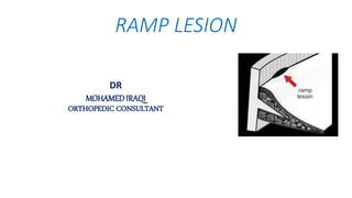
Roti bank chennai PPT [Autosaved].pptx1
Roti bank  chennai PPT [Autosaved].pptx1Roti bank  chennai PPT [Autosaved].pptx1
Roti bank chennai PPT [Autosaved].pptx1
 
Health Education on prevention of hypertension
Health Education on prevention of hypertensionHealth Education on prevention of hypertension
Health Education on prevention of hypertension
 
Demystifying-Gene-Editing-The-Promise-and-Peril-of-CRISPR.pdf
Demystifying-Gene-Editing-The-Promise-and-Peril-of-CRISPR.pdfDemystifying-Gene-Editing-The-Promise-and-Peril-of-CRISPR.pdf
Demystifying-Gene-Editing-The-Promise-and-Peril-of-CRISPR.pdf
 
India Clinical Trials Market: Industry Size and Growth Trends [2030] Analyzed...
India Clinical Trials Market: Industry Size and Growth Trends [2030] Analyzed...India Clinical Trials Market: Industry Size and Growth Trends [2030] Analyzed...
India Clinical Trials Market: Industry Size and Growth Trends [2030] Analyzed...
 
Antibiotic Stewardship by Anushri Srivastava.pptx
Antibiotic Stewardship by Anushri Srivastava.pptxAntibiotic Stewardship by Anushri Srivastava.pptx
Antibiotic Stewardship by Anushri Srivastava.pptx
 
The Docs PPG - 30.05.2024.pptx..........
The Docs PPG - 30.05.2024.pptx..........The Docs PPG - 30.05.2024.pptx..........
The Docs PPG - 30.05.2024.pptx..........
 
CHAPTER 1 SEMESTER V - ROLE OF PEADIATRIC NURSE.pdf
CHAPTER 1 SEMESTER V - ROLE OF PEADIATRIC NURSE.pdfCHAPTER 1 SEMESTER V - ROLE OF PEADIATRIC NURSE.pdf
CHAPTER 1 SEMESTER V - ROLE OF PEADIATRIC NURSE.pdf
 
.Metabolic.disordersYYSSSFFSSSSSSSSSSDDD
.Metabolic.disordersYYSSSFFSSSSSSSSSSDDD.Metabolic.disordersYYSSSFFSSSSSSSSSSDDD
.Metabolic.disordersYYSSSFFSSSSSSSSSSDDD
 
Contact ME {89011**83002} Haridwar ℂall Girls By Full Service Call Girl In Ha...
Contact ME {89011**83002} Haridwar ℂall Girls By Full Service Call Girl In Ha...Contact ME {89011**83002} Haridwar ℂall Girls By Full Service Call Girl In Ha...
Contact ME {89011**83002} Haridwar ℂall Girls By Full Service Call Girl In Ha...
 
BOWEL ELIMINATION BY ANUSHRI SRIVASTAVA.pptx
BOWEL ELIMINATION BY ANUSHRI SRIVASTAVA.pptxBOWEL ELIMINATION BY ANUSHRI SRIVASTAVA.pptx
BOWEL ELIMINATION BY ANUSHRI SRIVASTAVA.pptx
 
ABDOMINAL COMPARTMENT SYSNDROME
ABDOMINAL COMPARTMENT SYSNDROMEABDOMINAL COMPARTMENT SYSNDROME
ABDOMINAL COMPARTMENT SYSNDROME
 
Haridwar ❤CALL Girls 🔝 89011★83002 🔝 ❤ℂall Girls IN Haridwar ESCORT SERVICE❤
Haridwar ❤CALL Girls 🔝 89011★83002 🔝 ❤ℂall Girls IN Haridwar ESCORT SERVICE❤Haridwar ❤CALL Girls 🔝 89011★83002 🔝 ❤ℂall Girls IN Haridwar ESCORT SERVICE❤
Haridwar ❤CALL Girls 🔝 89011★83002 🔝 ❤ℂall Girls IN Haridwar ESCORT SERVICE❤
 
Navigating Women's Health: Understanding Prenatal Care and Beyond
Navigating Women's Health: Understanding Prenatal Care and BeyondNavigating Women's Health: Understanding Prenatal Care and Beyond
Navigating Women's Health: Understanding Prenatal Care and Beyond
 
Navigating Challenges: Mental Health, Legislation, and the Prison System in B...
Navigating Challenges: Mental Health, Legislation, and the Prison System in B...Navigating Challenges: Mental Health, Legislation, and the Prison System in B...
Navigating Challenges: Mental Health, Legislation, and the Prison System in B...
 
Global launch of the Healthy Ageing and Prevention Index 2nd wave – alongside...
Global launch of the Healthy Ageing and Prevention Index 2nd wave – alongside...Global launch of the Healthy Ageing and Prevention Index 2nd wave – alongside...
Global launch of the Healthy Ageing and Prevention Index 2nd wave – alongside...
 
ventilator, child on ventilator, newborn
ventilator, child on ventilator, newbornventilator, child on ventilator, newborn
ventilator, child on ventilator, newborn
 
A Community health , health for prisoners
A Community health  , health for prisonersA Community health  , health for prisoners
A Community health , health for prisoners
 
The Importance of Community Nursing Care.pdf
The Importance of Community Nursing Care.pdfThe Importance of Community Nursing Care.pdf
The Importance of Community Nursing Care.pdf
 
Medical Technology Tackles New Health Care Demand - Research Report - March 2...
Medical Technology Tackles New Health Care Demand - Research Report - March 2...Medical Technology Tackles New Health Care Demand - Research Report - March 2...
Medical Technology Tackles New Health Care Demand - Research Report - March 2...
 
Empowering ACOs: Leveraging Quality Management Tools for MIPS and Beyond
Empowering ACOs: Leveraging Quality Management Tools for MIPS and BeyondEmpowering ACOs: Leveraging Quality Management Tools for MIPS and Beyond
Empowering ACOs: Leveraging Quality Management Tools for MIPS and Beyond
 

Ramp lesion

  • 2. • Meniscal ramp lesions were studied by Hamberg and colleagues,in the 1980s but at that time were described only as “injuries of the posterior aspect of the medial meniscus.” • Later, Strobel, introduced the term ramp lesion and characterized the injury as a longitudinal tear, 2.5 cm in length, located at the posterior meniscocapsular junction of medial meniscus. • In 1991, Morgan ,described a surgical technique for arthroscopic repair of ramp lesions using a suture hook through a posteromedial portal. History
  • 3. Fig. 1 Arthroscopic trans-notch visualization of the posteromedial compartment of the left knee (* = posteromedial tibial plateau). Retraction of the ramp lesion using an arthroscopy probe placed through the posteromedial portal reveals the close anatomic relationship between the semimembranosus tendon and the meniscocapsular region.
  • 4. Anatomy • The anterior and posterior roots anchor the meniscus to the tibial plateau, and the body of the meniscus is attached to the adjacent joint capsule and to the tibia by the meniscotibial ligaments. • The reduced mobility of the medial meniscus makes it susceptible to injuries, especially in deep flexion and with rotational trauma when the pressure is increased in the posterior horn of the medial meniscus.
  • 5. • Capsular arm of the semimembranosus tendon insertion translates the posterior horn of the meniscus posteriorly during knee flexion.
  • 6. Why Ramp Lesion Is Important
  • 7. hidden lesion • A consequence of an underestimation of their incidence lead to a high rate of missed diagnoses. • The recent interest in these injuries heralds an increasing recognition of their importance and an emerging concept of their association with posteromedial knee instability. • The expression “hidden lesion,” also has been recently used to describe this injury, and the term refers to the difficulty in identifying ramp lesions from standard anterior arthroscopic portals, • and also with preoperative MRI, which has low sensititvity.
  • 8. • Meniscal ramp lesions are a “HOT TOPIC” because of increasing recognition that they have important biomechanical consequences and also that they occur much more frequently than was previously understood. • A systematic exploration of the posteromedial compartment of the knee via a trans-notch approach is needed to confirm the presence of a meniscal ramp lesion. • If left untreated, meniscal ramp lesions may contribute to residual antero-posterior instability & rotation laxity in the anterior cruciate ligament–reconstructed knee and may also result in failure of meniscal repair.
  • 10. • isolated ACL reconstruction fails to restore normal joint kinematics and results in residual laxity in the presence of a ramp lesion.
  • 11. • Ramp lesions are reported to occur frequently (9.3%–24.0%) in ACL-deficient knees, including in children and adolescents, • Thus, a significantly higher prevalence of ramp tears was reported in patients with chronic ACL tears compared with patients with acute ACL tears. • Papageorgiou et al reported that the forces on the medial meniscus were increased by 200% in response to anterior tibial loads with a transected ACL. Likewise, deficiency of the medial meniscus has been associated with ACLR failure Incidence
  • 12. Risk factors for the occurrence of ramp lesions in ACL-deficient knees • include male gender, patients younger than 30 years • revision ACL reconstruction • chronic injuries • preoperative side-to-side anteroposterior laxity difference of 6 mm or more • the presence of concomitant lateral meniscal tears • The presence of any of these factors should raise the index of suspicion for the existence of a ramp lesion.
  • 13. Objective diagnosis • there are no specific physical examination tests for ramp lesions • Recently, MRI have a broad range of sensitivities of both 1.5T and 3T • The most specific sign in the MRI evaluation of ramp lesions is the hyperintense signal that can be observed between the meniscus and the capsule. • the presence of PMTP edema demonstrated significantly greater odds for ramp lesions compared with meniscal body tears.
  • 16. Interesting Fact • All over the world, in orthopedic meetings, the meniscal root lesion is more extensively debated than the ramp lesion. This is despite the fact that ramp lesions are a lot more common (24% in ACLR in our experience). • This greater focus on root tears is perhaps because of the well-recognized loss of hoop force distribution and profound biomechanical consequences of root tears. However, it is increasingly recognized that ramp lesions also have important biomechanical consequences including persistent anteroposterior (AP) and rotational laxity and an association with higher ACL graft failure rates.
  • 17. • Arthroscopy is considered gold standard for diagnosis of ramp lesions. • 40 % percent of ramp lesions are not identified through standard anterior portal visualization and inspection of the posterior compartment via a trans-notch view, and posteromedial probing is required to identify them.
  • 18. Classification • Type 1: Meniscocapsular lesions. These lesions are very peripherally located in the synovial sheath. Mobility at probing is very low. • • Type 2: Partial superior lesions. These lesions are stable and can be diagnosed only by a trans-notch approach. Mobility at probing is low. • Type 3: Partial inferior or hidden lesions. The lesions are typically subtle or not immediately visible even with trans-notch visualization but can be strongly suggested by significant mobility on probing and also by identification of abnormal tissue quality on needling. • Type 4: A complete tear of the red-red zone. Mobility at probing is very high. • Type 5: A double tear involving the meniscocapsular junction and a second more anterior tear of the posterior horn.
  • 19. Fig. 3 Ramp lesion classification as proposed by Thaunat and colleagues. (A) Illustration demonstrating the posterior meniscus- capsular region and the areas of meniscus vascularization. (B) Type 1: Meniscocapsular lesion, located in the synovial sheath. Mobility at probing is very low. (C) Type 2: Upper partial lesion. Mobility at probing is low. These lesions are stable and can be diagnosed only by a trans-notch approach (D) Type 3: Lower lesion (“hidden lesion”). significant mobility on probing . not immediately visible even with trans-notch visualization (E) Type 4: Complete injury in the red-red area. Mobility at probing is very high. (F) Type 5: Double tear. involving the meniscocapsular junction and a second more anterior tear of the posterior horn
  • 20. Treatment options • isolated tears that are small (less than 10 mm) and stable may amenable to conservative treatment. abrasion and trephination of stable lesions (without repair). • posteromedial portal suture hook device. • all-inside suture technique Fig. 8 Image of a curved hook device, with the tip curvature pointing to the left side (ideal for suturing meniscal ramp lesion on a right knee).
  • 21. • The mobility of the meniscus at probing may lead the surgeon to suspect the presence of a posterior tear even if it is not visible by standard anterior viewing, because ramp lesions classified as types III, IV, and V may be highly mobile when pulled. • To facilitate the passage through this space, a valgus force is applied ,first in extension and then in flexion. If passage remains difficult, the use of a blunt trocar may be helpful. Tibial internal rotation may improve visualization of the tear by causing posterior tibial plateau subluxation and a posterior translation of the medial segment. Technical Tip
  • 22. systematic arthroscopic exploration of the knee joint • Step 1: Standard arthroscopic exploration • Step 2: Exploration of posteromedial compartment and probing the meniscocapsular junction with a needle • Step 3: Creation of a posteromedial portal • Step 4: Meniscal repair procedure
  • 23. Fig. 5 Arthroscopic view with a 30° scope of a right knee through the anterolateral portal. (A) Probe is inserted through the anteromedial portal in the space between the posterior cruciate ligament (PCL) and the medial femoral condyle (MFC). (B) Passage of the arthroscope through the space between the PCL and the MFC after a valgus force is applied. (C) Image obtained via trans-notch visualization of the posteromedial region of the knee, observing the posteromedial capsule and the meniscal ramp lesion. TS, tibial spine. Two Postero-medial PortalTecnique
  • 24. Fig. 6 External image of the knee demonstrating how transillumination with the arthroscope positioned in the trans-notch view & The knee is positioned at 90° of flexion to minimize the risk of injury to neurovascular structures and a needle is introduced above the hamstring tendons, 1 cm posterior to the medial tibiofemoral joint line, pointing toward the lesion
  • 25. Fig. 7 Arthroscopic image through trans-notch vision showing 2 steps for the creation of the posteromedial portal. (A)Insertion of the needle to verify the correct positioning of the portal and (B) creation of the posteromedial portal with a no. 11 scalpel. MFC, medial femoral condyle.
  • 26. Fig. 9 Detailing of a meniscal ramp suture procedure. (A)Revitalization of the edges of the lesion using a shaver. (B) Suture device inserted through the posteromedial portal. (C) Penetration of the peripheral edge of the lesion. (D) Penetration of the suture device through the inner portion of the tear, encompassing both ends of the lesion. (E) Progression of the suture, which is then captured out of the knee with the aid of a grasper. (F) First stitch being completed using a knot pusher. Internal rotation of the foot helps keep the medial femoral condyle away from the posterior segment of the meniscus and facilitates the procedure.
  • 27. Fig. 10 Final arthroscopic aspect of a meniscal ramp repair, as seen by trans-notch view, in which 3 stitches were required to close the lesion.
  • 28. Fig 1(A) Arthroscopic image of right knee viewing through the anterolateral portal. Tight medial compartment, measured as 4 mm, results in difficult arthroscopic probe examination of the PHMM. (B) Fenestration of the medial collateral ligament, proximal to the meniscus and directly on the femur, is performed by an 18-g spinal needle. (C) This provides an increased medial joint space, measured as 11 mm, and adequate visualization of the meniscus without damaging the cartilage. (MFC, medial femoral condyle; MTP, medial tibial plateu; PHMM, posterior horn of the medial meniscus.) Trans-Septal Portal Tecnique
  • 29. Fig 2(A) Right knee: a posteromedial portal is established 2.5 cm distal from the bony prominence of medial epicondyle and 2.5 cm posterior from this measured point with the knee in 90° of flexion. (B) Right knee: the posterolateral portal is located anterior to the long head of the biceps and proximal and posterior to the LCL. (LCL, lateral collateral ligament; ME, medial epicondyle; PL, posterolateral portal; PM, posteromedial portal.)
  • 30. Fig 3 Posteromedial portal establishment on the right knee with the “nick and spread” technique. With the knee in 90° of flexion, modified Gillquist maneuver is performed into the posteromedial compartment. This helps to visualize portal establishment with a needle A) and the portal is enlarged with a scalpel B) . A double dam flexible flanged cannula (Arthrex) is placed into the portal C) . Adequate visualization of the PHMM is obtained with the cannula nicely retracting the capsule medially . (MFC, medial femoral condyle; PHMM, posterior horn of medial meniscus; PM, posteromedial portal.)
  • 31. Fig 4 Posterolateral portal establishment on the right knee. (B). and then the portal is enlarged with a scalpel. (D). Arthroscopic image of the shaver through the posteromedial portal with direct visualization during trans-septal portal/window creation. Note that the opening of the shaver is oriented anterior to avoid popliteal artery injury. (LCL, lateral collateral ligament; LFC, lateral femoral condyle; PL, posterolateral portal.)
  • 32. Fig 5 With the help of a switching stick (A), a standard long arthroscopic cannula is placed into the posterolateral portal to facilitate instrument use through this portal (B). (PL, posterolateral portal.)
  • 33. Fig 6 (A) Right knee: with the knee in 90° of flexion, the arthroscope is then placed through the posteromedial portal to view laterally, and the arthroscopic shaver is gently advanced from the posterolateral portal to extend the hole in the septum for facilitating the passage of the arthroscope or instrument insertion and manipulation. (B) Arthroscopic image viewing from the PM portal: the trans-septal portal (dashed borders) has been established without damaging the PCL. (LFC, lateral femoral condyle; MFC, medial femoral condyle; PCL, posterior cruciate ligament; PL, posterolateral portal; PM, posteromedial portal.)
  • 34. Fig 7 (A) Right knee arthroscopic image: the arthroscope is placed through the PL portal and advanced through the trans-septal portal to directly visualize the ramp lesion. An arthroscopic rasp determines the margins of the lesion after removing overlying fibrotic tissue. (B) Right knee arthroscopic image: the edges of the lesion are freshened by an arthroscopic shaver. (MFC, medial femoral condyle; PL, posterolateral.)
  • 35. Fig 8 (A) Right knee arthroscopic image from the trans-septal portal: a 45° right suture passer (Depuy) is penetrating the right PMMH through the posteromedial portal, after already shuttling a single limb of a UHMPE suture through the capsular component. (B) Eyelet suture shuttle advanced into the joint to be retrieved by an atraumatic grasper through the posteromedial portal. (MFC, medial femoral condyle; PMMH, posterior horn of medial meniscus; UHMPE, ultra- high molecular weight polyethylene fiber.)
  • 36. Fig 9 (A) Right knee arthroscopic image: the sutures are passed in a vertical mattress fashion, adequately reducing the torn tissue and reconstituting the ramp of the PHMM. (B) Right knee arthroscopic image through a trans-septal portal: sutures are placed in 5-7 mm intervals and knots are kept on the capsular side when tying arthroscopically to limit abrasion to the medial femoral condyle. (MFC, medial femoral condyle; PHMM, posterior horn of medial meniscus.)
  • 37. a meniscal ramp lesion arthroscopic repair with an all-inside technique with the Fast-Fix 360 device, detailing the use of the accessory posteromedial portal, and the addition of an arthroscopic grasper that raises the retracted menisco-tibial ligament, to allow correct fixation. Although this technique seems technically less challenging for most surgeons All-inside suture technique
  • 38. Rehabilitation protocol • Most protocols agree that early knee motion is beneficial; however, knee hyperflexion is associated with anterior tibial translation, which can increase the stress on the repair. • In our practice, the postoperative management of an ACL reconstruction associated with ramp repair is modified by restriction of the active and passive range of motion to 0 to 90° of flexion during the first 6 weeks. • Progression to full weight bearing is allowed by postoperative week 3. Jogging is permitted after week 12 to 16, pivot activity at 6 months, and full activity at 9 months for all patients.
  • 39. Take Home Message • Meniscal ramp lesions are common but frequently under-recognized in the ACL-injured knee. • It is important to highlight that both preoperative MRI and arthroscopic evaluation via classic anterior portals are associated with high rates of missed diagnoses. • For that reason, a systematic exploration of the posteromedial compartment is advocated in all ACL-injured knees. • Failure to recognize and repair ramp lesions is associated with persistent anterior and rotational knee laxity, suggesting it makes part of a posteromedial instability, • in the setting of an ACL tear. Concomitant suture repair of these lesions, via a posteromedial portal, can restore normal biomechanics and is associated with excellent clinical outcomes.