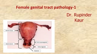
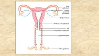
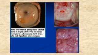
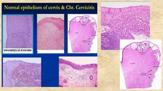
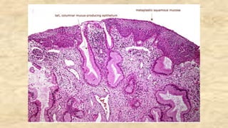
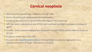
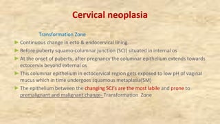
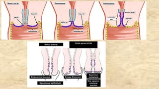
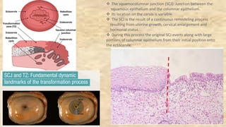
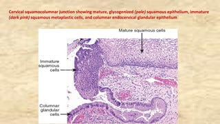
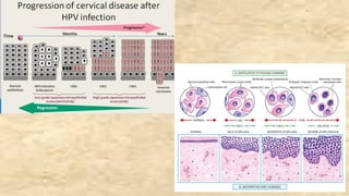
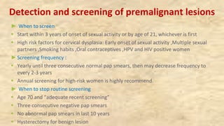
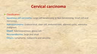
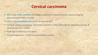
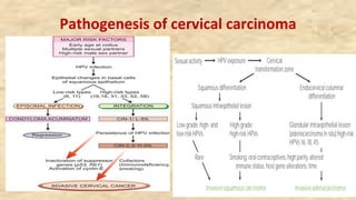
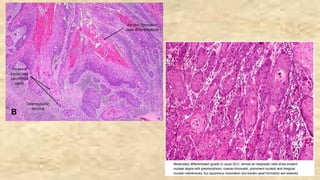
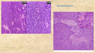
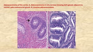
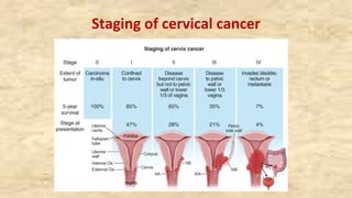
Female genital tract pathology-1
Dr. Rupinder
Kaur
At the end of this session medical student should be able to:
►Discuss the etiology and morphological features of cervicitis.
► Discuss the epidemiology of carcinoma cervix
► Enumerate the etiological factors for the development of carcinoma cervix.
►Describe the pathogenesis of carcinoma cervix.
►Describe the progression of carcinoma cervix.
►Discuss the pathological features of carcinoma cervix
►Discuss the methods of diagnosis of carcinoma of cervix.
►Enumerate and discuss various screening methods and programs for early detection of
carcinoma cervix
v The endocervix is lined by a simple columnar epithelium that
secretes mucus.
v Mucinous columnar epithelium lines the surface and the
underlying glands
v The ectocervix is covered by non-keratinizing, stratified
squamous epithelium, either native or metaplastic in
continuity with the vaginal epithelium.
v The squamous epithelium is composed by multiple layers:
basal, parabasal, intermediate and superficial layer
Cervix
►It is both a sentinel for potentially serious upper genital tract infections and a target
for viruses and other carcinogens, which may lead to invasive carcinoma
►Regular Pap smears have proved beneficial in detecting cancer at the early stage with
increase in the survival
►Pathology
v Metaplasia, Inflammatory (Cervicitis)
v Cervical neoplasia: Benign ( Polyps, Microglandular hyperplasia) ; Malignant
Cervicitis
►Inflammation commonly due to constant exposure to vaginal bacteria
►Found in almost all reproductive as well as post menopausal females
►Etiological agent: Streptococcus, HSV-2, staphylococcus, Chlamydia trachomatis (50-
75%), Neisseria gonorrhea
►May be associated with endometritis, salpingitis, pelvic inflammatory disease (PID) or
chorioamnionitis
► Can be acute and chronic (most common)
Cervicitis
►Morphology:
Gross- Eversion of ectocervix with hyperemia, edema and granular surface
Nabothian(retention cysts) may be grossly visible as pearly grey vesicles.
Histopathology :
- Inflammation of cervical mucosa with increased lymphocytes, plasma cells, neutrophils
( acute) and tingible body macrophages,
- Columnar cell proliferation (micro- glandular change)
- Nabothian cysts/ follicles (due to occlusion of cervical gland ducts ) &
- Squamous metaplasia
Cervical neoplasia
►Most common gynecologic malignancy world wide
►Ranks 3rd among all malignancies among females
►Incidence has reduced to almost 80% with regular PAP screening
►HPV has been implicated as one of the most common and highly oncogenic etiological
agent
►In India –most common malignancy; along with breast it forms almost 40% of cancer of
all sites
►Incidence varies from 21%-50%
►Occurs in the transformation zone as intraepithelial neoplasia progressing on to
invasive carcinoma through carcinoma in situ(CIS)
Cervical neoplasia
Transformation Zone
►Continuous change in ecto & endocervical lining
►Before puberty squamo-columnar junction (SCJ) situated in internal os
►At the onset of puberty, after pregnancy the columnar epithelium extends towards
ectocervix beyond external os
►This columnar epithelium in ectocervical region gets exposed to low pH of vaginal
mucus which in time undergoes squamous metaplasia(SM)
►The epithelium between the changing SCJ’s are the most labile and prone to
premalignant and malignant change- Transformation Zone
v The squamocolumnar junction (SCJ): Junction between the
squamous epithelium and the columnar epithelium.
v Its location on the cervix is variable.
v The SCJ is the result of a continuous remodeling process
resulting from uterine growth, cervical enlargement and
hormonal status.
v During this process the original SCJ everts along with large
portions of columnar epithelium from their initial position onto
the ectocervix.
Cervical squamocolumnar junction showing mature, glycogenized (pale) squamous epithelium, immature
(dark pink) squamous metaplastic cells, and columnar endocervical glandular epithelium
Cervical neoplasia- etiopathogenesis
►Multiple etiologic agents
v Persistent infection with Human papilloma virus- 16, 18
v Early age of marriage & intercourse
v Multiple sexual partners
v Dietary deficiency: Vit A, Folic acid
v Cigarette smoking: polycyclic aromatic hydrocarbons are carcinogenic and forms
damaging DNA adducts in cervical epithelium
v Multiparity
v Lack of circumcision of male partner: accumulation of smegma which is carcinogenic
and might induce malignant transformation
v Oral contraceptive usage
v Immunosupression
Cervical neoplasia- etiopathogenesis
Human papilloma virus ( HPV):
v High risk: subtypes 16,18 (MC), 31,33,35
v Low risk: 6,11,42,44 ( cause condyloma)
v HPV exposure occurs through sexual transmission
v The epithelial cells get transformed to premalignant change (CIN/ IEN)
v In malignant transformation HPV-DNA gets covalently linked and gets incorporated
within the host genome
v HPV 16& 18 and its gene E 6 produces protein which binds to cancer supressor gene
(p53) in the host and causes proteolytic digestion of p53
v Other HPV’s through E7 gene binds to another tumor suppressor gene (Rb)
v This destroys the ability of both p53 and Rb gene to suppress carcinogenic
transformation by affecting cell cycle regulation
v Similar transformation can also occur in endocervical cells leading to adenocarcinoma
Cervical intraepithelial neoplasia(CIN)
►Spectrum of dysplasia (atypical change) confined within the epithelium (intraepithelial)
that begins in basal layers and progressively involves other layers to form carcinoma in
situ
►Classification based upon the cellular atypia and involvement of different thickness of
the epithelium
►Earlier were classified in 3 grades: CIN I, CIN II, CIN III; Conventionally graded as mild,
moderate, & severe dysplasia
v CIN I: involvement of basal one third of epithelium (mild)
v CIN II: lower two third involvement ( mod. Dysplasia)
v CIN III: > than two third but not involving entire thickness (severe dysplasia)
v Carcinoma in situ (CIS): involvement of complete thickness of the epithelium with intact
basement membrane
►The atypical changes includes:
- Increased nuclear size, hyperchromasia and increased mitotic rate, Increased N/C ratio
and loss of polarity
Squamous intrepithelial neoplasia (SIL)
►National Cancer Institute proposed this terminology in Bethesda system for cervical
and vaginal cytology reporting
►3 grades (# TIER system)of CIN have been adjusted in 2 grades (2 TIER system)of
squamous intraepithelial lesion
►Low grade SIL (LSIL) and High grade SIL (HSIL)
►LSIL corresponds to CIN I; is a flat condyloma with koilocytic atypia, related to HPV
6& 11 infection
►HSIL corresponds to CIN II & III with abnormal pleomorphic atypical squamous cells.
HPV 16 & 18 have been implicated in its etiology
► LSIL: 60% of them regress and around 10% progresses to HSIL
►HSIL: 30 % pf the lesion regress and about 10% progresses to invasive carcinoma
Nuclear atypia in LSIL. : The most significant feature of LSIL
is nuclear atypia. This is characterized by nuclear
enlargement, hyperchromasia, nuclear irregularity, and
variation in nuclear size
Koilocytosis in LSIL. : The cytological features of a
productive HPV infection include multinucleation and
perinuclear cytoplasmic cavitation or halos. The
combination of nuclear atypia and cytoplasmic halos is
referred to as koilocytosis.
HSIL with marked variability in nuclear size
(anisonucleosis). Anisonucleosis is a variable feature of
HSIL
HSIL: Undifferentiated neoplastic cells replace 50–70% of the
epithelium. The nuclear : cytoplasmic ratio is high, and
the cytoplasmic membranes and the basal layer are indistinct
Detection and screening of premalignant lesions
►Criteria for screening test: Simple, specific, cost effective, acceptable, accurate,
repeatable, can be performed by paramedics/ staff
►Benefits of early detection: Improve prognosis ( decrease mortality & morbidity ,
Increased survival), less radical treatment
►SCREENING: Methods of prevention
v Primary : Avoid precipitating/ risk factors, vaccination, counselling
v Secondary: Screening, early detection
v Tertiary: Treatment or mitigation of damage
►Secondary: Systemic application of a test in an asymptomatic person to detect early
lesions; Can be selective ( high risk group) or Mass screening
Prevention of cervical cancer
Detection and screening of premalignant lesions
►Methods of cervical screening:
v Cytological: Conventional PAP smear, Liquid based cytology; automated cytological
screening
v HPV testing:
v Visual: VIC, Colposcopy, cervicography
►PAP Smear: Named after Dr. George Papanicolaou (1883-1962)
►Usefulness of PAP smear
Ø Effective screening method to detect premalignant and malignant lesions
Ø Long latent period of 10-15 years between CIN and invasive cancer allows adequate
treatment of CIN and prevention of invasive cancer has been proved successful in
reducing the incidence of invasive cancer by 80% and the mortality by 70%
Ø It is important to detect at early stages as low grade lesions are usually reversible with
adequate early treatment
Detection and screening of premalignant lesions
► When to screen
§ Start within 3 years of onset of sexual activity or by age of 21, whichever is first
§ High risk factors for cervical dysplasia: Early onset of sexual activity ,Multiple sexual
partners ,Smoking habits ,Oral contraceptives ,HPV and HIV positive women
►Screening frequency :
§ Yearly until three consecutive normal pap smears, then may decrease frequency to
every 2-3 years
§ Annual screening for high-risk women is highly recommend.
► When to stop routine screening
§ Age 70 and “adequate recent screening”
§ Three consecutive negative pap smears
§ No abnormal pap smears in last 10 years
§ Hysterectomy for benign lesion
Cervical carcinoma
►Classification:
- Squamous cell carcinoma: Large cell keratinizing or Non-keratinizing, Small cell and
Verrucous
- Adenocarcinoma: Endocervical, clear cell, endometrioid, adenoid cystic, adenoma
malignum
- Mixed: Adenosquamous, glassy cell
- Neuroendocrine: large and small
- Others: Lymphoma, melanoma and sarcoma
Cervical carcinoma
►SCC is the most common histologic subtype of cervical cancer, accounting for
approximately 80% of cases
►HSIL is an immediate precursor of cervical SCC
►Cervical adenocarcinoma- 2nd most common (15%) followed by adenosquamous &
neuroendocrine (<5%)
►Peak age incidence is 45 years
►Etiopathogenesis – same as preinvasive cancers
Pathogenesis of cervical carcinoma
Invasive cervical carcinoma- morphology
►GROSS: 3 distinct types:
§ Fungating, ulcerating and infiltrating
§ Fungating: cauliflower like growth protruding into the vaginal cavity; most common; can
be endophytic ( within the endocervical canal)
►M/E: 4 types
§ Squamous ( epidermoid) cell carcinoma- most common can be large cell (keratinizing and
non keratinizing) & small cell type
§ Adenocarcinoma ( papillary & glandular pattern)
§ Adenosquamous
§ undifferentiated
Squamous cell carcinoma (SCC)
►Large cell Keratinizing
- Large cells with moderate to abundant pink keratinized cytoplasm
- Cells are arranged in sheets, clusters
- Severe degree of pleomorphism with high mitotic rate and frequent mitosis
- At places keratinized epithelium are present in concentric whorls- keratin pearls
►Non –keratinizing- no keratin pearls ( mod diff SCC)
►Small-cell non-keratinizing: small cell with large hyperchromatic nuclei and scant
eosinophilic cytoplasm; has poor prognosis
Squamous cell carcinoma of the cervix. A, Microinvasive squamous cell carcinoma with invasive nest
breaking through the basement membrane of HSIL. B, Invasive squamous cell carcinoma
Non keratinizing SCC
Adenocarcinoma of the cervix. A, Adenocarcinoma in situ (arrow) showing dark glands adjacent to
normal, pale endocervcial glands. B, Invasive adenocarcinoma.
Staging of cervical cancer
Spread and clinical staging of cervical carcinoma
► SPREAD:
v Direct spread: urinary bladder, peritoneum, ureter, rectum vagina
v Lymphatic spread: paracervical lymph nodes, obturator LN’s, external iliac group of LN’s
v Hematogenous spread: liver, lung bones
► STAGING:
v Stage 0: carcinoma in situ (CIS)
v Stage I: carcinoma confined to cervix
v Stage II: carcinoma involving parametrium ( but not upto pelvic wall) or upper 2/3rd of vagina
v Stage III: carcinoma extending to pelvic wall and or lower 2/3rd of vagina
v Stage IV: carcinoma spreading beyond pelvis or involving mucosa of urinary bladder or rectum
along with distant metastasis
Treatment
Depends upon the stage of the disease
Cervical carcinoma: clinical features &
complications
►Bleeding per vaginum: irregular, intermenstrual, post coital, post menopausal
►Vaginal discharge; copious, purulent
►Urinary symptoms: dysuria, hematuria, frequency
►Cachexia
►Low back pain
►COMPLICATIONS: Pyometra, vesicovaginal fistula, uretric obstruction leading to hydro-
nephrosis, uremia
►D/D: Cervical tuberculosis, Syphilitic cervicitis- Cervical ulcers
Polyps: mucus, cervical, fibroid
Endometrial carcinoma
Cervical cancer: Investigations
►Pap smear: if no obvious lesion
►Colposcopy
►Cervical biopsy ( obvious lesions)
►Pretreatment investigations: Routine CBC, RFT,LFT, urine R/E,CXR, abdomen and pelvic
U/s , MRI, IVU, PET CT
►Cystoscopy, Proctoscopy
►Molecular testing for CIN: HPV,p 16, p 21, Ki-67 ( proliferative index), pRb & p 53 gene
mutational studies
At the end of this session medical student should be able to:
►Discuss the etiology and morphological features of cervicitis.
► Discuss the epidemiology of carcinoma cervix
► Enumerate the etiological factors for the development of carcinoma cervix.
►Describe the pathogenesis of carcinoma cervix.
►Describe the progression of carcinoma cervix.
►Discuss the pathological features of carcinoma cervix
►Discuss the methods of diagnosis of carcinoma of cervix.
►Enumerate and discuss various screening methods and programs for early detection of
carcinoma cervix
Lecture  1 Pathology: Cervix pdf

Lecture 1 Pathology: Cervix pdf

  • 1.
    Female genital tractpathology-1 Dr. Rupinder Kaur
  • 2.
    At the endof this session medical student should be able to: ►Discuss the etiology and morphological features of cervicitis. ► Discuss the epidemiology of carcinoma cervix ► Enumerate the etiological factors for the development of carcinoma cervix. ►Describe the pathogenesis of carcinoma cervix. ►Describe the progression of carcinoma cervix. ►Discuss the pathological features of carcinoma cervix ►Discuss the methods of diagnosis of carcinoma of cervix. ►Enumerate and discuss various screening methods and programs for early detection of carcinoma cervix
  • 5.
    v The endocervixis lined by a simple columnar epithelium that secretes mucus. v Mucinous columnar epithelium lines the surface and the underlying glands v The ectocervix is covered by non-keratinizing, stratified squamous epithelium, either native or metaplastic in continuity with the vaginal epithelium. v The squamous epithelium is composed by multiple layers: basal, parabasal, intermediate and superficial layer
  • 6.
    Cervix ►It is botha sentinel for potentially serious upper genital tract infections and a target for viruses and other carcinogens, which may lead to invasive carcinoma ►Regular Pap smears have proved beneficial in detecting cancer at the early stage with increase in the survival ►Pathology v Metaplasia, Inflammatory (Cervicitis) v Cervical neoplasia: Benign ( Polyps, Microglandular hyperplasia) ; Malignant
  • 7.
    Cervicitis ►Inflammation commonly dueto constant exposure to vaginal bacteria ►Found in almost all reproductive as well as post menopausal females ►Etiological agent: Streptococcus, HSV-2, staphylococcus, Chlamydia trachomatis (50- 75%), Neisseria gonorrhea ►May be associated with endometritis, salpingitis, pelvic inflammatory disease (PID) or chorioamnionitis ► Can be acute and chronic (most common)
  • 8.
    Cervicitis ►Morphology: Gross- Eversion ofectocervix with hyperemia, edema and granular surface Nabothian(retention cysts) may be grossly visible as pearly grey vesicles. Histopathology : - Inflammation of cervical mucosa with increased lymphocytes, plasma cells, neutrophils ( acute) and tingible body macrophages, - Columnar cell proliferation (micro- glandular change) - Nabothian cysts/ follicles (due to occlusion of cervical gland ducts ) & - Squamous metaplasia
  • 12.
    Cervical neoplasia ►Most commongynecologic malignancy world wide ►Ranks 3rd among all malignancies among females ►Incidence has reduced to almost 80% with regular PAP screening ►HPV has been implicated as one of the most common and highly oncogenic etiological agent ►In India –most common malignancy; along with breast it forms almost 40% of cancer of all sites ►Incidence varies from 21%-50% ►Occurs in the transformation zone as intraepithelial neoplasia progressing on to invasive carcinoma through carcinoma in situ(CIS)
  • 13.
    Cervical neoplasia Transformation Zone ►Continuouschange in ecto & endocervical lining ►Before puberty squamo-columnar junction (SCJ) situated in internal os ►At the onset of puberty, after pregnancy the columnar epithelium extends towards ectocervix beyond external os ►This columnar epithelium in ectocervical region gets exposed to low pH of vaginal mucus which in time undergoes squamous metaplasia(SM) ►The epithelium between the changing SCJ’s are the most labile and prone to premalignant and malignant change- Transformation Zone
  • 15.
    v The squamocolumnarjunction (SCJ): Junction between the squamous epithelium and the columnar epithelium. v Its location on the cervix is variable. v The SCJ is the result of a continuous remodeling process resulting from uterine growth, cervical enlargement and hormonal status. v During this process the original SCJ everts along with large portions of columnar epithelium from their initial position onto the ectocervix.
  • 16.
    Cervical squamocolumnar junctionshowing mature, glycogenized (pale) squamous epithelium, immature (dark pink) squamous metaplastic cells, and columnar endocervical glandular epithelium
  • 17.
    Cervical neoplasia- etiopathogenesis ►Multipleetiologic agents v Persistent infection with Human papilloma virus- 16, 18 v Early age of marriage & intercourse v Multiple sexual partners v Dietary deficiency: Vit A, Folic acid v Cigarette smoking: polycyclic aromatic hydrocarbons are carcinogenic and forms damaging DNA adducts in cervical epithelium v Multiparity v Lack of circumcision of male partner: accumulation of smegma which is carcinogenic and might induce malignant transformation v Oral contraceptive usage v Immunosupression
  • 18.
    Cervical neoplasia- etiopathogenesis Humanpapilloma virus ( HPV): v High risk: subtypes 16,18 (MC), 31,33,35 v Low risk: 6,11,42,44 ( cause condyloma) v HPV exposure occurs through sexual transmission v The epithelial cells get transformed to premalignant change (CIN/ IEN) v In malignant transformation HPV-DNA gets covalently linked and gets incorporated within the host genome v HPV 16& 18 and its gene E 6 produces protein which binds to cancer supressor gene (p53) in the host and causes proteolytic digestion of p53 v Other HPV’s through E7 gene binds to another tumor suppressor gene (Rb) v This destroys the ability of both p53 and Rb gene to suppress carcinogenic transformation by affecting cell cycle regulation v Similar transformation can also occur in endocervical cells leading to adenocarcinoma
  • 21.
    Cervical intraepithelial neoplasia(CIN) ►Spectrumof dysplasia (atypical change) confined within the epithelium (intraepithelial) that begins in basal layers and progressively involves other layers to form carcinoma in situ ►Classification based upon the cellular atypia and involvement of different thickness of the epithelium ►Earlier were classified in 3 grades: CIN I, CIN II, CIN III; Conventionally graded as mild, moderate, & severe dysplasia v CIN I: involvement of basal one third of epithelium (mild) v CIN II: lower two third involvement ( mod. Dysplasia) v CIN III: > than two third but not involving entire thickness (severe dysplasia) v Carcinoma in situ (CIS): involvement of complete thickness of the epithelium with intact basement membrane ►The atypical changes includes: - Increased nuclear size, hyperchromasia and increased mitotic rate, Increased N/C ratio and loss of polarity
  • 22.
    Squamous intrepithelial neoplasia(SIL) ►National Cancer Institute proposed this terminology in Bethesda system for cervical and vaginal cytology reporting ►3 grades (# TIER system)of CIN have been adjusted in 2 grades (2 TIER system)of squamous intraepithelial lesion ►Low grade SIL (LSIL) and High grade SIL (HSIL) ►LSIL corresponds to CIN I; is a flat condyloma with koilocytic atypia, related to HPV 6& 11 infection ►HSIL corresponds to CIN II & III with abnormal pleomorphic atypical squamous cells. HPV 16 & 18 have been implicated in its etiology ► LSIL: 60% of them regress and around 10% progresses to HSIL ►HSIL: 30 % pf the lesion regress and about 10% progresses to invasive carcinoma
  • 25.
    Nuclear atypia inLSIL. : The most significant feature of LSIL is nuclear atypia. This is characterized by nuclear enlargement, hyperchromasia, nuclear irregularity, and variation in nuclear size Koilocytosis in LSIL. : The cytological features of a productive HPV infection include multinucleation and perinuclear cytoplasmic cavitation or halos. The combination of nuclear atypia and cytoplasmic halos is referred to as koilocytosis.
  • 26.
    HSIL with markedvariability in nuclear size (anisonucleosis). Anisonucleosis is a variable feature of HSIL HSIL: Undifferentiated neoplastic cells replace 50–70% of the epithelium. The nuclear : cytoplasmic ratio is high, and the cytoplasmic membranes and the basal layer are indistinct
  • 27.
    Detection and screeningof premalignant lesions ►Criteria for screening test: Simple, specific, cost effective, acceptable, accurate, repeatable, can be performed by paramedics/ staff ►Benefits of early detection: Improve prognosis ( decrease mortality & morbidity , Increased survival), less radical treatment ►SCREENING: Methods of prevention v Primary : Avoid precipitating/ risk factors, vaccination, counselling v Secondary: Screening, early detection v Tertiary: Treatment or mitigation of damage ►Secondary: Systemic application of a test in an asymptomatic person to detect early lesions; Can be selective ( high risk group) or Mass screening
  • 28.
  • 29.
    Detection and screeningof premalignant lesions ►Methods of cervical screening: v Cytological: Conventional PAP smear, Liquid based cytology; automated cytological screening v HPV testing: v Visual: VIC, Colposcopy, cervicography ►PAP Smear: Named after Dr. George Papanicolaou (1883-1962) ►Usefulness of PAP smear Ø Effective screening method to detect premalignant and malignant lesions Ø Long latent period of 10-15 years between CIN and invasive cancer allows adequate treatment of CIN and prevention of invasive cancer has been proved successful in reducing the incidence of invasive cancer by 80% and the mortality by 70% Ø It is important to detect at early stages as low grade lesions are usually reversible with adequate early treatment
  • 30.
    Detection and screeningof premalignant lesions ► When to screen § Start within 3 years of onset of sexual activity or by age of 21, whichever is first § High risk factors for cervical dysplasia: Early onset of sexual activity ,Multiple sexual partners ,Smoking habits ,Oral contraceptives ,HPV and HIV positive women ►Screening frequency : § Yearly until three consecutive normal pap smears, then may decrease frequency to every 2-3 years § Annual screening for high-risk women is highly recommend. ► When to stop routine screening § Age 70 and “adequate recent screening” § Three consecutive negative pap smears § No abnormal pap smears in last 10 years § Hysterectomy for benign lesion
  • 32.
    Cervical carcinoma ►Classification: - Squamouscell carcinoma: Large cell keratinizing or Non-keratinizing, Small cell and Verrucous - Adenocarcinoma: Endocervical, clear cell, endometrioid, adenoid cystic, adenoma malignum - Mixed: Adenosquamous, glassy cell - Neuroendocrine: large and small - Others: Lymphoma, melanoma and sarcoma
  • 33.
    Cervical carcinoma ►SCC isthe most common histologic subtype of cervical cancer, accounting for approximately 80% of cases ►HSIL is an immediate precursor of cervical SCC ►Cervical adenocarcinoma- 2nd most common (15%) followed by adenosquamous & neuroendocrine (<5%) ►Peak age incidence is 45 years ►Etiopathogenesis – same as preinvasive cancers
  • 34.
  • 35.
    Invasive cervical carcinoma-morphology ►GROSS: 3 distinct types: § Fungating, ulcerating and infiltrating § Fungating: cauliflower like growth protruding into the vaginal cavity; most common; can be endophytic ( within the endocervical canal) ►M/E: 4 types § Squamous ( epidermoid) cell carcinoma- most common can be large cell (keratinizing and non keratinizing) & small cell type § Adenocarcinoma ( papillary & glandular pattern) § Adenosquamous § undifferentiated
  • 37.
    Squamous cell carcinoma(SCC) ►Large cell Keratinizing - Large cells with moderate to abundant pink keratinized cytoplasm - Cells are arranged in sheets, clusters - Severe degree of pleomorphism with high mitotic rate and frequent mitosis - At places keratinized epithelium are present in concentric whorls- keratin pearls ►Non –keratinizing- no keratin pearls ( mod diff SCC) ►Small-cell non-keratinizing: small cell with large hyperchromatic nuclei and scant eosinophilic cytoplasm; has poor prognosis
  • 38.
    Squamous cell carcinomaof the cervix. A, Microinvasive squamous cell carcinoma with invasive nest breaking through the basement membrane of HSIL. B, Invasive squamous cell carcinoma
  • 40.
  • 41.
    Adenocarcinoma of thecervix. A, Adenocarcinoma in situ (arrow) showing dark glands adjacent to normal, pale endocervcial glands. B, Invasive adenocarcinoma.
  • 42.
  • 43.
    Spread and clinicalstaging of cervical carcinoma ► SPREAD: v Direct spread: urinary bladder, peritoneum, ureter, rectum vagina v Lymphatic spread: paracervical lymph nodes, obturator LN’s, external iliac group of LN’s v Hematogenous spread: liver, lung bones ► STAGING: v Stage 0: carcinoma in situ (CIS) v Stage I: carcinoma confined to cervix v Stage II: carcinoma involving parametrium ( but not upto pelvic wall) or upper 2/3rd of vagina v Stage III: carcinoma extending to pelvic wall and or lower 2/3rd of vagina v Stage IV: carcinoma spreading beyond pelvis or involving mucosa of urinary bladder or rectum along with distant metastasis
  • 44.
    Treatment Depends upon thestage of the disease
  • 45.
    Cervical carcinoma: clinicalfeatures & complications ►Bleeding per vaginum: irregular, intermenstrual, post coital, post menopausal ►Vaginal discharge; copious, purulent ►Urinary symptoms: dysuria, hematuria, frequency ►Cachexia ►Low back pain ►COMPLICATIONS: Pyometra, vesicovaginal fistula, uretric obstruction leading to hydro- nephrosis, uremia ►D/D: Cervical tuberculosis, Syphilitic cervicitis- Cervical ulcers Polyps: mucus, cervical, fibroid Endometrial carcinoma
  • 46.
    Cervical cancer: Investigations ►Papsmear: if no obvious lesion ►Colposcopy ►Cervical biopsy ( obvious lesions) ►Pretreatment investigations: Routine CBC, RFT,LFT, urine R/E,CXR, abdomen and pelvic U/s , MRI, IVU, PET CT ►Cystoscopy, Proctoscopy ►Molecular testing for CIN: HPV,p 16, p 21, Ki-67 ( proliferative index), pRb & p 53 gene mutational studies
  • 47.
    At the endof this session medical student should be able to: ►Discuss the etiology and morphological features of cervicitis. ► Discuss the epidemiology of carcinoma cervix ► Enumerate the etiological factors for the development of carcinoma cervix. ►Describe the pathogenesis of carcinoma cervix. ►Describe the progression of carcinoma cervix. ►Discuss the pathological features of carcinoma cervix ►Discuss the methods of diagnosis of carcinoma of cervix. ►Enumerate and discuss various screening methods and programs for early detection of carcinoma cervix