
Recommended
More Related Content
What's hot
What's hot (20)
Similar to Physiology of pain pathway
Similar to Physiology of pain pathway (20)
Recently uploaded
Recently uploaded (20)
Physiology of pain pathway
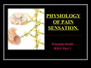
- 1. Priyanka Doshi M.D.S. Part 1 PHYSIOLOGY OF PAIN SENSATION.
- 2. Definition The sensation of pain is defined as theThe sensation of pain is defined as the physical adjunct of an imperativephysical adjunct of an imperative protective reflex.protective reflex. pain is an, unpleasant sensory and emotional experience associated with actual or potential tissue damage - TheThe International Association for the Study ofInternational Association for the Study of Pain (IASP)Pain (IASP)
- 3. Sunday, June 10, 2018
- 4. Neuron
- 6. Types of pain Fast pain is due to activity of myelinated A δ fibres and it is appreciated as sharp bright and localized sensation. Slow pain is due to activity of unmyelinated C fibres and it is appreciated as dull aching and more diffuse. PeripheralPeripheral NerveNerve C-FiberC-Fiber A-delta FiberA-delta Fiber
- 7. Receptors for Pain The receptors in the skin and other tissues are free nerve endings of small myelinated A δ and non myelinated C fibres. They are widespread in the superficial layers of skin as well as in certain internal tissues such as the periosteum, arterial walls, joint surfaces, falx and tentorium of cranial vault.
- 8. Sunday, June 10, 2018
- 10. Sunday, June 10, 2018
- 11. Sunday, June 10, 2018
- 12. Sunday, June 10, 2018
- 13. NEUROTRANSMITTERS It is a chemical substance that acts as the mediator for the transmission of nerve impulse from one neuron to another neuron through a synapse. Sunday, June 10, 2018
- 14. classification 1. Depending upon chemical nature: Amino acids: GABA, Glycerine, Glutamate,Aspartate. Amines: Noradrenaline,Adrenaline,Dopamine, Serotonin,Histamine Others: Acetylcholine, NO Sunday, June 10, 2018
- 15. Sunday, June 10, 2018
- 17. Transduction: Pain stimuli is converted to electrical energy.This electrical energy is known as Transduction. This stimulus sends an impulse across a peripheral nerve fiber (nociceptor).
- 18. Transmission A delta fibers (myelinated) send sharp, localized and distinct sensations. C fibers (unmyelinated) relay impulses that are poorly localized, burning and persistent pain. Pain stimuli travel- spinothalamic tracts.
- 19. Perception Person is aware of pain – somatosensory cortex identifies the location and intensity of pain Person unfolds a complex reaction- physiological and behavioral responses is perceived.
- 20. Modulation Inhibitory neurotransmitters like endogenous opioids work to hinder the pain transmission. This inhibition of the pain impulse is known as modulation
- 21. GATE CONTROL THEORY Melzack & Wall, 1965 Substantia Gelatinosa (SG) in dorsal horn of spinal cord acts as a ‘gate’ – only allows one type of impulses to connect with the brain. If A-beta neurons are stimulated – SG is activated which closes the gate to A-delta & C neurons If A-delta & C neurons are stimulated – SG is blocked which closes the gate to A-beta neurons
- 22. GATE CONTROL THEORY Gate - located in the dorsal horn of the spinal cord Smaller, slower n. fibers carry pain impulses Larger, faster n. fibers carry other sensations Impulses from faster fibers arriving @ gate 1st inhibit pain impulses (acupuncture/pressure, cold, heat, chem. skin irritation). Brain Pain Heat, Cold, Mechanical Gate (T cells/ SG)
- 23. Gate control theory When pain sensation is produced-- other afferents particularly the touch fibres reaching the posterior column of spinal cord are also activated These dorsal column fibres send collaterals to the cells of substantia gelatinosa in the dorsal grey horn. Thus impulses ascending via dorsal column fibres pass through the collaterals and reach substantia gelatinosa.
- 24. Here these impulses inhibit the release of substance P by the pain nerve endings. So that the pain sensation is suppressed. Thus the gating of pain in dorsal grey horn level is similar to presynaptic inhibition. Sunday, June 10, 2018
- 25. How pain is transmitted to brain
- 26. Dual pathways for pain transmission From peripheral receptors to spinal cord: Aδ fibers (fast fibers) – for fast pain C fibers (slow fibers) – for slow pain From spinal cord to brain: via Anterolateral (Spinothalamic) tract Neo-spinothalamic tract – for fast pain Paleo-spinothalamic tract – for slow pain
- 27. •Thalamus – ventrobasal complex •Reticular formation Spinothalamic tract Spinal cord (lamina I – lamina marginalis) Peripheral fibers Aδ fibers Pain receptor (Free nerve endings) •Somatosensory cortex •Other basal areas of brain
- 28. •Reticular nuclei,Tectal area & periaqueduvtal grey region •Thalamus Spinothalamic tract Spinal cord (lamina II & III – substantia gelatinosa) Peripheral fibers C fibers Pain receptor (Free nerve endings) •Thalamus (IL & VL nuclei) •Hypothalamus •Other basal areas of brain
- 29. PHYSIOLOGY OF PAIN PERCEPTION InjuryInjury DescendingDescending PathwayPathway PeripheralPeripheral NerveNerve DorsalDorsal RootRoot GanglionGanglion C-FiberC-Fiber A-beta FiberA-beta Fiber A-delta FiberA-delta Fiber AscendingAscending PathwaysPathways DorsalDorsal HornHorn BrainBrain Spinal CordSpinal Cord 9
- 30. •Periaqueductal grey •Periventricular nuclei Raphe magnus nucleus Nucleus reticularis paragigantocellularis Spinal cord (pain inhibiting complex in dorsal horn) Hypothalamus (periventricular nucleus & MFB) Pain suppression (Analgesia) system of brain & spinal cord NeurotransmittersNeurotransmitters SerotoninSerotonin Opiates (enkephalins)Opiates (enkephalins)
- 31. Mechanism of action of LA
- 32. In its resting state the nerve membrane is : • slightly permeable to Na+ • freely permeable to K+ • freely permeable to Cl- ions
- 33. Depolarization: Excitation of a nerve segment leads to an increase in permeability of the cell membrane to Na+ . This whole process takes 0.3msec.
- 34. Repolarization: The action potential is terminated when the membrane repolarizes. • Inactivation of increased permeability to Na+ •Na+ and K+ move along concentration gradients. • After reaching original level of -70mV slight excess of Na+ so now sodium pump works to pump Na+ out and K+ in. whole process takes about 0.7 msec.
- 35. Impulse propagation: After initiation of action potential by a stimulus, the new electrical equilibrium in this segment of nerve produces local currents that begin flowing between the depolarized and the adjacent resting area.
- 36. Theories of local anaesthetic agents 1. Acetylcholine theory 2. Calcium displacement theory 3. Surface charge repulsion theory. 4. Membrane expansion theory 5. Specific receptor theory
- 38. Mechanism of action of local anesthetics 1. Displacement of calcium ions from the sodium channel receptor site 2. Binding of the L.A. Molecule to this receptor 3. Blockade of the sodium channel 4. Decrease in sodium conductance 5. Depression of the rate of electrical depolarization 6. Failure to achieve firing potential 7. Lack of development of action potential. 8. Conduction blockade
- 39. Thank You
Editor's Notes
- Structure of brain nd spinal cord are arranged in 2 layers. Gray matter: nerve cell bodies and proximal parts of nerve fibers arisinf from nerve cell body White matter: nerve fibers Brain: white matter centrally placed Outer dura mater, middel arachnoid mater nd pia mater Sub arachnoid space: csf
- Neuron: structural and functional unit of nervous system. Classification: No of poles: unipolar, bipolar, multipolar Function: motor E (long axon ), sensory A (short axon) Length of axon: golgi type 1 nd 2. Node of ranvier Myelin sheath formed by proteins alternating with lipids lipids are cholesterol,lectithin,cerebroside Non mylineted covered by neurilemma
- Myelin sheath formed by proteins alternating with lipids lipids are cholesterol,lectithin,cerebroside Non mylineted covered by neurilemma
- Classification of nerve fibers 1.structure: mylinated, unmylinated 2.distribution: somatic , autonomic 3. origin: cranial, spinal 4. function: sensory, motor 5.Secretion of neurotransmitter: adrenergic, cholinergic 6. Diameter nd conduction of impulse: a,b,c
- Exteroceptors: Gives response to stimuli arising from outside of the body.
- Interoceptors: gives response to stimuli arising from within the body Visceroceptors: situated in the viscera. Proprioceptors: gives response to change in the position of different parts of the body
- Synapse is the junction between the 2 neurons.\
- Vesicle Receptors: g proteins protein kinase or ligand gated receptors. Removed by macrophages (astrocytes)
- Center for pain sensation is the postcentral gyrus of parietal cortex. Fibers reaching hypothalamus are due to pain stimulus. 1st : are the cells in the post. Nerve root ganglia from here impulses r transmitted to spinal cord . 2nd:neurons of marginal nucleus nd substantia gelatinosa forms it. fibers from these neurons ascend in the form of the lateral spinothalamic tract 3rd: neurons in: thalamic nucleus, reticular formation, tectum, axons from these neurons reach the sensory area of cerebral cortex. Nd some reach to hypothalamus.
- Pain that manifests in diverse diseases may operate through common mechanisms. No pain mechanism is an inevitable consequence of a particular disease process. A given pain mechanism could be responsible for many different symptoms. More than one mechanism can operate in a single patient, and these may change over time. The main neurotransmitter in primary afferents is the excitatory amino acid glutamate. Activation of nociceptors causes the release of glutamate from central terminals; this release acts on the ionotropic glutamate receptor amino-3-hydroxy-5-methylisoxazole-4-proprionic acid postsynaptically to cause a rapid depolarization of dorsal horn neurones and, if threshold is reached, action potential discharge. Transduction: noxious stimuli cause ion channels in the membranes of thermal, mechanical, and chemical receptors located in the skin and tissue to open. Ions enter the receptor and depolarize it. Transmission: a wave of depolarization, or action potential, travels toward the spinal cord via A-beta (thinly myelinated) fibers and C (unmyelinated) fibers and up the ascending pathway. A-beta (light touch) fibers may become sensitized by CNS mechanisms to produce allodynia. Modulation/Perception: the ascending pain pathway carries impulses from the nociceptor to the sensory cortex; thus the sensation of pain is perceived. Interpretation: impulses are carried by 1st, 2nd, and 3rd order neurons. 1st order neurons carry impulses from the nociceptor to the dorsal horn of the spinal cord. 2nd order neurons carry impulses from the spinal cord to the thalamus, while 3rd order neurons carry the impulse from the thalamus to the primary sensory cortex. Crossman AR, Neary D. Neuroanatomy, 2nd ed. Churchill Livingstone, 2000. Galer B, Gammaitoni A, Alvarez N. 6. Immunology [XIV. Pain]. In: Dale DC, Federman DD, eds. WebMD Scientific American® Medicine. New York, NY:WebMD Corporation; 2003. Guyton AC, Hall J. Textbook of Medical Physiology, 10th Ed. Saunders, 2000. Woolf CJ, Mannion RJ. Neuropathic pain: aetiology, symptoms, mechanisms, and management. Lancet. 1999;353:1959-1964.
- Ach is involved in nerve conduction. block is produced by displacment of ca from some membrane site nd control permiability if na La acted by binding to the nerve membrane nd changing the electrical potential at the membrane surface La molecules binds to the hydrophobic part of exitable mebrane. They are highly lipic soluble so absorb into membrane nd cause expansion of some critical part of membrane due to which diameter decreses of sodium channel. La acts by binding to specific receptor of sodium channel
- Possible Methods Of Interference Of Excitation Process – Local Anesthetics Altering the basic resting potential of the nerve membrane Altering the threshold potential Decreasing the rate of depolarization Prolonging the rate of repolarization