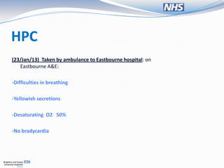
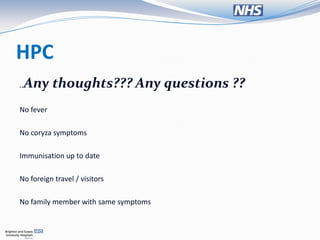
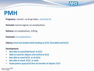
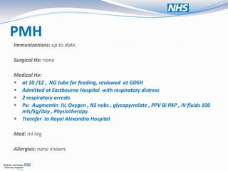
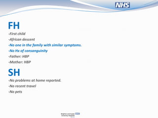
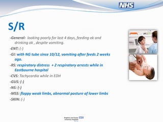
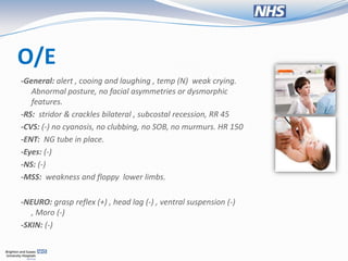
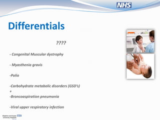
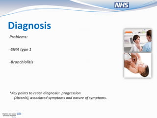
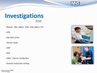
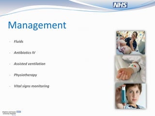
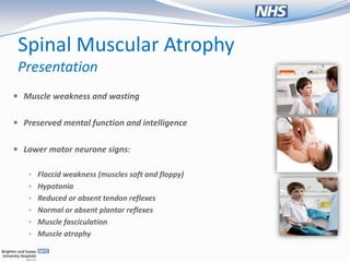
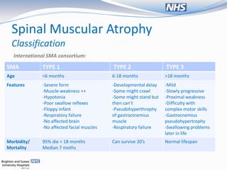
Objectives
 Case presentation.


 Brief review of the topic.


 Review of literature – ethical issues
  surrounding the case
Case presentation
• T.A. , 13/12 female

PC
 Respiratory distress + uncontrolled
  sputum production
HPC
2 weeks ago: feeding problems / vomiting after feeding + increased sputum production.
-Sputum yellow in colour and with foul smell
- GP , Px Hyoscine patch.

Next day:
-Sputum production worse and continues.


2nd day (23/Jan/13):
-Baby has an episode “funny turn”:   ???

-Perioral Cyanosis
-No jerking or limb movement
-Rolling back of eyes
-> 5 mins
-Difficulties in breathing
HPC
(23/Jan/13) Taken by ambulance to Eastbourne hospital: on
   Eastbourne A&E:

-Difficulties in breathing

-Yellowish secretions

-Desaturating O2 50%

-No bradycardia
HPC
 Any thoughts??? Any questions ??
..


No fever

No coryza symptoms

Immunisation up to date

No foreign travel / visitors

No family member with same symptoms
PMH
Pregnancy : normal , no drugs taken , antenatal ok

Perinatal: normal vaginal, no complications.

Delivery: no complications, 3.09 kg

Postnatal: no complications.

Infancy: tired and choked while feeding at 2/12. Breastfed until 6/12

Development:
 Not able to raise/hold head at 2/12
 Able to track for objects and smiled at 3/12
 Not able to crawl 6/12 or sit 6/12
 Not able to stand 9/12 or walk
 Good palmar grasp 6/12 but no transfer of objects 7/12
PMH
Immunizations: up to date.

Surgical Hx: none

Medical Hx:
 at 10 /12 , NG tube for feeding, reviewed at GOSH
 Admitted at Eastbourne Hospital with respiratory distress
 2 respiratory arrests
 Px: Augmentin IV, Oxygen , NS nebs , glycopyrrolate , PPV Bi PAP , IV fluids 100
  mls/kg/day , Physiotherapy.
 Transfer to Royal Alexandra Hospital

Med: nil reg

Allergies: none known.
FH
-First child
-African descent
-No one in the family with similar symptoms.
-No Hx of consanguinity
-Father: HBP
-Mother: HBP


SH
-No problems at home reported.
-No recent travel
-No pets
S/R
-General: looking poorly for last 4 days, feeding ok and
   drinking ok , despite vomiting.
-ENT: (-)
-GI: with NG tube since 10/12, vomiting after feeds 2 weeks
   ago.
-RS: respiratory distress + 2 respiratory arrests while in
   Eastbourne hospital
-CVS: Tachycardia while in EDH
-GUS: (-)
-NS: (-)
-MSS: floppy weak limbs, abnormal posture of lower limbs
-SKIN: (-)
O/E
-General: alert , cooing and laughing , temp (N) weak crying.
   Abnormal posture, no facial asymmetries or dysmorphic
   features.
-RS: stridor & crackles bilateral , subcostal recession, RR 45
-CVS: (-) no cyanosis, no clubbing, no SOB, no murmurs. HR 150
-ENT: NG tube in place.
-Eyes: (-)
-NS: (-)
-MSS: weakness and floppy lower limbs.

-NEURO: grasp reflex (+) , head lag (-) , ventral suspension (-)
   , Moro (-)
-SKIN: (-)
Differentials
                             ????

- Congenital Muscular dystrophy

- Myasthenia gravis

-Polio

-Carbohydrate metabolic disorders (GSD’s)
+
-Broncoaspiration pneumonia

-Viral upper respiratory infection
Diagnosis
Problems:

-SMA type 1

-Bronchiolitis


*Key points to reach diagnosis: progression
  (chronic), associated symptoms and nature of symptoms.
Investigations
                                ????
-Bloods: FBC, ABG’s, CRP, ESR, U&E’s, LFT

-CPK

-Dip stick Urine

-Throat swab

-CXR

-ECG

-EMG / Nerve conduction

-Genetic molecular testing
Management
- Fluids


- Antibiotics IV


- Assisted ventilation


- Physiotherapy


- Vital signs monitoring
Spinal Muscular Atrophy
 General points
 Genetic disease


 Causes muscle weakness and progressive loss of
  movement

 There are 4 types
Spinal Muscular Atrophy
 Presentation
 Muscle weakness and wasting


 Preserved mental function and intelligence


 Lower motor neurone signs:

    Flaccid weakness (muscles soft and floppy)
    Hypotonia
    Reduced or absent tendon reflexes
    Normal or absent plantar reflexes
    Muscle fasciculation
    Muscle atrophy
Spinal Muscular Atrophy
 Presentation
 Feeding well few weeks post-natal


 Early sign of a tiring infant that doesn't finish feeds


 Fasciculation of the tongue


 Symmetrical proximal weakness
Spinal Muscular Atrophy
  Classification
   International SMA consortium:
SMA           TYPE 1                        TYPE 2                  TYPE 3
Age           <6 months                     6-18 months             >18 months
Features      -Severe form                  -Developmental delay    -Mild
              -Muscle weakness ++           -Some might crawl       -Slowly progressive
              -Hypotonia                    -Some might stand but   -Proximal weakness
              -Poor swallow reflexes        then can’t              -Difficulty with
              -Floppy infant                -Pseudohyperthrophy     complex motor skills
              -Respiratory failure          of gastroctnemius       -Gastrocnemius
              -No affected brain            muscle                  pseudohypertrophy
              -No affected facial muscles   -Respiratory failure    -Swallowing problems
                                                                    later in life
Morbidity/    95% die < 18 months           Can survive 20’s        Normal lifespan
Mortality     Median 7 moths
Spinal Muscular Atrophy
Epidemiology
 Is the second most common lethal autosomal recessive
  disease in Caucasians1

 In the UK, carrier frequency case per 60-80,000 individuals2


 SMA type 2 most common


            1Wirth B; An update of the mutation spectrum of the survival motor neuron gene (SMN1) in autosomal
            recessive spinal muscular atrophy (SMA). Hum Mutat. 2000;15(3):228-37.
            2 Tsao B & Stojic AS; Spinal muscular atrophy. emedicine, January 2009
Spinal Muscular Atrophy
Pathophysiology

 Autosomic recessive disorder - affected individuals
  carry both.


 Mutation SMN gene on 5q13


 95% of infants type 1 SMA homozygously deleted
  for exon 7 SMN 1 gene.
Spinal Muscular Atrophy
 Pathophysiology

 Loss of this gene – loss of function in proteins for
  RNA processing

 Toxic effect on lower motor neurones


 Anterior horn cells affected


 CN affected V, VII, IX and XII
Spinal Muscular Atrophy
Differentials
 Congenital myotonic dystrophy


 Duchenne muscular dystrophy


 Disorders of carbohydrate metabolism


 Myasthenia gravis


 Polio
Spinal Muscular Atrophy
Investigations
 Bloods:
-creatinine , normal in SMA type 1

 Genetic testing:
-prenatally or postnatally
-molecular genetic testing

 Electrophysiology:
-diminished nerve signals
-helps differentiate
-sensory nerve conduction normal

 Muscle biopsy:
-atrophy of muscle
-differentiate with other neuromuscular disorders
Spinal Muscular Atrophy
Management
 No treatment / cure


 Invasive ventilation for type 1


 Multidisciplinary approach for palliative and supportive care:


-Physiotherapy
-Respiratory medicine – ventilatory support
-Dietician – NG / gastrostomy
-Neurology
-Psychological support
Spinal Muscular Atrophy
Prevention
 In families with previous child w/SMA


 Genetic diagnosis


 IVF and pre-implantation


 Transferring non-affected embryos
Spinal Muscular Atrophy
Ethical issues
 Offer ventilatory support when no current cure for the disease
  and considering quality of life?

 Any study that guides decision making in ventilatory support in
  SMA type 1 ?
Spinal Muscular Atrophy
Ethical issues
  Pediatrics. 2002 Aug;110(2 Pt 1):e24.

  Respiratory support in spinal muscular atrophy type I: a survey of
  physician
  practices and attitudes.
  Hardart MK, Burns JP, Truog RD. Department of Anesthesia and Critical Care, Children's Hospital, Harvard Medical
  School, Boston, Massachusetts


 This study suggests a wide variation not only in what is recommended but also in
  what is actually offered to families of these children.

 Study suggests that physician training and attitudes affect recommendations
  regarding mechanical ventilation and ultimately family decision making.
Spinal Muscular Atrophy
Ethical issues
  Paediatr Respir Rev. 2008 Mar;9(1):45-50;
  The use of mechanical ventilation is appropriate in children
  with genetically proven spinal muscular atrophy type 1: the motion for.
  Bach JR. Department of Physical Medicine and Rehabilitation, UMDNJ-New Jersey Medical School, University Hospital, Newark


 The purpose of this paper is to report prolongation of survival for SMA type 1 :
  trachostomy vs non-invasive ventilation

 Tracheostomy might prolong survival over 20 yrs , but patients do not develop speech
  and lose ability to breathe

 The majority of non-invasively managed SMA 1 patients develop ability to
  communicate verbally and maintain some autonomous breathing ability
Spinal Muscular Atrophy
Ethical issues
Paediatr Respir Rev. 2008 Mar;9(1):45-50;
The use of mechanical ventilation is appropriate in children
with genetically proven spinal muscular atrophy type 1: the motion for.
Bach JR. Department of Physical Medicine and Rehabilitation, UMDNJ-New Jersey Medical School, University Hospital, Newark


 Clinicians significantly underestimate the care providers' view of patient's quality of life.
  As a result, they rarely offer non-invasive means to prolong life

 Non-invasive aids & tracheostomy can prolong survival for SMA 1 patients


 Should be left up to the family to decide which, if either, they would like to use
Spinal Muscular Atrophy
Ethical issues
IN CONCLUSION..

 Should affected patients be offered ventilatory support when no
  current cure for the disease?

 Clinicians often underestimate carer’s views


 Anecdote & clinical judgement still guide doctor's decision making
  in ventilatory support in SMA type 1
References
   Tsao B & Stojic AS; Spinal muscular atrophy. emedicine, January 2009

   Wirth B; An update of the mutation spectrum of the survival motor neuron gene (SMN1) in
    autosomal recessive spinal muscular atrophy (SMA). Hum Mutat. 2000;15(3):228-37.

   Sarnat HB. Spinal muscular atrophies. In: Kliegman RM, Behrman RE, Jenson HB, Stanton BF.
    Nelson Textbook of Pediatrics. 19th ed. Philadelphia, Pa: Elsevier; 2011:chap 604.2

   Rudolf M, Lee T, Levene M. Paediatrics and Child Health. Wiley Blackwell, 2001; 3rd ed.

   Lissauer T, Clayden G. Illustrated textbook of Paediatrics. UK: Mosby Elsevier, 2007; 3rd ed.

   Tasker R, McClure R, Acerini C. Oxford handbook of Paediatrics. Oxford: Oxford University
    press, 2008.
The end

Paediatrics - Case presentation: respiratory distress + developmental delay

  • 2.
    Objectives  Case presentation. Brief review of the topic.  Review of literature – ethical issues surrounding the case
  • 3.
    Case presentation • T.A., 13/12 female PC  Respiratory distress + uncontrolled sputum production
  • 4.
    HPC 2 weeks ago:feeding problems / vomiting after feeding + increased sputum production. -Sputum yellow in colour and with foul smell - GP , Px Hyoscine patch. Next day: -Sputum production worse and continues. 2nd day (23/Jan/13): -Baby has an episode “funny turn”: ??? -Perioral Cyanosis -No jerking or limb movement -Rolling back of eyes -> 5 mins -Difficulties in breathing
  • 5.
    HPC (23/Jan/13) Taken byambulance to Eastbourne hospital: on Eastbourne A&E: -Difficulties in breathing -Yellowish secretions -Desaturating O2 50% -No bradycardia
  • 6.
    HPC Any thoughts???Any questions ?? .. No fever No coryza symptoms Immunisation up to date No foreign travel / visitors No family member with same symptoms
  • 7.
    PMH Pregnancy : normal, no drugs taken , antenatal ok Perinatal: normal vaginal, no complications. Delivery: no complications, 3.09 kg Postnatal: no complications. Infancy: tired and choked while feeding at 2/12. Breastfed until 6/12 Development:  Not able to raise/hold head at 2/12  Able to track for objects and smiled at 3/12  Not able to crawl 6/12 or sit 6/12  Not able to stand 9/12 or walk  Good palmar grasp 6/12 but no transfer of objects 7/12
  • 8.
    PMH Immunizations: up todate. Surgical Hx: none Medical Hx:  at 10 /12 , NG tube for feeding, reviewed at GOSH  Admitted at Eastbourne Hospital with respiratory distress  2 respiratory arrests  Px: Augmentin IV, Oxygen , NS nebs , glycopyrrolate , PPV Bi PAP , IV fluids 100 mls/kg/day , Physiotherapy.  Transfer to Royal Alexandra Hospital Med: nil reg Allergies: none known.
  • 9.
    FH -First child -African descent -Noone in the family with similar symptoms. -No Hx of consanguinity -Father: HBP -Mother: HBP SH -No problems at home reported. -No recent travel -No pets
  • 10.
    S/R -General: looking poorlyfor last 4 days, feeding ok and drinking ok , despite vomiting. -ENT: (-) -GI: with NG tube since 10/12, vomiting after feeds 2 weeks ago. -RS: respiratory distress + 2 respiratory arrests while in Eastbourne hospital -CVS: Tachycardia while in EDH -GUS: (-) -NS: (-) -MSS: floppy weak limbs, abnormal posture of lower limbs -SKIN: (-)
  • 11.
    O/E -General: alert ,cooing and laughing , temp (N) weak crying. Abnormal posture, no facial asymmetries or dysmorphic features. -RS: stridor & crackles bilateral , subcostal recession, RR 45 -CVS: (-) no cyanosis, no clubbing, no SOB, no murmurs. HR 150 -ENT: NG tube in place. -Eyes: (-) -NS: (-) -MSS: weakness and floppy lower limbs. -NEURO: grasp reflex (+) , head lag (-) , ventral suspension (-) , Moro (-) -SKIN: (-)
  • 12.
    Differentials ???? - Congenital Muscular dystrophy - Myasthenia gravis -Polio -Carbohydrate metabolic disorders (GSD’s) + -Broncoaspiration pneumonia -Viral upper respiratory infection
  • 13.
    Diagnosis Problems: -SMA type 1 -Bronchiolitis *Keypoints to reach diagnosis: progression (chronic), associated symptoms and nature of symptoms.
  • 14.
    Investigations ???? -Bloods: FBC, ABG’s, CRP, ESR, U&E’s, LFT -CPK -Dip stick Urine -Throat swab -CXR -ECG -EMG / Nerve conduction -Genetic molecular testing
  • 15.
    Management - Fluids - AntibioticsIV - Assisted ventilation - Physiotherapy - Vital signs monitoring
  • 16.
    Spinal Muscular Atrophy General points  Genetic disease  Causes muscle weakness and progressive loss of movement  There are 4 types
  • 17.
    Spinal Muscular Atrophy Presentation  Muscle weakness and wasting  Preserved mental function and intelligence  Lower motor neurone signs:  Flaccid weakness (muscles soft and floppy)  Hypotonia  Reduced or absent tendon reflexes  Normal or absent plantar reflexes  Muscle fasciculation  Muscle atrophy
  • 18.
    Spinal Muscular Atrophy Presentation  Feeding well few weeks post-natal  Early sign of a tiring infant that doesn't finish feeds  Fasciculation of the tongue  Symmetrical proximal weakness
  • 19.
    Spinal Muscular Atrophy Classification International SMA consortium: SMA TYPE 1 TYPE 2 TYPE 3 Age <6 months 6-18 months >18 months Features -Severe form -Developmental delay -Mild -Muscle weakness ++ -Some might crawl -Slowly progressive -Hypotonia -Some might stand but -Proximal weakness -Poor swallow reflexes then can’t -Difficulty with -Floppy infant -Pseudohyperthrophy complex motor skills -Respiratory failure of gastroctnemius -Gastrocnemius -No affected brain muscle pseudohypertrophy -No affected facial muscles -Respiratory failure -Swallowing problems later in life Morbidity/ 95% die < 18 months Can survive 20’s Normal lifespan Mortality Median 7 moths
  • 20.
    Spinal Muscular Atrophy Epidemiology Is the second most common lethal autosomal recessive disease in Caucasians1  In the UK, carrier frequency case per 60-80,000 individuals2  SMA type 2 most common 1Wirth B; An update of the mutation spectrum of the survival motor neuron gene (SMN1) in autosomal recessive spinal muscular atrophy (SMA). Hum Mutat. 2000;15(3):228-37. 2 Tsao B & Stojic AS; Spinal muscular atrophy. emedicine, January 2009
  • 21.
    Spinal Muscular Atrophy Pathophysiology Autosomic recessive disorder - affected individuals carry both.  Mutation SMN gene on 5q13  95% of infants type 1 SMA homozygously deleted for exon 7 SMN 1 gene.
  • 22.
    Spinal Muscular Atrophy Pathophysiology  Loss of this gene – loss of function in proteins for RNA processing  Toxic effect on lower motor neurones  Anterior horn cells affected  CN affected V, VII, IX and XII
  • 23.
    Spinal Muscular Atrophy Differentials Congenital myotonic dystrophy  Duchenne muscular dystrophy  Disorders of carbohydrate metabolism  Myasthenia gravis  Polio
  • 24.
    Spinal Muscular Atrophy Investigations Bloods: -creatinine , normal in SMA type 1  Genetic testing: -prenatally or postnatally -molecular genetic testing  Electrophysiology: -diminished nerve signals -helps differentiate -sensory nerve conduction normal  Muscle biopsy: -atrophy of muscle -differentiate with other neuromuscular disorders
  • 25.
    Spinal Muscular Atrophy Management No treatment / cure  Invasive ventilation for type 1  Multidisciplinary approach for palliative and supportive care: -Physiotherapy -Respiratory medicine – ventilatory support -Dietician – NG / gastrostomy -Neurology -Psychological support
  • 26.
    Spinal Muscular Atrophy Prevention In families with previous child w/SMA  Genetic diagnosis  IVF and pre-implantation  Transferring non-affected embryos
  • 27.
    Spinal Muscular Atrophy Ethicalissues  Offer ventilatory support when no current cure for the disease and considering quality of life?  Any study that guides decision making in ventilatory support in SMA type 1 ?
  • 28.
    Spinal Muscular Atrophy Ethicalissues Pediatrics. 2002 Aug;110(2 Pt 1):e24. Respiratory support in spinal muscular atrophy type I: a survey of physician practices and attitudes. Hardart MK, Burns JP, Truog RD. Department of Anesthesia and Critical Care, Children's Hospital, Harvard Medical School, Boston, Massachusetts  This study suggests a wide variation not only in what is recommended but also in what is actually offered to families of these children.  Study suggests that physician training and attitudes affect recommendations regarding mechanical ventilation and ultimately family decision making.
  • 29.
    Spinal Muscular Atrophy Ethicalissues Paediatr Respir Rev. 2008 Mar;9(1):45-50; The use of mechanical ventilation is appropriate in children with genetically proven spinal muscular atrophy type 1: the motion for. Bach JR. Department of Physical Medicine and Rehabilitation, UMDNJ-New Jersey Medical School, University Hospital, Newark  The purpose of this paper is to report prolongation of survival for SMA type 1 : trachostomy vs non-invasive ventilation  Tracheostomy might prolong survival over 20 yrs , but patients do not develop speech and lose ability to breathe  The majority of non-invasively managed SMA 1 patients develop ability to communicate verbally and maintain some autonomous breathing ability
  • 30.
    Spinal Muscular Atrophy Ethicalissues Paediatr Respir Rev. 2008 Mar;9(1):45-50; The use of mechanical ventilation is appropriate in children with genetically proven spinal muscular atrophy type 1: the motion for. Bach JR. Department of Physical Medicine and Rehabilitation, UMDNJ-New Jersey Medical School, University Hospital, Newark  Clinicians significantly underestimate the care providers' view of patient's quality of life. As a result, they rarely offer non-invasive means to prolong life  Non-invasive aids & tracheostomy can prolong survival for SMA 1 patients  Should be left up to the family to decide which, if either, they would like to use
  • 31.
    Spinal Muscular Atrophy Ethicalissues IN CONCLUSION..  Should affected patients be offered ventilatory support when no current cure for the disease?  Clinicians often underestimate carer’s views  Anecdote & clinical judgement still guide doctor's decision making in ventilatory support in SMA type 1
  • 32.
    References  Tsao B & Stojic AS; Spinal muscular atrophy. emedicine, January 2009  Wirth B; An update of the mutation spectrum of the survival motor neuron gene (SMN1) in autosomal recessive spinal muscular atrophy (SMA). Hum Mutat. 2000;15(3):228-37.  Sarnat HB. Spinal muscular atrophies. In: Kliegman RM, Behrman RE, Jenson HB, Stanton BF. Nelson Textbook of Pediatrics. 19th ed. Philadelphia, Pa: Elsevier; 2011:chap 604.2  Rudolf M, Lee T, Levene M. Paediatrics and Child Health. Wiley Blackwell, 2001; 3rd ed.  Lissauer T, Clayden G. Illustrated textbook of Paediatrics. UK: Mosby Elsevier, 2007; 3rd ed.  Tasker R, McClure R, Acerini C. Oxford handbook of Paediatrics. Oxford: Oxford University press, 2008.
  • 33.