The document summarizes key information about the phylum Basidiomycota. Some key points:
- Basidiomycota includes about 30% of all fungi and has over 15,000 species, including mushrooms, puffballs, rusts, and smuts. Most are saprophytic.
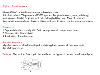
- They have septate mycelium usually with dolipore septa and clamp connections. They undergo a dikaryophase and produce basidiospores.
- Asexual reproduction occurs via fragmentation, budding, or sporidia. Sexual reproduction involves the dikaryophase and plasmogamy between compatible nuclei, ending with karyogamy and meiosis in the bas