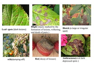
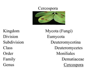
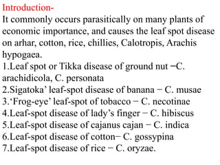
1. Cercospora is a genus of fungi that commonly causes leaf spot diseases on many economically important plants.
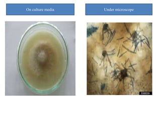
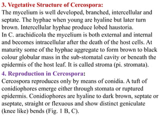
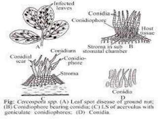
2. It reproduces asexually through the production of conidiophores and conidia. Conidiophores emerge through stomata or ruptured leaf epidermis and produce single conidia at their tips.
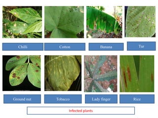
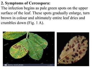
3. Cercospora species can cause significant damage and yield loss to crops like groundnuts, banana, tobacco, and cotton through the leaf spot diseases they produce.


![PAT201_PPT[1].pptx fundamentals of plant pathology](https://cdn.slidesharecdn.com/ss_thumbnails/pat201ppt1-250924180330-8bd732e5-thumbnail.jpg?width=640&height=640&fit=bounds)


