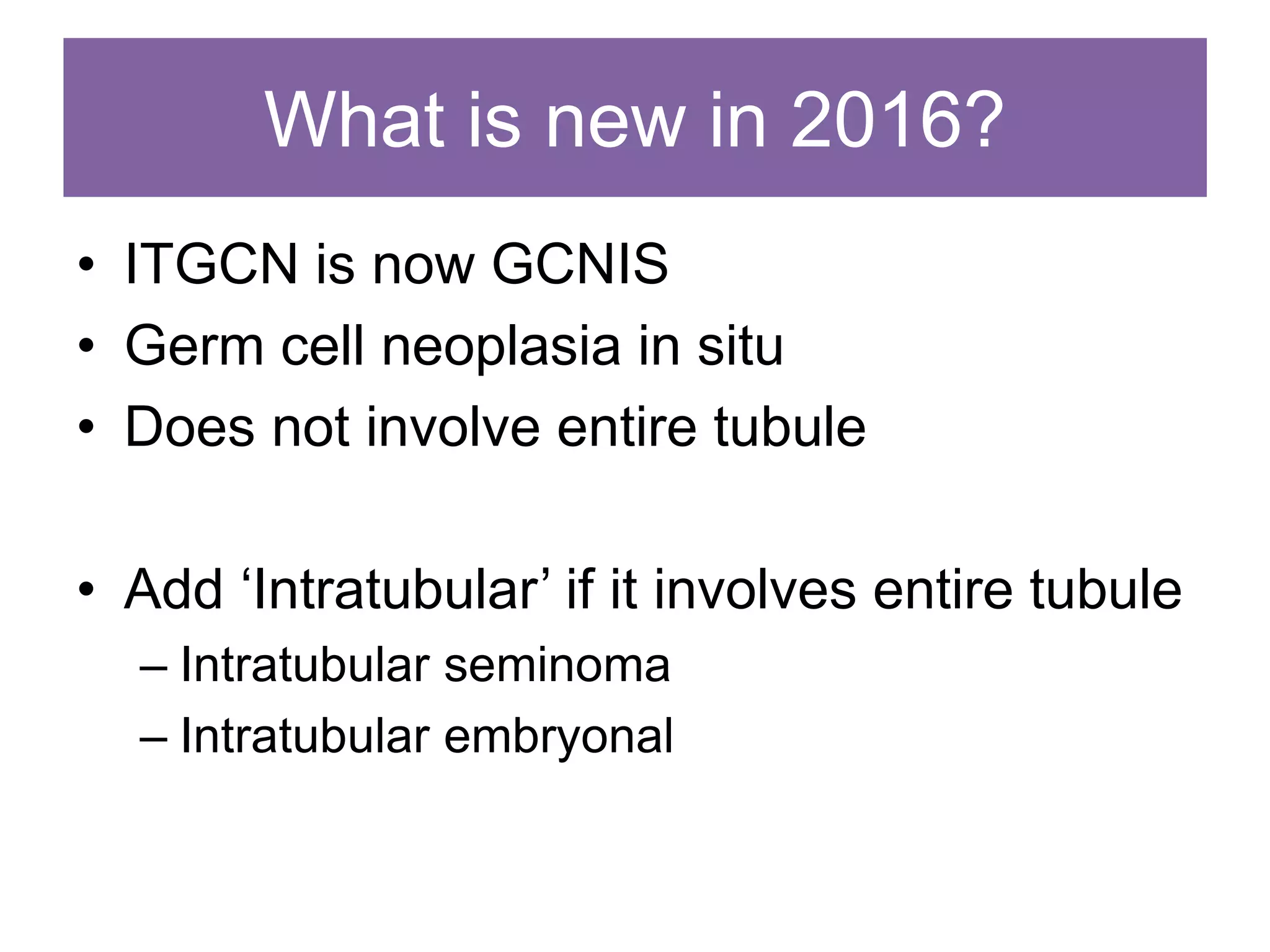
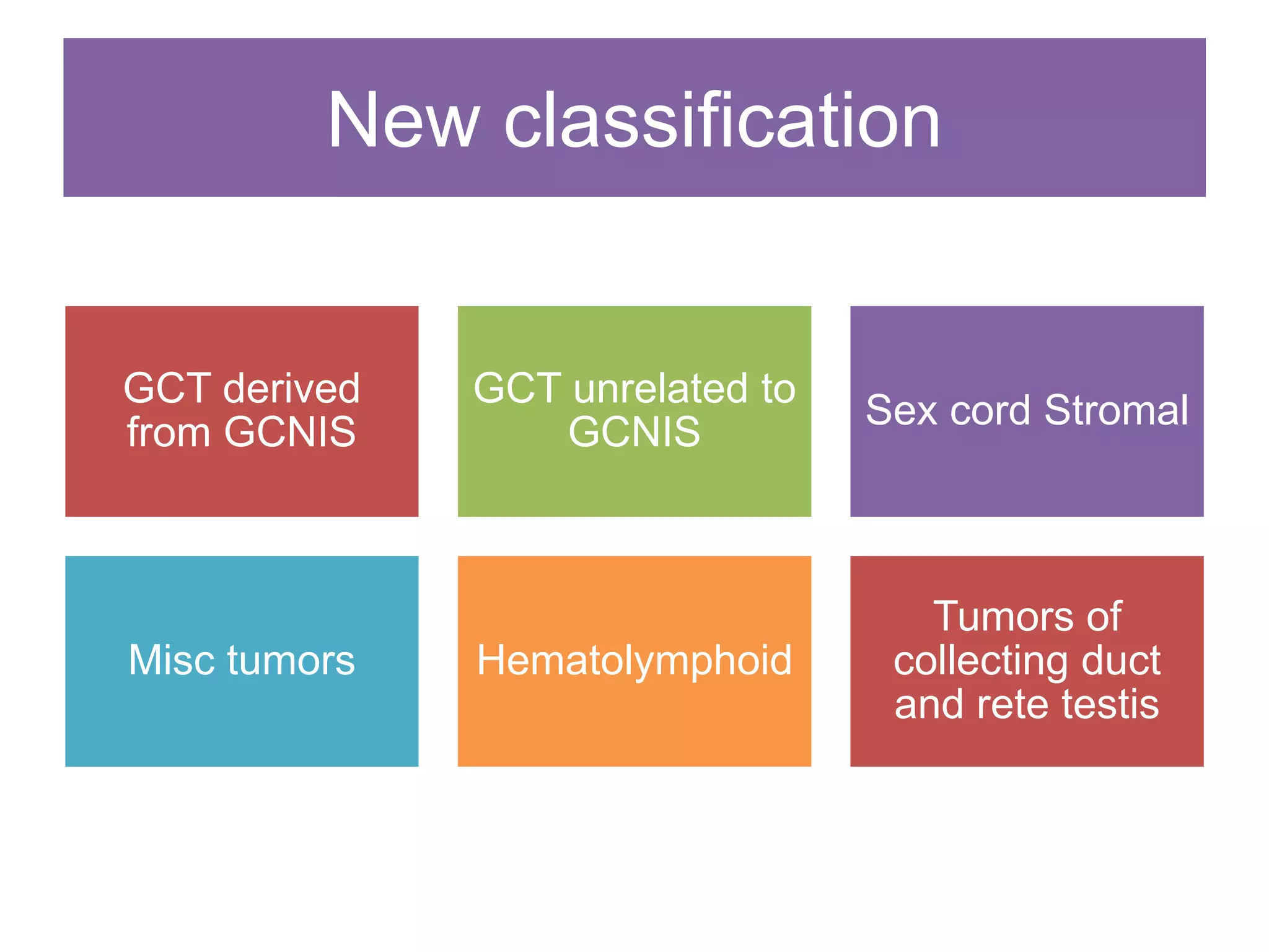
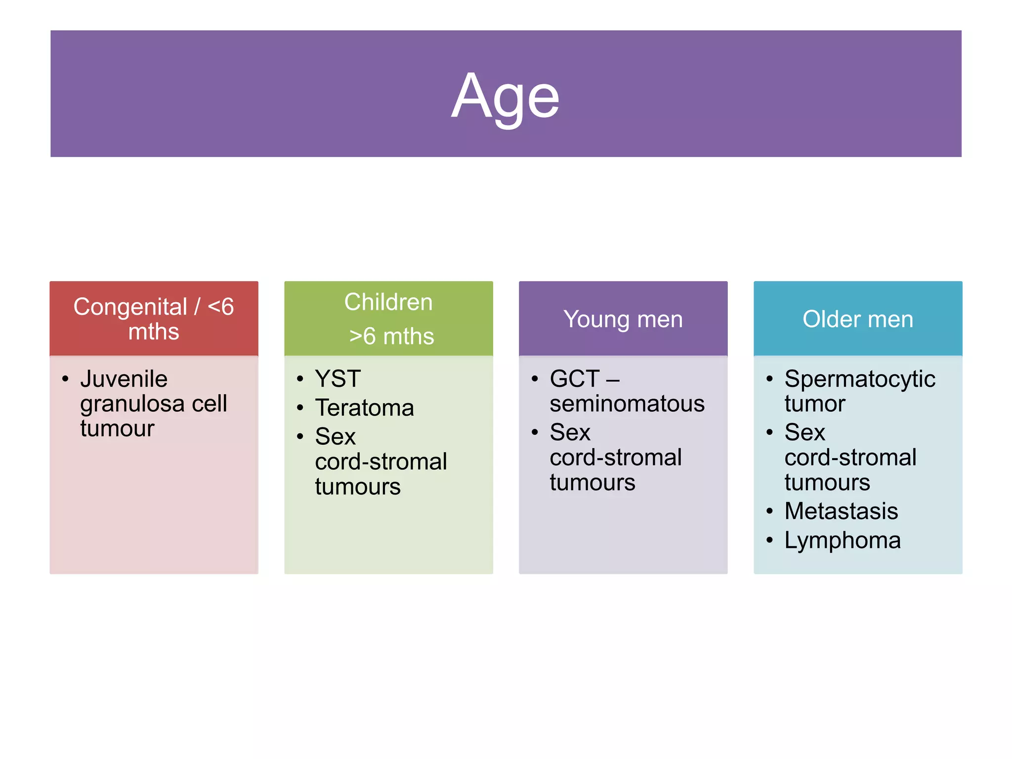
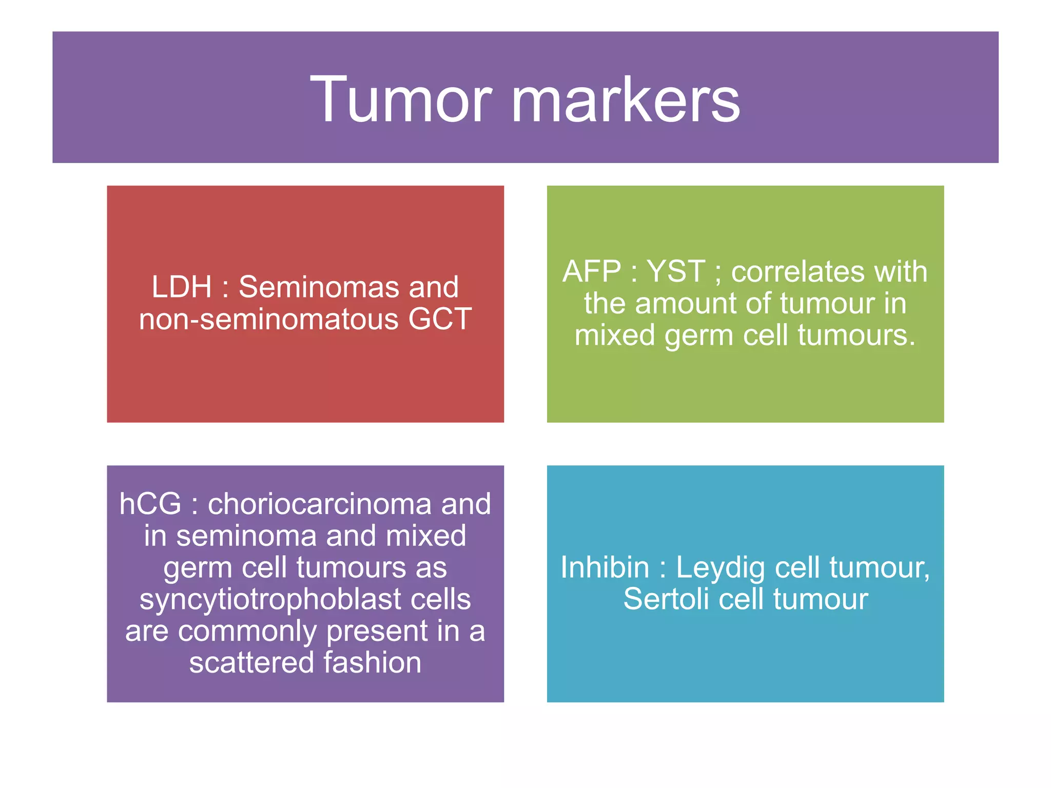
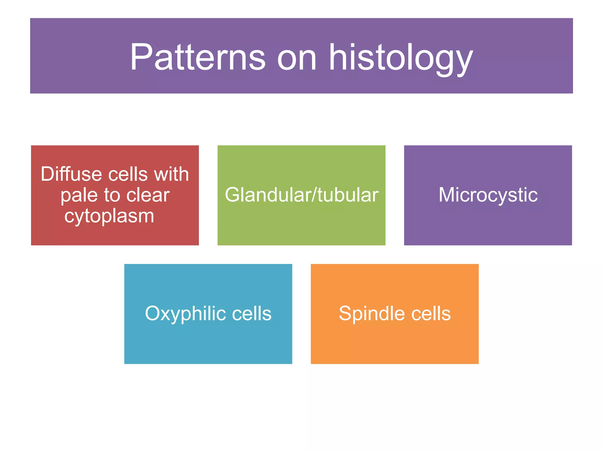
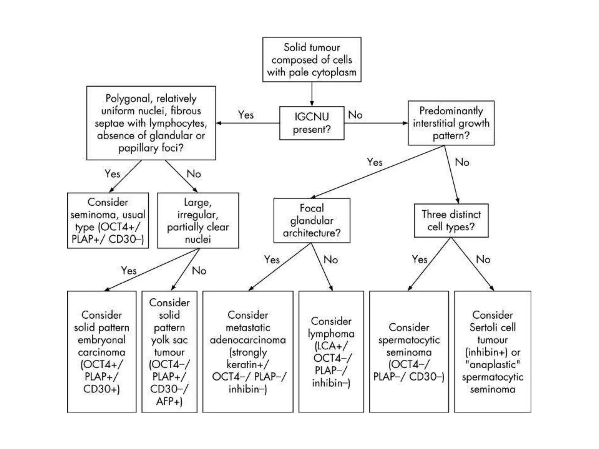
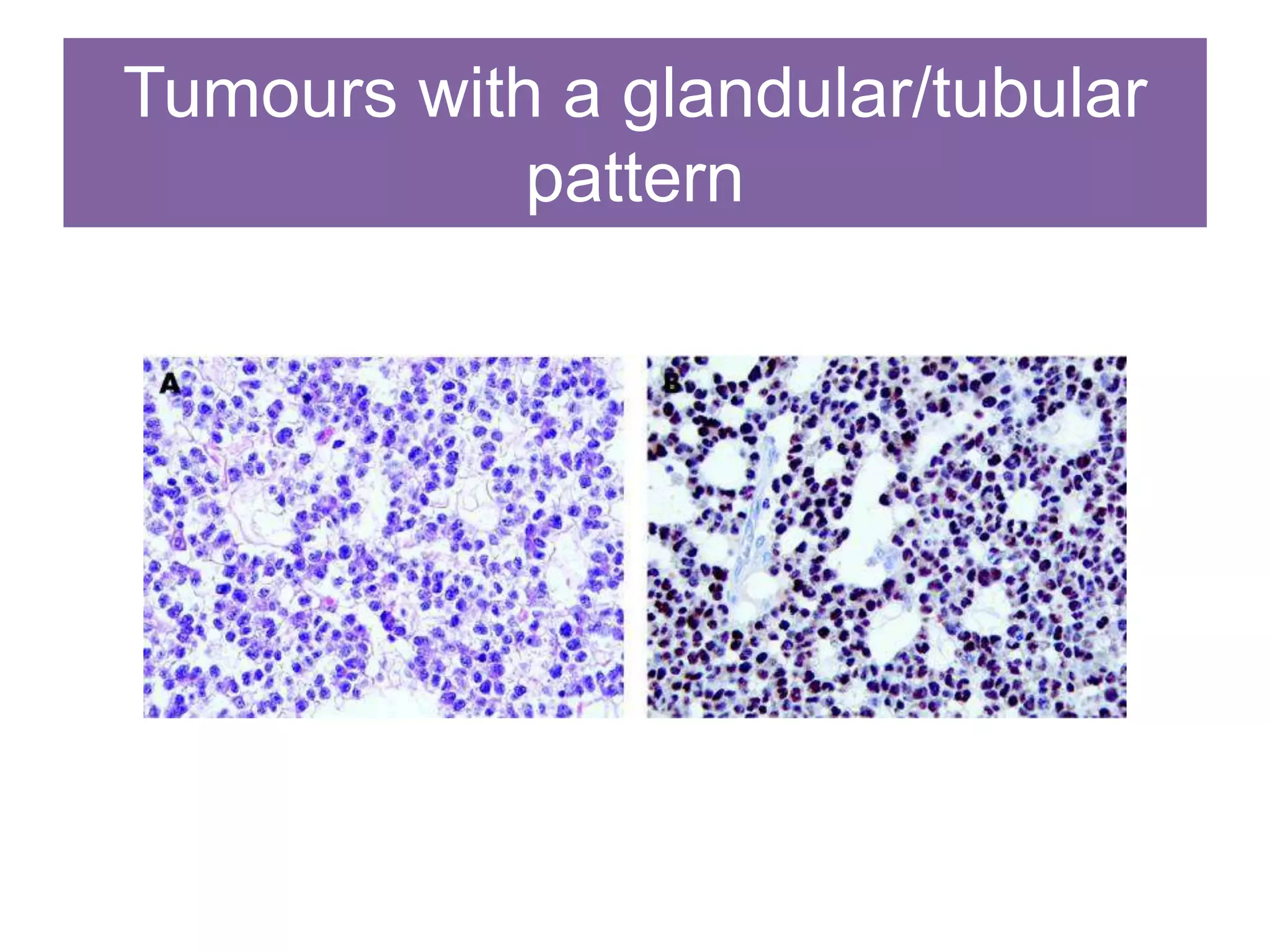
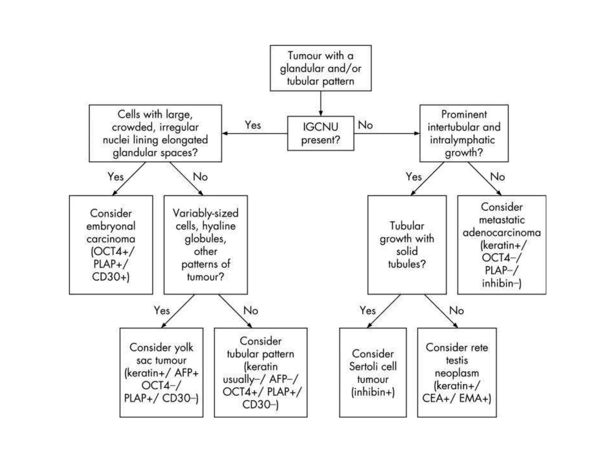
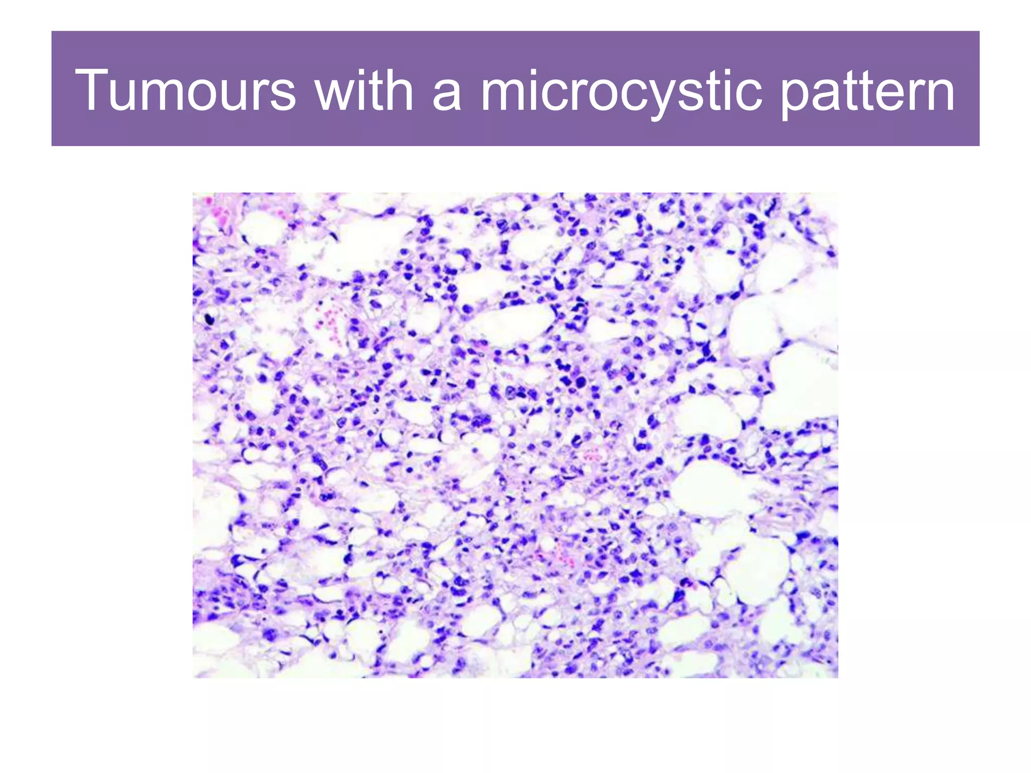
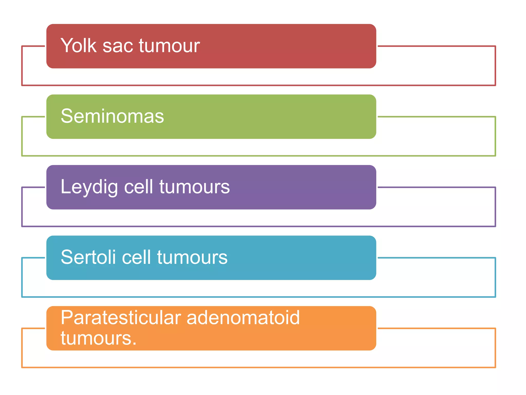
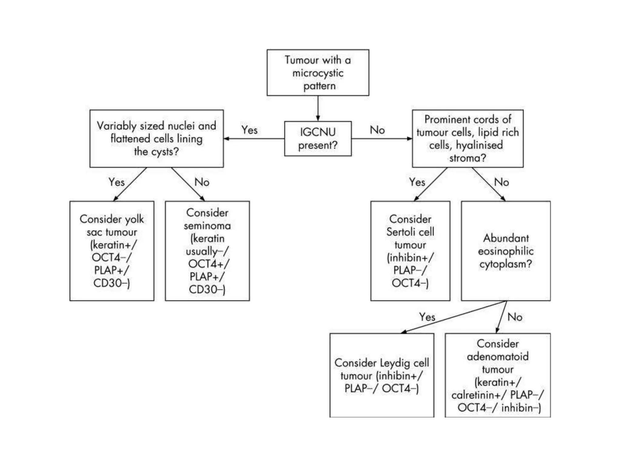
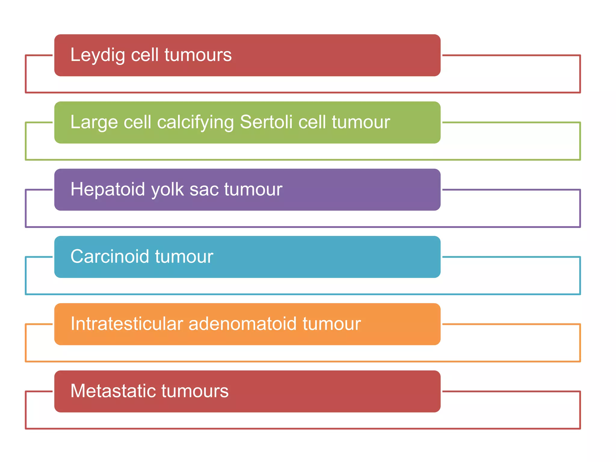
Approach to Testicular Tumors
What is new in 2016?
• ITGCN is now GCNIS
• Germ cell neoplasia in situ
• Does not involve entire tubule
• Add ‘Intratubular’ if it involves entire tubule
– Intratubular seminoma
– Intratubular embryonal
New classification
GCT derived
from GCNIS
GCT unrelated to
GCNIS
Sex cord Stromal
Misc tumors Hematolymphoid
Tumors of
collecting duct
and rete testis
Most neoplastic scrotal masses
ultimately prove to be germ cell
tumours and are recognisable
with routine haematoxylin and
eosin‐stained sections
Age
Medical
history
Tumour site
(testicular vs
paratesticular)
Gross
Before we see the slide !
Age
Congenital / <6
mths
• Juvenile
granulosa cell
tumour
Children
>6 mths
• YST
• Teratoma
• Sex
cord‐stromal
tumours
Young men
• GCT –
seminomatous
• Sex
cord‐stromal
tumours
Older men
• Spermatocytic
tumor
• Sex
cord‐stromal
tumours
• Metastasis
• Lymphoma
History
Undescended testis
Previous diagnosis of a germ
cell tumour or GCNIS or IT
GCT
Possibility of a GCT very high
Previous or current
carcinoma, lymphoma or
leukaemia
Likely secondary tumour
Gross
Seminomas
• Nodules of
homogenous white
or tan tissue
Non‐seminomatous
germ cell tumours
• Zones of
haemorrhage or
necrosis
Teratoma
• Cystic
• Tooth
• Cartilage
Sex cord stromal
• Yellow or tan
Tumor markers
LDH : Seminomas and
non‐seminomatous GCT
AFP : YST ; correlates with
the amount of tumour in
mixed germ cell tumours.
hCG : choriocarcinoma and
in seminoma and mixed
germ cell tumours as
syncytiotrophoblast cells
are commonly present in a
scattered fashion
Inhibin : Leydig cell tumour,
Sertoli cell tumour
Hormones
Small minority
Rare androgen
excess or
gynaecomastia
resulting from hCG
hCG also has thyroid
stimulating activity -
hyperthyroidism
Hypercalcaemia:
parathyroid
hormone‐like
substance –
seminoma
Steroid producing :
leydig –
Pseudopuberty
Gynecomastia : Sex
cord stromal
Patterns on histology
Diffuse cells with
pale to clear
cytoplasm
Glandular/tubular Microcystic
Oxyphilic cells Spindle cells
Cells with pale to clear cytoplasm
in a diffuse arrangement
Seminoma
Spermatocytic tumor
Solid variant of embryonal carcinoma
Solid pattern yolk sac tumour
Sertoli cell tumour and Leydig cell tumour
Metastases
Lymphoma
Tumours with a glandular/tubular
pattern
Embryonal carcinoma and yolk sac tumour
Occasional cases of seminoma
Sertoli cell tumours usually have tubular
architecture in at least some foci of the tumour
Rete testis
Metastases
Tumours with a microcystic pattern
Yolk sac tumour
Seminomas
Leydig cell tumours
Sertoli cell tumours
Paratesticular adenomatoid
tumours.
Oxyphilic tumours
Leydig cell tumours
Large cell calcifying Sertoli cell tumour
Hepatoid yolk sac tumour
Carcinoid tumour
Intratesticular adenomatoid tumour
Metastatic tumours
Tumours in children
Yolk sac tumour
Juvenile granulosa
cell tumour
Intratubular atypical cells
Paratesticular tumours with a glandular‐like,
tubular or papillary pattern
Metastatic adenocarcinoma
Mullerian‐type epithelial
tumours
Adenomatoid tumour
Mesothelioma
Tumours with spindle cell morphology
Leydig cell tumours
Gonadal stromal fibromas
Malignant mesotheliomas
Rhabdomyosarcoma
Renike crystals
Hyaline globules
Schiller Duval bodies
Call Exner bodies
Synchiotrophoblasts
Summarise
Thank You

Approach to testicular tumors