
Recommended
Recommended
More Related Content
What's hot
What's hot (20)
Similar to Parturition in domestic animals.pptx
Similar to Parturition in domestic animals.pptx (20)
Recently uploaded
Recently uploaded (20)
Parturition in domestic animals.pptx
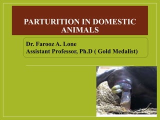
- 1. PARTURITION IN DOMESTIC ANIMALS Dr. Farooz A. Lone Assistant Professor, Ph.D ( Gold Medalist)
- 2. Parturition: Delivery of fetus through the birth canal on completion of gestation period. or Physiological process by which the pregnant uterus delivers the fetus and placenta from the maternal animal. The muscular canal that forms from the uterus to the outside of the body through which the fetus passes during birth. Birth canal is formed by the uterus, cervix and vagina within the pelvic bones and their attachments.
- 3. Symptoms of approaching parturition Cow, Ewe, Goat Separation from herd increased walking/searching Increased transition from standing to lying and vice versa Licking own body and attention toward abdomen Scraping or pawing the ground Less feeding behaviour Relaxed ligaments 24 - 48 h prepartum raised tail head frequent urination restlessness Bitch Vomition, Drop in 1-2°C rectal temp 24 h prepartum Nesting behavior. Mare……………….Sweating, Waxing of teats, Restlessness
- 4. Symptoms of approaching parturition
- 5. Stages of parturition Stages Stage 1: cervical dilatation Stage 3: Expulsion of fetus Stage 2: Delivery of the fetus
- 6. 1st Stage: Cervical dilatation • Changes not visible externally • Preparation of birth canal and foetus for expulsion • Signs of discomfort, mild colic, restlessness with elevated pulse and respiratory rate, body temperature falls • Structure of cervix changes • Onset of myometrial contractions • Foetus assumes the disposition for expulsion
- 7. Cervical Changes • Loosening of the ground substance due to changes in the composition of collagen components. • Increased incorporation of water which permits collagen fibres to separate under extension forces. • Cervix dilates: external os opening before internal os and becomes cone shaped due to wide dilation of external os. • Simultaneous shortening and internal os dilation. • Vagina and uterus form continuous canal that becomes tightly engaged by the distended allantochorion
- 9. Onset of myometrial contractions Cervi-cotubular contractions prevent the premature displacement of fetuses, thus ensuring orderly expulsion from the horns Isolated ,uncoordinated waves changes to regular and coordinated peristaltic type. Frequency increases from 12-24 per hour in last 2 hours to 48 per hour just before expulsion (30 per hour in ewe). Presence of cervico-tubular and tubular-cervical contractions. Placental attachment becomes less intimate. Superficial cells undergo fatty degeneration .
- 10. Fetal orientation/disposition • Separation of margins with haemorrhage in deciduate placenta. • Becomes more active and disposes itself. • Progressive rotation from ventral to dorsal position and fore limbs, head and neck extended in foal and puppy. • In calf and lamb extension only. • Flexed knees of calf first occupy dilating cervix; 30 minutes later digits are in cervix and it extends carpal joints in its efforts to ‘stand up in utero’. .
- 12. Duration . Animal 1st Stage (hrs) Cow, Buffalo 2-6 Mare 1-4 Ewe 2-6 Sow 2-12 Camel 2-7 Bitch 12
- 13. 2nd. Stage: Delivery of fetus . • Refers to expulsion of foetus • In polytocous species stage cannot be separated from 3rd stage. Sign: • Appearance of abdominal contractions, superimposed upon onset of each myometrial contractions. • Disappearance of cervico-tubular contractions. • Ferguson’s reflex • Allanto-chorionic sac ruptures and gush of urine like fluid escapes from vulva. • Amnion traverses vagina and appears at vulva as ‘water-bag’ with foetal limbs. • Foetal head next occupies vulva, contractions of uterine and abdominal muscles reach climax of expulsive effort, maximum effort coinciding with the birth of the foetal occiput. • Further straining causes foetal thorax to pass through vulva. • Usually, birth of hips quickly follows and hind limbs may be expelled.
- 14. . • Foetus is born in amnion and quick movement causes its rupture; respirations, then begin. • In mare, cow and ewe (when monotocous) foetus is usually delivered in anterior presentation, dorsal position and extended posture. • In polytocous bitch and sow up to 40–45% of foetuses may be normally delivered in posterior presentation. • Duration Animal 2nd Stage Cow, Buffalo 70 min Mare 17 min Ewe 1 hr Sow 4 hrs Camel 30 min Bitch 6-12 hrs
- 15. . 3rd. Stage: Expulsion of fetal membranes • After 2nd stage, regular abdominal contractions largely • cease. (Gillette and Holm, 1963) • Myometrial contractions persist; decrease in amplitude. • These contractions are important for dehiscence and expulsion of fetal membranes. • Waves of contractions passing from uterine tube to cervix persist, but in cow and sow reappearance of contractions in reverse direction. • Weakening of acellular layer of adhesive protein, ‘glue line’ between cotyledonary and caruncular epithelium, is probably important in ensuring placental separation. • In last 5 days of gestation collagenisation of placentome and flattening of maternal crypt epithelium in cow. • Foetal villi shrink, owing mainly to sudden loss of turgidity related to escape of blood from foetal side of placenta when umbilical cord ruptures.
- 16. . • Early degenerative or maturational changes which are seen in caruncles of ewe and cow, cause separation of fetal membrane. • Apex of allanto-chorionic sac becomes inverted and as sac is ‘rolled’ down cornua the fetal villi are drawn out of crypts. • This forms a mass within maternal pelvis which stimulates reflex contractions of abdominal muscles leads to expulsion of fetal membranes. • Domestic animals normally eat afterbirth except Mare. • In polytocous species, dehiscence and expulsion of fetal membranes are interspersed with fetal births. • Stimulus of suckling causes release of oxytocin, which promotes ‘letdown’ of milk and augmentation of myometrial contractions. • Suckling resulted in greater synchrony of contractions and increase in number of tubo-cervical contractions. • Suckling exerts a favourable influence on expulsion of afterbirth.
- 17. . Animal 3rd Stage Cow, Buffalo 6-12 hrs Mare 30min-3hrs Ewe 2-3 hrs Sow 4 hrs Camel 1 hr Bitch Along with fetus or shortly
- 18. . Cow
- 19. Theories of parturition initiation Physical factors Increase in fetal size: this increases uterine irritability Uterine distension : reversal of progesterone block Fatty degeneration of placenta and presence of infarcts: leads to interference in fetal nutrition Biochemical factors: Increase in CO2 tension in maternal blood due to increased fetal activity…………………..this ↑uterine contractility Release of fetal antigens: →serotonin →release of collagenase and stoppage of blood supply to cotyledons.
- 20. Neuroendocrine factors: Fetal factors •↑ in CRH in hypothalamus →stimulate ACTH •↑ in ACTH by pituitary →stimulate cortisol release •↑ in cortisol from adrenals→ convert P4 to E2 & release of PG
- 21. Maternal factors Reversal of P4 block → ↑ myometrial contractility Release of relaxin → dilation of the birth canal Placental estrogen rise →release of PG ↑ myometrial contractility Pro-inflammatory cytokines → dilation of the birth canal Release of PG → softening of cervix, contractions ↑ Release of Oxytocin → ↑ myometrial contractions
- 22. Stage 1 (Initiation of Parturition) Stage 1 (Initiation of Parturition) Fetal Stress Fetal Stress Due increase in size and limited space Fetal Adrenal Gland Fetal Adrenal Gland Corticoids (cortisol) Corticoids (cortisol) Corticoids (cortisol) Corticoids (cortisol) Release of pituitary ACTH (adreno-corticotropic hormone) Release of pituitary ACTH (adreno-corticotropic hormone) 1) Removal of progesterone block 2) Elevation of repro. tract secretion 1) Removal of progesterone block 2) Elevation of repro. tract secretion
- 23. Removal of Progesterone Block Removal of Progesterone Block How does progesterone secretion is inhibited? Elevated cortisol promotes the synthesis of 3 enzymes These 3 enzymes convert progesterone to estradiol Elevated cortisol promotes the synthesis of 3 enzymes These 3 enzymes convert progesterone to estradiol 17 hydoxylase 17 hydoxylase 17-20 lyase 17-20 lyase Aromatase Aromatase
- 24. Pregnenolone (C21) Estrogen (C18) Progesterone (C21) Testosterone (C19)
- 25. Fetal hypothalamus ↓ Fetal Pituitary ↓ ACTH ↓ Fetal adrenal ↓ Adrenal corticosteroids ↓ Convert progesterone to estrogen ← Feto-placental estrogens → Relaxin ↓ ↓ → →→ → →→→ → →→↓ Cervical softening ← ← ← ← ↓ Cotyledons / Myometrium ↑ Proinflamatory cytokines ↓ Luteolysis ← Release of PGF →oxytocin ↓ ↓ ↓ ↓ Decrease in serum progesterone ↓ ↓→ ↓ ↑ Abdominal contractions Myometrial contractions → Fetal Expulsion ↑ Oxytocin ↑ Posterior Pituitary Possible endocrine changes that occur during the peri-parturient period in the sow, ewe, cow and their effects
- 27. Parturition Fetal Nutritional Demands Placental Insufficiency Hypothalamus Anterior Pituitary Adrenal Cortex CRH ACTH Fetal Corticosteroids (Cortisol) Lung (surfactant) Liver (glycogen) Thyroid (metabolism) Progesterone Estrogen PGF2a Uterine Contractions PGF2a Relaxin Oxytocin Uterine Endometrium » Oxytocin receptors Ovary (CL) Cow, Sow Triggers CL Regression Placentome Cervical Ripening
- 28. Final Role of Oxytocin Ferguson’s reflex Sensory Neurons in Cervix Oxytocin from Posterior Pituitary Myometrial Contractions
- 29. Orientation of Fetus •Fetus must reorient prior to parturition •It occurs during first stage of parturition •Initially on back •Reorient so that feet and head will exit first •Breech ………..Rear of fetus comes first • Orientation not important in pig •Abnormal orientation results in dystocia
- 32. Prepartum fetal changes: •Changes do occur in the fetus before delivery and these are essential because the fetus has to prepare itself for the external environment outside the uterus. The changes include •Maturation of fetal lungs: The surfactants in the lungs increase which reduce the surface tension within the alveoli. •Increased output of tri-iodothyronine and catecholamines to meet the increased metabolic demands. •Closure of the ductus arteriosus and the closure of the foramen ovale. •Increased glycogen reserves in the liver to meet the demands •on delivery by the production of glucose as a source of energy post delivery.
- 33. Sequence of parturition events in the cow
- 34. Parturition in the buffalo
- 35. 1 2 2 2 3
- 36. Stages of labor in a goat 1 2 3 • The first water bag(allantochorion) is protruded • The second water bag (amnion) and the fetus are protruding through the vulvar lips • The placenta is being dropped in the third stage of labor