Clinical Cases in Cardiology  Dr. Ihab Suliman  http://clinicalcases.org/2005/07/cardiology-cases.html
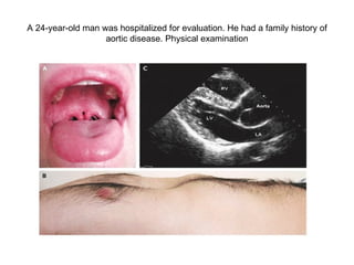
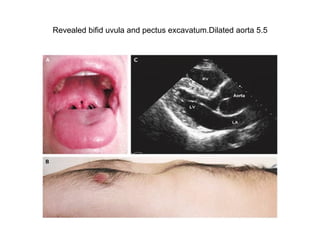
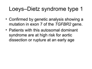
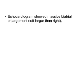
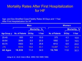
A 24-year-old man was hospitalized for evaluation. He had a family history of aortic disease. Physical examination
Revealed bifid uvula and pectus excavatum.Dilated aorta 5.5
Loeys–Dietz syndrome type 1 Confirmed by genetic analysis showing a mutation in exon 7 of the  TGFBR2  gene.  Patients with this autosomal dominant syndrome are at high risk for aortic dissection or rupture at an early age
 
Pectus Excavatum Pectus excavatum  is present at birth or within the 1st year of life in most patients. It is reported 1/( 300 to 400) of lives birth and rare in blacks. M:F=4:1 Etiology is unknown. Associated withScoliosis,Kyphosis,Myopathy,Marfan’s syndrome,Cerebral palsy,Prune-belly syndrome,Tuberous sclerosis
A 59-year-old man with a medical history of hypertension, hyperlipidemia, and coronary artery disease presented with transient, painless visual obscuration in the left eye,
he had undergone placement of a stent in the left carotid artery for severe stenosis   Retinal examination of the left eye showed multiple, tiny refractile retinal arteriolar cholesterol emboli and a saddle embolus superior to the optic nerve (Panel A, arrow).  Two months later, repeat examination showed an increase in the number of cholesterol emboli (Panel B). The patient's visual acuity was unchanged (20/25 bilaterally).  Four weeks later, a sudden, painless loss of the left superior visual field occurred. Examination revealed whitening in the inferior macular region (Panel C, arrow), a finding that was consistent with an occlusion at the second major bifurcation of the inferior temporal branch of the retinal artery. After carotid stenting, ongoing deposition of retinal emboli may occur. The patient was left with a deficit in the superior visual field.
83 years old lady chronic AF , Dysphagia
An esophagogram obtained to evaluate dysphagia for solid food revealed a prominent impression of the left atrium on the esophagus , without evidence of obstruction.
CXR findings ?
Chest radiography (Panel A) revealed cardiomegaly (cardiothoracic ratio, 0.86), splaying of the carina, and an elevated left main bronchus (arrows). Plus PPM DDD
 
Echocardiogram showed massive biatrial enlargement (left larger than right),
An 83-year-old woman with long-standing atrial fibrillation who had previously undergone atrioventricular nodal ablation and pacemaker placement presented with symptoms of progressive heart failure. The patient was discharged home on medical management after prolonged diuresis.
What is the mortality for elederly people with heart failure after first admission ? Male ? Female ?
Mortality Rates After First Hospitalization  for HF  Jong et al.  Arch Intern Med.  2002;162:1689-1694. Age- and Sex-Stratified Case-Fatality Rates 30 Days and 1 Year  After First Hospitalization for HF Men    Women Mortality, %    Mortality, % Age Group, y  No. of Patients  30-Day  1-Year  No. of Patients  30-Day  1-Year 20-49 50-64 65-74 ≥ 75 All Ages 655 3048 5923 9310 18,936 4.6 5.5 8.6 15.6 11.4 15.0 20.5 28.8 43.1 34.0 375 1892 4412 13,087 19,766 4.3 5.4 6.8 14.7 11.8 10.9 19.5 23.0 37.9 32.3
Type of the valve ?
http://content.nejm.org/cgi/content/full/358/21/e24/DC1
In 1960, Dr. Albert Starr and Lowell Edwards, an electrical engineer, achieved successful implantation of the Starr–Edwards valve in the mitral position.
CXR ??
 
 
A biopsy confirmed the presence of bronchogenic adenocarcinoma, which was inoperable .
A 56-year-old obese man comes to the emergency department because of crushing chest pain that has been present for 3 hours. The pain radiates to his left arm and neck. He also complains of nausea. On physical examination, the patient is found to be sweating and his blood pressure is 164/122 mm Hg. Laboratory analysis reveals that his cardiac enzyme levels are elevated. His ECG is abnormal with ST-segment depression. Which of the following is the pathology underlying the correct diagnosis? A. Coronary artery vasospasm caused by cigarettes and cocaine B. Complete occlusion of the coronary arteries by a mural thrombus C. Increased cardiac demand with coronary arteries that are greater than 75% occluded D. Ischemic necrosis of 30% of the ventricular wall E. Ischemic necrosis of 70% of the venricular wall
The correct answer is D. The patient has a subendocardial infarction, which is caused by ischemic necrosis of <50% of the ventricular wall.  This area of the myocardium is the last section of the myocardium to be perfused and, as a result, is the first to undergo necrosis from prolonged ischemia. Ischemia is typically due to diffuse atherosclerosis or a transient thrombosis.
A 62-year-old breast cancer survivor visits her physician because of weakness, fatigue, fever, and weight gain 5 years following her radiation therapy. The physician also elicits complaints about abdominal discomfort and exertional dyspnea. Physical examination reveals hepatomegaly and jugular venous distention that fails to subside on inspiration, but shows no evidence of hypotension or pulsus paradoxus. An echocardiogram shows reduced end-diastolic volumes and elevated diastolic pressures in both ventricles. Which of the following is the most likely diagnosis? A. Cardiac tamponade B. Congestive heart failure C. Constrictive pericarditis D. Dilated cardiomyopathy E. Recurrence of breast cancer
The correct answer is C. Constrictive pericarditis interferes with the filling of the ventricles because of granulation tissue formation in the pericardium.  It can follow purulent viral infections, trauma, neoplastic diseases, mediastinal irradiation, and other chronic diseases. Pericardial thickening and calcification are sometimes apparent on CT and MRI.
A 76-year-old man receives a pacemaker to treat a dangerous form of heart block. This form of heart block is characterized by a constant PR interval with randomly blocked QRS complexes. The patient’s ECG prior to treatment is shown in the image. Which of the following is the abnormality responsible for this type of heart block? A. Atrioventricular nodal abnormality B. Defect in the His-Purkinje system C. Independently contracting atria and ventricles D. Retrograde conduction E. Sinoatrial nodal abnormality
The correct answer is B. This is a Mobitz type II second-degree heart block. A defect in the His-Purkinje system is responsible for this type of heart block defect. Answer A is not correct. In contrast to this patient’s findings, atrioventricular nodal abnormalities lengthen the PR interval and are responsible for first-degree heart block and Mobitz type I second-degree heart block. Answer C is not correct. Independently contracting atria and ventricles occur in the complete absence or ablation of the His-Purkinje system, not simply a defect in the system. Answer D is not correct. Retrograde conductions would result in an increase in the number of P waves and a decrease in the PR interval. Answer E is not correct. Sinoatrial nodal abnormalities are responsible for problems in automaticity and would not result in randomly dropped QRS complexes.
 
Pneumopericardium
 

Clinical Cardiology

  • 1.
    Clinical Cases inCardiology Dr. Ihab Suliman http://clinicalcases.org/2005/07/cardiology-cases.html
  • 2.
    A 24-year-old manwas hospitalized for evaluation. He had a family history of aortic disease. Physical examination
  • 3.
    Revealed bifid uvulaand pectus excavatum.Dilated aorta 5.5
  • 4.
    Loeys–Dietz syndrome type1 Confirmed by genetic analysis showing a mutation in exon 7 of the TGFBR2 gene. Patients with this autosomal dominant syndrome are at high risk for aortic dissection or rupture at an early age
  • 5.
  • 6.
    Pectus Excavatum Pectusexcavatum is present at birth or within the 1st year of life in most patients. It is reported 1/( 300 to 400) of lives birth and rare in blacks. M:F=4:1 Etiology is unknown. Associated withScoliosis,Kyphosis,Myopathy,Marfan’s syndrome,Cerebral palsy,Prune-belly syndrome,Tuberous sclerosis
  • 7.
    A 59-year-old manwith a medical history of hypertension, hyperlipidemia, and coronary artery disease presented with transient, painless visual obscuration in the left eye,
  • 8.
    he had undergoneplacement of a stent in the left carotid artery for severe stenosis Retinal examination of the left eye showed multiple, tiny refractile retinal arteriolar cholesterol emboli and a saddle embolus superior to the optic nerve (Panel A, arrow). Two months later, repeat examination showed an increase in the number of cholesterol emboli (Panel B). The patient's visual acuity was unchanged (20/25 bilaterally). Four weeks later, a sudden, painless loss of the left superior visual field occurred. Examination revealed whitening in the inferior macular region (Panel C, arrow), a finding that was consistent with an occlusion at the second major bifurcation of the inferior temporal branch of the retinal artery. After carotid stenting, ongoing deposition of retinal emboli may occur. The patient was left with a deficit in the superior visual field.
  • 9.
    83 years oldlady chronic AF , Dysphagia
  • 10.
    An esophagogram obtainedto evaluate dysphagia for solid food revealed a prominent impression of the left atrium on the esophagus , without evidence of obstruction.
  • 11.
  • 12.
    Chest radiography (PanelA) revealed cardiomegaly (cardiothoracic ratio, 0.86), splaying of the carina, and an elevated left main bronchus (arrows). Plus PPM DDD
  • 13.
  • 14.
    Echocardiogram showed massivebiatrial enlargement (left larger than right),
  • 15.
    An 83-year-old womanwith long-standing atrial fibrillation who had previously undergone atrioventricular nodal ablation and pacemaker placement presented with symptoms of progressive heart failure. The patient was discharged home on medical management after prolonged diuresis.
  • 16.
    What is themortality for elederly people with heart failure after first admission ? Male ? Female ?
  • 17.
    Mortality Rates AfterFirst Hospitalization for HF Jong et al. Arch Intern Med. 2002;162:1689-1694. Age- and Sex-Stratified Case-Fatality Rates 30 Days and 1 Year After First Hospitalization for HF Men Women Mortality, % Mortality, % Age Group, y No. of Patients 30-Day 1-Year No. of Patients 30-Day 1-Year 20-49 50-64 65-74 ≥ 75 All Ages 655 3048 5923 9310 18,936 4.6 5.5 8.6 15.6 11.4 15.0 20.5 28.8 43.1 34.0 375 1892 4412 13,087 19,766 4.3 5.4 6.8 14.7 11.8 10.9 19.5 23.0 37.9 32.3
  • 18.
    Type of thevalve ?
  • 19.
  • 20.
    In 1960, Dr.Albert Starr and Lowell Edwards, an electrical engineer, achieved successful implantation of the Starr–Edwards valve in the mitral position.
  • 21.
  • 22.
  • 23.
  • 24.
    A biopsy confirmedthe presence of bronchogenic adenocarcinoma, which was inoperable .
  • 25.
    A 56-year-old obeseman comes to the emergency department because of crushing chest pain that has been present for 3 hours. The pain radiates to his left arm and neck. He also complains of nausea. On physical examination, the patient is found to be sweating and his blood pressure is 164/122 mm Hg. Laboratory analysis reveals that his cardiac enzyme levels are elevated. His ECG is abnormal with ST-segment depression. Which of the following is the pathology underlying the correct diagnosis? A. Coronary artery vasospasm caused by cigarettes and cocaine B. Complete occlusion of the coronary arteries by a mural thrombus C. Increased cardiac demand with coronary arteries that are greater than 75% occluded D. Ischemic necrosis of 30% of the ventricular wall E. Ischemic necrosis of 70% of the venricular wall
  • 26.
    The correct answeris D. The patient has a subendocardial infarction, which is caused by ischemic necrosis of <50% of the ventricular wall. This area of the myocardium is the last section of the myocardium to be perfused and, as a result, is the first to undergo necrosis from prolonged ischemia. Ischemia is typically due to diffuse atherosclerosis or a transient thrombosis.
  • 27.
    A 62-year-old breastcancer survivor visits her physician because of weakness, fatigue, fever, and weight gain 5 years following her radiation therapy. The physician also elicits complaints about abdominal discomfort and exertional dyspnea. Physical examination reveals hepatomegaly and jugular venous distention that fails to subside on inspiration, but shows no evidence of hypotension or pulsus paradoxus. An echocardiogram shows reduced end-diastolic volumes and elevated diastolic pressures in both ventricles. Which of the following is the most likely diagnosis? A. Cardiac tamponade B. Congestive heart failure C. Constrictive pericarditis D. Dilated cardiomyopathy E. Recurrence of breast cancer
  • 28.
    The correct answeris C. Constrictive pericarditis interferes with the filling of the ventricles because of granulation tissue formation in the pericardium. It can follow purulent viral infections, trauma, neoplastic diseases, mediastinal irradiation, and other chronic diseases. Pericardial thickening and calcification are sometimes apparent on CT and MRI.
  • 29.
    A 76-year-old manreceives a pacemaker to treat a dangerous form of heart block. This form of heart block is characterized by a constant PR interval with randomly blocked QRS complexes. The patient’s ECG prior to treatment is shown in the image. Which of the following is the abnormality responsible for this type of heart block? A. Atrioventricular nodal abnormality B. Defect in the His-Purkinje system C. Independently contracting atria and ventricles D. Retrograde conduction E. Sinoatrial nodal abnormality
  • 30.
    The correct answeris B. This is a Mobitz type II second-degree heart block. A defect in the His-Purkinje system is responsible for this type of heart block defect. Answer A is not correct. In contrast to this patient’s findings, atrioventricular nodal abnormalities lengthen the PR interval and are responsible for first-degree heart block and Mobitz type I second-degree heart block. Answer C is not correct. Independently contracting atria and ventricles occur in the complete absence or ablation of the His-Purkinje system, not simply a defect in the system. Answer D is not correct. Retrograde conductions would result in an increase in the number of P waves and a decrease in the PR interval. Answer E is not correct. Sinoatrial nodal abnormalities are responsible for problems in automaticity and would not result in randomly dropped QRS complexes.
  • 31.
  • 32.
  • 33.