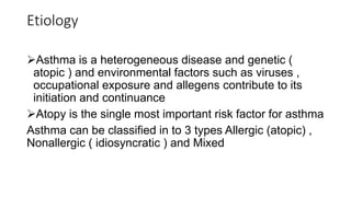
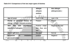
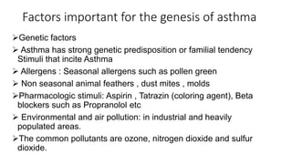
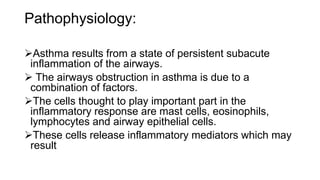
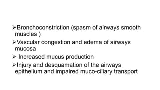
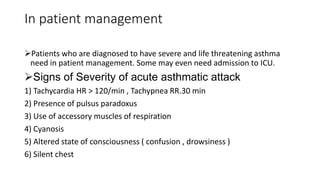
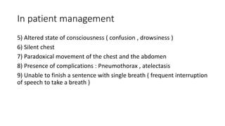
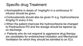
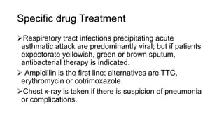
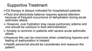
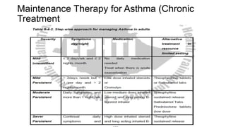
Bronchial Asthma
• Learning Objective: At the end of this unit the student will be able to
• 1. Define bronchial asthma
• 2. Understand the epidemiology of bronchial asthma.
• 3. Describe the etiology of bronchial asthma.
• 4. Understand the pathophysiology of bronchial asthma .
• 5. Identify the clinical manifestations of bronchial asthma.
• 6. List the signs of severity of bronchial asthma.
• 7. Make an accurate diagnosis of bronchial asthma.
• 8. Manage most cases of bronchial asthma.
• 9. Refer complicated cases of bronchial asthma.
Definition:
Bronchial asthma is defined as chronic inflammatory
disease of airways
characterized by increased responsiveness of the
tracheobronchial tree to a multiplicity of stimuli.
It is associated with widespread airway obstruction that
is reversible (but not completely in some patients),
either spontaneously or with treatment
Epidemiology:
Asthma is a common disease The prevalence of
asthma is rising in different parts of the world.
 It can occur at any age; but it usually starts early in
life. About 50% of patients develop asthma before the
age of 10 and another 35% before the age of 40.
Males are affected twice as common as females in
early life; this sex difference equalizes by age 30.
Most cases of asthma are associated with personal
or family history of allergic disease such as eczema,
rhinitis and urticaria.
Etiology
Asthma is a heterogeneous disease and genetic (
atopic ) and environmental factors such as viruses ,
occupational exposure and allegens contribute to its
initiation and continuance
Atopy is the single most important risk factor for asthma
Asthma can be classified in to 3 types Allergic (atopic) ,
Nonallergic ( idiosyncratic ) and Mixed
Factors important for the genesis of asthma
Genetic factors
 Asthma has strong genetic predisposition or familial tendency
Stimuli that incite Asthma
 Allergens : Seasonal allergens such as pollen green
 Non seasonal animal feathers , dust mites , molds
Pharmacologic stimuli: Aspirin , Tatrazin (coloring agent), Beta
blockers such as Propranolol etc
 Environmental and air pollution: in industrial and heavily
populated areas.
The common pollutants are ozone, nitrogen dioxide and sulfur
dioxide.
Pathophysiology:
Asthma results from a state of persistent subacute
inflammation of the airways.
 The airways obstruction in asthma is due to a
combination of factors.
The cells thought to play important part in the
inflammatory response are mast cells, eosinophils,
lymphocytes and airway epithelial cells.
These cells release inflammatory mediators which may
result
Bronchoconstriction (spasm of airways smooth
muscles )
Vascular congestion and edema of airways
mucosa
 Increased mucus production
Injury and desquamation of the airways
epithelium and impaired muco-ciliary transport
Symptom and Signs
The symptoms of each asthmatic patient differ greatly
in frequency and degree.
Some asthmatics are symptom free, with an occasional
episode that is mild and brief; others have mild
coughing and wheezing much of the time, punctuated
by severe
exacerbations of symptoms following exposure to known
allergens, viral infection, exercise etc.
 Psychological factors particularly those associated with
crying, screaming or hard laughing may precipitate
symptoms.
Symptom and Signs
An attack usually begins acutely with paroxysms
of wheezing, coughing, and shortness of breath,
or insidiously with slowly increasing
manifestations of respiratory distress.
The asthmatic first notices dyspnea, tachypnea,
cough and tightness in the chest and may even
notice audible wheezes.
On physical examination
Varying degrees of respiratory distress tachypnea,
tachycardia, and audible wheezes are often present.
Dehydration may be present because of sweating
and tachypnea.
 Chest examination shows a prolonged expiratory
phase with relatively high pitched wheezes
throughout inspiration and most of expiration.
In more severe episodes, patients may be unable to
speak more than a few words without stopping for
breath.
On physical examination
Cyanosis is usually a late sign of hypoxia.
 Confusion and lethargy may indicate the onset of
progressive respiratory failure.
Less wheezing (silent chest) might indicate mucous
plug or patient fatigue with less airflow.
 And it is a sign of impending respiratory failure.
 The presence, absence, or prominence of wheezes
does not correlate precisely with the severity of the
attack.
On physical examination
The most reliable clinical signs include the degree of
dyspnea at rest, cyanosis, difficulty in speaking and use
of accessory muscles of respiration.
 This is confirmed by arterial blood gas analysis.
 Between acute attacks, breath sounds may be normal
during quiet respiration.
However, low grade wheezing maybe heard at any time
in some patients, even when they claim to be
completely asymptomatic.
Complications during an Acute Attack of
Asthma
Pneumothorax: It may present as sudden worsening
of respiratory distress, accompanied by sharp chest
pain and on examination, hyperresonant lung with a
shift of mediastinum. Chest x-ray confirms the
diagnosis.
Mediastinal and subcutaneous emphysema due to
alveolar rupture Atelectasis due to obstruction
 Dilated right heart chambers (Corpulmonale) : from
chronic hypoxemia and pulmonary hypertension
Respiratory failure
Laboratory Findings
Eosinophilia is a common finding.
Sputum is tenacious, rubbery and whitish or may be
yellowish; eosinophils are presentin the sputum.
Chest x-ray: varies from normal to hyperinflation.
Atelectasis and pneumothorax may be seen in
complicated cases.
 Pulmonary function tests are valuable in differential
diagnosis and in known patients to assess the degree
of airways obstruction.
Diagnosis
Asthma should be considered in anyone who wheezes. A family
history of allergy, rhinitis or asthma can be elicited in most
asthmatics.
Differential diagnosis includes:
In children: foreign body obstruction, viral URTI involving the
epiglottis (croup), and bronchiolitis (RSV infection);
In adults: COPD, heart failure, endobronchial TB, and
malignancies.
Physical examination should search for heart failure and signs of
chronic hypoxemia (clubbing).
Unilateral wheezes usually indicate obstruction by foreign bodies
Prevention of attacks
The role of environmental factors (e.g. animal dander,
dust, airborne moulds, and pollens) in acute
exacerbations is clear.
Allergens that can be controlled by avoidance should
be eliminated.
Nonspecific exacerbating factors (e.g. cigarette smoke,
odors, irritant fumes, and change in temperature,
atmospheric pressure, and humidity) should also be
investigated and avoided if possible.
Treatment
General principles
Assessing the severity of the attack is paramount
in deciding management
Bronchodilators should be used in orderly
progression
 Decide when to start corticosteroids
Treatment of the Acute Attack
 Salbutamol aerosol (Ventolin ) two puffs every 20 minutes
for three doses is the 1st line of treatment.
 Adrenaline 1:1000 can be given in doses up to a maximum
of 0.2 ml in children and 0.3 ml in adults, repeated once or
twice in 20 to 30 min (if there is no hypertension or any other
contra indication).
If the initial treatment fails, Aminophylline 250 mg IV diluted in
dextrose in water should be given slowly over 10-15 minutes,
once.
In patient management
Patients who are diagnosed to have severe and life threatening asthma
need in patient management. Some may even need admission to ICU.
Signs of Severity of acute asthmatic attack
1) Tachycardia HR > 120/min , Tachypnea RR.30 min
2) Presence of pulsus paradoxus
3) Use of accessory muscles of respiration
4) Cyanosis
5) Altered state of consciousness ( confusion , drowsiness )
6) Silent chest
In patient management
5) Altered state of consciousness ( confusion , drowsiness )
6) Silent chest
7) Paradoxical movement of the chest and the abdomen
8) Presence of complications : Pneumothorax , atelectasis
9) Unable to finish a sentence with single breath ( frequent interruption
of speech to take a breath )
Specific drug Treatment
Aminoplylline in doses of 1mg/kg/hr in a continuous IV
infusion should be given.
Corticosteroids should also be given IV e.g. Hydrocortisone
4mg/kg IV every 4 hrs.
When the patient improves the hydrocortisone be changed
to Prednisolone PO and the dosage should be tapered up
on discharge.
 Patients who do not respond to aggressive drug therapy
are candidates for endotracheal intubation and Mechanical
Ventilation for which they should be admitted to an ICU.
Specific drug Treatment
Respiratory tract infections precipitating acute
asthmatic attack are predominantly viral; but if patients
expectorate yellowish, green or brown sputum,
antibacterial therapy is indicated.
 Ampicillin is the first line; alternatives are TTC,
erythromycin or cotrimoxazole.
Chest x-ray is taken if there is suspicion of pneumonia
or complications.
Supportive Treatment
O2 therapy is always indicated for hospitalized patients
Fluid and electrolyte balance requires special attention
because of frequent occurrence of dehydration during acute
asthmatic attack.
 However, over hydration may cause pulmonary edema and
one should be cautious in fluid administration.
 Anxiety is common in patients with severe acute asthmatic
attack.
However this can be overcome when underlying hypoxia and
feeling of asphyxiation is treated.
Health personnel should be considerate and reassure the
patient.
Maintenance Therapy for Asthma (Chronic
Treatment
Chronic obstructive pulmonary diseases
(COPD)
• Learning Objective: At the end of this unit the student will be able to
• 1. Define chronic obstructive pulmonary diseases (COPD
• 2. List the etiologies of COPD
• 3. Explain the epidemiology of COPD
• 4. Describe the pathophysiology of COPD
• 5. Identify the clinical manifestations of COPD
• 6. Outline the main differences between c. bronchitis and emphysema
• 7. Describe the most commonly used tests for the diagnosis of COPD
• 8. Make a diagnosis of COPD
Definition:
Chronic obstructive pulmonary diseases are conditions
characterized by chronic irreversible airway obstruction
causing an increased resistance to outflow of air due to
chronic bronchitis and emphysema.
Both these diseases occur together in the same
individual in a variable proportion but the manifestations
of one often predominates the clinical picture.
1) Chronic bronchitis: is a condition associated with
excessive tracheobronchial mucus production sufficient
to cause cough with expectoration of sputum for at least
3 months in a year for over 2 consecutive years.
Etiology
Emphysema: Any factor leading to chronic alveolar
inflammation would encourage development of an
emphysematous lesion.
Smoking has adverse effects on lung defenses, leading to
emphysematous change.
Congenital enzyme defects such as α1- antitrypsin deficiency
are also risk factors for the disease.
 Chronic bronchitis: with sufficient exposure to bronchial
irritants, particularly cigarette smoke, most persons develop
some degree of chronic bronchitis with signs of inflammation of
the airways.
In developing countries household smoke from fire wood is
said to be a major contributing factor.
Prevalence:
COPD is a major health problem especially in western
societies because of the effect of cigarette smoking and
aging.
Males are affected more than females which could be
attributed to the higher prevalence of smoking in males.
Nowadays, the incidence of this disease in females is
increasing because of the increasing smoking habit.
Pathological changes and pathophysiology
Chronic bronchitis is characterized by hypertrophy of
mucus glands in both large and small airways with
thickening of walls and accompanying excess
production of mucus and narrowing of airway lumen.
In C. bronchitis, alveoli are often spared, and no
vessel loss and lung perfusion remains normal but
ventilation is very much reduced.
This leads to abnormal V/Q (arteriovenous shunt)
and patients usually suffer from hypoxemia
(manifested with cyanosis) and acidosis, which
causes pulmonary hypertension and right heart failure
in the long term.
Pathological changes and pathophysiology
 On the other hand emphysema is characterized by destruction of
alveolar septa and distension of alveoli resulting in reduced
surface area and loss of vessels, the later causing reduced
perfusion.
 Moreover, emphysema causes mucus production and airway
narrowing with accompanying reduction in ventilation.
This leads to retention of carbon dioxide in the blood and severe
dyspnea from reduced tissue perfusion.
However, these patients don’t suffer from hypoxia and acidosis,
and have less chance of development of pulmonary hypertension
and cor-pulmonale.
Clinical features:
COPD is thought to begin early in adult life but
significant symptoms and disability do not appear until
middle age.
A mild "smoker's cough" is often present many years
before onset of exertional dyspnea.
Gradual progressive exertional dyspnea is the most
common presenting complaint.
 Cough, wheezing, recurrent respiratory infections or,
occasionally weakness, weight loss, or reduced libido
may also be initial manifestations.
Physical findings:
in COPD are very variable especially in early stages.
A consistent abnormality is obstruction to expiratory
airflow manifested by prolonged forced expiration
(normally < 4 seconds).
 Typical findings including gross pulmonary
hyperinflation, prolonged expiration during quiet
breathing, pursed-lip breathing, stooped posture, and
marked use of accessory muscles of respiration are
seen in later stages of COPD.
Physical findings:
Other findings are rhonchi, diminished vesicular breath
sounds, tachycardia, distant heart sounds, and
decreased diaphragmatic excursion.
The chest may be remarkably "quiet" in advanced
stages of emphysema but is usually "noisy" in patients
with chronic bronchitis.
In advanced cases, frank cyanosis may be there from
hypoxemia; a plethoric appearance associated with
secondary erythrocytosis and, signs of right-sided heart
failure in patients with cor-pulmonale.
 Mild edema may be there even without heart failure.
Diagnosis:
COPD should be suspected in any patient with chronic
productive cough and/or exertional dyspnea of uncertain
etiology, or whose physical examination reveals
evidence of prolonged forced expiration.
Pulmonary function tests (spirometric testing) are done
at specialized hospitals to determine the type of
pulmonary obstruction.
Red cell counts may reveal erythrocytosis and elevated
hematochrit in chronic hypoxemic patients.
Diagnosis:
The pattern of physiologic abnormality in each
patient depends to some extent on the relative
severity of intrinsic bronchial disease and
emphysema.
In patients with severe emphysema, resting
hypoxemia is usually mild (i.e. no or less
cyanosis). In patients with chronic bronchitis,
severe hypoxemia may be noted relatively early.
Course and Prognosis
In the early stage of COPD, some reversal of
airway obstruction and considerable
symptomatic improvement can often be obtained
with therapy, but the long-term prognosis is
less favorable in such patients.
Course and Prognosis
Treatment: so far no curative treatment.
Therapy is directed at relieving symptoms,
controlling potentially fatal exacerbations, and
slowing of the progression of the disorder.
Avoidance of bronchial irritants, especially
cessation of smoking, is of primary importance.
• Thus, the treatment is outlined as follows:
1) Treatment of infection: COPD patients with
purulent sputum should be treated with a broad
spectrum antibiotic. Commonly used drugs include
co-trimoxazole 980mg POBID or TTC or Ampicillin
250 - 500mg four times a day for 10 days. The
course can be repeated at the first sign of
recurrence of bronchial infection.
2) Control of bronchospasm: β-agonists like
salbutamol, or one of the theophyllines can be used.
Corticosteroids do not have a major role in
maintenance treatment.
3) Facilitation of drainage of bronchial secretion:
adequate hydration with oral fluids is essential to
prevent drying of secretions. Inhalation of mist,
postural drainage, and chest exercise may help.
4) Hypoxemia: this will lead to cor-pulmonale in
patients with predominant c.bronchitis.
Oxygen should be given in such patients with hypoxia,
and in severe cases a portable oxygen therapy ( 16 hrs
/day) for home use is recommended.
5) Control of heart failure: the most important measures
are correction of hypoxemia, administration of diuretics
and restriction of sodium intake.
6) Exercise: prolonged inactivity leads to exercise
intolerance. Regular exercise as long as there is no
severe heart disease is recommended.
• Thank you

ASTHMA AND COPD .pptx

  • 1.
  • 2.
    • Learning Objective:At the end of this unit the student will be able to • 1. Define bronchial asthma • 2. Understand the epidemiology of bronchial asthma. • 3. Describe the etiology of bronchial asthma. • 4. Understand the pathophysiology of bronchial asthma . • 5. Identify the clinical manifestations of bronchial asthma. • 6. List the signs of severity of bronchial asthma. • 7. Make an accurate diagnosis of bronchial asthma. • 8. Manage most cases of bronchial asthma. • 9. Refer complicated cases of bronchial asthma.
  • 3.
    Definition: Bronchial asthma isdefined as chronic inflammatory disease of airways characterized by increased responsiveness of the tracheobronchial tree to a multiplicity of stimuli. It is associated with widespread airway obstruction that is reversible (but not completely in some patients), either spontaneously or with treatment
  • 4.
    Epidemiology: Asthma is acommon disease The prevalence of asthma is rising in different parts of the world.  It can occur at any age; but it usually starts early in life. About 50% of patients develop asthma before the age of 10 and another 35% before the age of 40. Males are affected twice as common as females in early life; this sex difference equalizes by age 30. Most cases of asthma are associated with personal or family history of allergic disease such as eczema, rhinitis and urticaria.
  • 5.
    Etiology Asthma is aheterogeneous disease and genetic ( atopic ) and environmental factors such as viruses , occupational exposure and allegens contribute to its initiation and continuance Atopy is the single most important risk factor for asthma Asthma can be classified in to 3 types Allergic (atopic) , Nonallergic ( idiosyncratic ) and Mixed
  • 7.
    Factors important forthe genesis of asthma Genetic factors  Asthma has strong genetic predisposition or familial tendency Stimuli that incite Asthma  Allergens : Seasonal allergens such as pollen green  Non seasonal animal feathers , dust mites , molds Pharmacologic stimuli: Aspirin , Tatrazin (coloring agent), Beta blockers such as Propranolol etc  Environmental and air pollution: in industrial and heavily populated areas. The common pollutants are ozone, nitrogen dioxide and sulfur dioxide.
  • 8.
    Pathophysiology: Asthma results froma state of persistent subacute inflammation of the airways.  The airways obstruction in asthma is due to a combination of factors. The cells thought to play important part in the inflammatory response are mast cells, eosinophils, lymphocytes and airway epithelial cells. These cells release inflammatory mediators which may result
  • 9.
    Bronchoconstriction (spasm ofairways smooth muscles ) Vascular congestion and edema of airways mucosa  Increased mucus production Injury and desquamation of the airways epithelium and impaired muco-ciliary transport
  • 10.
    Symptom and Signs Thesymptoms of each asthmatic patient differ greatly in frequency and degree. Some asthmatics are symptom free, with an occasional episode that is mild and brief; others have mild coughing and wheezing much of the time, punctuated by severe exacerbations of symptoms following exposure to known allergens, viral infection, exercise etc.  Psychological factors particularly those associated with crying, screaming or hard laughing may precipitate symptoms.
  • 11.
    Symptom and Signs Anattack usually begins acutely with paroxysms of wheezing, coughing, and shortness of breath, or insidiously with slowly increasing manifestations of respiratory distress. The asthmatic first notices dyspnea, tachypnea, cough and tightness in the chest and may even notice audible wheezes.
  • 12.
    On physical examination Varyingdegrees of respiratory distress tachypnea, tachycardia, and audible wheezes are often present. Dehydration may be present because of sweating and tachypnea.  Chest examination shows a prolonged expiratory phase with relatively high pitched wheezes throughout inspiration and most of expiration. In more severe episodes, patients may be unable to speak more than a few words without stopping for breath.
  • 13.
    On physical examination Cyanosisis usually a late sign of hypoxia.  Confusion and lethargy may indicate the onset of progressive respiratory failure. Less wheezing (silent chest) might indicate mucous plug or patient fatigue with less airflow.  And it is a sign of impending respiratory failure.  The presence, absence, or prominence of wheezes does not correlate precisely with the severity of the attack.
  • 14.
    On physical examination Themost reliable clinical signs include the degree of dyspnea at rest, cyanosis, difficulty in speaking and use of accessory muscles of respiration.  This is confirmed by arterial blood gas analysis.  Between acute attacks, breath sounds may be normal during quiet respiration. However, low grade wheezing maybe heard at any time in some patients, even when they claim to be completely asymptomatic.
  • 15.
    Complications during anAcute Attack of Asthma Pneumothorax: It may present as sudden worsening of respiratory distress, accompanied by sharp chest pain and on examination, hyperresonant lung with a shift of mediastinum. Chest x-ray confirms the diagnosis. Mediastinal and subcutaneous emphysema due to alveolar rupture Atelectasis due to obstruction  Dilated right heart chambers (Corpulmonale) : from chronic hypoxemia and pulmonary hypertension Respiratory failure
  • 16.
    Laboratory Findings Eosinophilia isa common finding. Sputum is tenacious, rubbery and whitish or may be yellowish; eosinophils are presentin the sputum. Chest x-ray: varies from normal to hyperinflation. Atelectasis and pneumothorax may be seen in complicated cases.  Pulmonary function tests are valuable in differential diagnosis and in known patients to assess the degree of airways obstruction.
  • 17.
    Diagnosis Asthma should beconsidered in anyone who wheezes. A family history of allergy, rhinitis or asthma can be elicited in most asthmatics. Differential diagnosis includes: In children: foreign body obstruction, viral URTI involving the epiglottis (croup), and bronchiolitis (RSV infection); In adults: COPD, heart failure, endobronchial TB, and malignancies. Physical examination should search for heart failure and signs of chronic hypoxemia (clubbing). Unilateral wheezes usually indicate obstruction by foreign bodies
  • 18.
    Prevention of attacks Therole of environmental factors (e.g. animal dander, dust, airborne moulds, and pollens) in acute exacerbations is clear. Allergens that can be controlled by avoidance should be eliminated. Nonspecific exacerbating factors (e.g. cigarette smoke, odors, irritant fumes, and change in temperature, atmospheric pressure, and humidity) should also be investigated and avoided if possible.
  • 19.
    Treatment General principles Assessing theseverity of the attack is paramount in deciding management Bronchodilators should be used in orderly progression  Decide when to start corticosteroids
  • 20.
    Treatment of theAcute Attack  Salbutamol aerosol (Ventolin ) two puffs every 20 minutes for three doses is the 1st line of treatment.  Adrenaline 1:1000 can be given in doses up to a maximum of 0.2 ml in children and 0.3 ml in adults, repeated once or twice in 20 to 30 min (if there is no hypertension or any other contra indication). If the initial treatment fails, Aminophylline 250 mg IV diluted in dextrose in water should be given slowly over 10-15 minutes, once.
  • 21.
    In patient management Patientswho are diagnosed to have severe and life threatening asthma need in patient management. Some may even need admission to ICU. Signs of Severity of acute asthmatic attack 1) Tachycardia HR > 120/min , Tachypnea RR.30 min 2) Presence of pulsus paradoxus 3) Use of accessory muscles of respiration 4) Cyanosis 5) Altered state of consciousness ( confusion , drowsiness ) 6) Silent chest
  • 22.
    In patient management 5)Altered state of consciousness ( confusion , drowsiness ) 6) Silent chest 7) Paradoxical movement of the chest and the abdomen 8) Presence of complications : Pneumothorax , atelectasis 9) Unable to finish a sentence with single breath ( frequent interruption of speech to take a breath )
  • 23.
    Specific drug Treatment Aminoplyllinein doses of 1mg/kg/hr in a continuous IV infusion should be given. Corticosteroids should also be given IV e.g. Hydrocortisone 4mg/kg IV every 4 hrs. When the patient improves the hydrocortisone be changed to Prednisolone PO and the dosage should be tapered up on discharge.  Patients who do not respond to aggressive drug therapy are candidates for endotracheal intubation and Mechanical Ventilation for which they should be admitted to an ICU.
  • 24.
    Specific drug Treatment Respiratorytract infections precipitating acute asthmatic attack are predominantly viral; but if patients expectorate yellowish, green or brown sputum, antibacterial therapy is indicated.  Ampicillin is the first line; alternatives are TTC, erythromycin or cotrimoxazole. Chest x-ray is taken if there is suspicion of pneumonia or complications.
  • 25.
    Supportive Treatment O2 therapyis always indicated for hospitalized patients Fluid and electrolyte balance requires special attention because of frequent occurrence of dehydration during acute asthmatic attack.  However, over hydration may cause pulmonary edema and one should be cautious in fluid administration.  Anxiety is common in patients with severe acute asthmatic attack. However this can be overcome when underlying hypoxia and feeling of asphyxiation is treated. Health personnel should be considerate and reassure the patient.
  • 26.
    Maintenance Therapy forAsthma (Chronic Treatment
  • 27.
    Chronic obstructive pulmonarydiseases (COPD) • Learning Objective: At the end of this unit the student will be able to • 1. Define chronic obstructive pulmonary diseases (COPD • 2. List the etiologies of COPD • 3. Explain the epidemiology of COPD • 4. Describe the pathophysiology of COPD • 5. Identify the clinical manifestations of COPD • 6. Outline the main differences between c. bronchitis and emphysema • 7. Describe the most commonly used tests for the diagnosis of COPD • 8. Make a diagnosis of COPD
  • 28.
    Definition: Chronic obstructive pulmonarydiseases are conditions characterized by chronic irreversible airway obstruction causing an increased resistance to outflow of air due to chronic bronchitis and emphysema. Both these diseases occur together in the same individual in a variable proportion but the manifestations of one often predominates the clinical picture. 1) Chronic bronchitis: is a condition associated with excessive tracheobronchial mucus production sufficient to cause cough with expectoration of sputum for at least 3 months in a year for over 2 consecutive years.
  • 29.
    Etiology Emphysema: Any factorleading to chronic alveolar inflammation would encourage development of an emphysematous lesion. Smoking has adverse effects on lung defenses, leading to emphysematous change. Congenital enzyme defects such as α1- antitrypsin deficiency are also risk factors for the disease.  Chronic bronchitis: with sufficient exposure to bronchial irritants, particularly cigarette smoke, most persons develop some degree of chronic bronchitis with signs of inflammation of the airways. In developing countries household smoke from fire wood is said to be a major contributing factor.
  • 30.
    Prevalence: COPD is amajor health problem especially in western societies because of the effect of cigarette smoking and aging. Males are affected more than females which could be attributed to the higher prevalence of smoking in males. Nowadays, the incidence of this disease in females is increasing because of the increasing smoking habit.
  • 31.
    Pathological changes andpathophysiology Chronic bronchitis is characterized by hypertrophy of mucus glands in both large and small airways with thickening of walls and accompanying excess production of mucus and narrowing of airway lumen. In C. bronchitis, alveoli are often spared, and no vessel loss and lung perfusion remains normal but ventilation is very much reduced. This leads to abnormal V/Q (arteriovenous shunt) and patients usually suffer from hypoxemia (manifested with cyanosis) and acidosis, which causes pulmonary hypertension and right heart failure in the long term.
  • 32.
    Pathological changes andpathophysiology  On the other hand emphysema is characterized by destruction of alveolar septa and distension of alveoli resulting in reduced surface area and loss of vessels, the later causing reduced perfusion.  Moreover, emphysema causes mucus production and airway narrowing with accompanying reduction in ventilation. This leads to retention of carbon dioxide in the blood and severe dyspnea from reduced tissue perfusion. However, these patients don’t suffer from hypoxia and acidosis, and have less chance of development of pulmonary hypertension and cor-pulmonale.
  • 33.
    Clinical features: COPD isthought to begin early in adult life but significant symptoms and disability do not appear until middle age. A mild "smoker's cough" is often present many years before onset of exertional dyspnea. Gradual progressive exertional dyspnea is the most common presenting complaint.  Cough, wheezing, recurrent respiratory infections or, occasionally weakness, weight loss, or reduced libido may also be initial manifestations.
  • 34.
    Physical findings: in COPDare very variable especially in early stages. A consistent abnormality is obstruction to expiratory airflow manifested by prolonged forced expiration (normally < 4 seconds).  Typical findings including gross pulmonary hyperinflation, prolonged expiration during quiet breathing, pursed-lip breathing, stooped posture, and marked use of accessory muscles of respiration are seen in later stages of COPD.
  • 35.
    Physical findings: Other findingsare rhonchi, diminished vesicular breath sounds, tachycardia, distant heart sounds, and decreased diaphragmatic excursion. The chest may be remarkably "quiet" in advanced stages of emphysema but is usually "noisy" in patients with chronic bronchitis. In advanced cases, frank cyanosis may be there from hypoxemia; a plethoric appearance associated with secondary erythrocytosis and, signs of right-sided heart failure in patients with cor-pulmonale.  Mild edema may be there even without heart failure.
  • 36.
    Diagnosis: COPD should besuspected in any patient with chronic productive cough and/or exertional dyspnea of uncertain etiology, or whose physical examination reveals evidence of prolonged forced expiration. Pulmonary function tests (spirometric testing) are done at specialized hospitals to determine the type of pulmonary obstruction. Red cell counts may reveal erythrocytosis and elevated hematochrit in chronic hypoxemic patients.
  • 37.
    Diagnosis: The pattern ofphysiologic abnormality in each patient depends to some extent on the relative severity of intrinsic bronchial disease and emphysema. In patients with severe emphysema, resting hypoxemia is usually mild (i.e. no or less cyanosis). In patients with chronic bronchitis, severe hypoxemia may be noted relatively early.
  • 40.
    Course and Prognosis Inthe early stage of COPD, some reversal of airway obstruction and considerable symptomatic improvement can often be obtained with therapy, but the long-term prognosis is less favorable in such patients.
  • 41.
    Course and Prognosis Treatment:so far no curative treatment. Therapy is directed at relieving symptoms, controlling potentially fatal exacerbations, and slowing of the progression of the disorder. Avoidance of bronchial irritants, especially cessation of smoking, is of primary importance. • Thus, the treatment is outlined as follows:
  • 42.
    1) Treatment ofinfection: COPD patients with purulent sputum should be treated with a broad spectrum antibiotic. Commonly used drugs include co-trimoxazole 980mg POBID or TTC or Ampicillin 250 - 500mg four times a day for 10 days. The course can be repeated at the first sign of recurrence of bronchial infection. 2) Control of bronchospasm: β-agonists like salbutamol, or one of the theophyllines can be used. Corticosteroids do not have a major role in maintenance treatment. 3) Facilitation of drainage of bronchial secretion: adequate hydration with oral fluids is essential to prevent drying of secretions. Inhalation of mist, postural drainage, and chest exercise may help.
  • 43.
    4) Hypoxemia: thiswill lead to cor-pulmonale in patients with predominant c.bronchitis. Oxygen should be given in such patients with hypoxia, and in severe cases a portable oxygen therapy ( 16 hrs /day) for home use is recommended. 5) Control of heart failure: the most important measures are correction of hypoxemia, administration of diuretics and restriction of sodium intake. 6) Exercise: prolonged inactivity leads to exercise intolerance. Regular exercise as long as there is no severe heart disease is recommended.
  • 44.