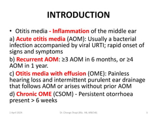
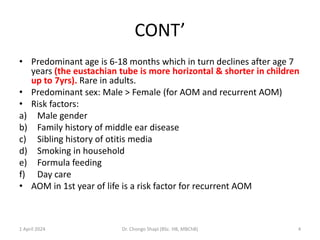
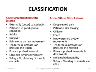
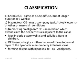
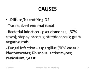
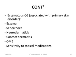
3. INTRODUCTION
• Otitis media - Inflammation of the middle ear
a) Acute otitis media (AOM): Usually a bacterial
infection accompanied by viral URTI; rapid onset of
signs and symptoms
b) Recurrent AOM: ≥3 AOM in 6 months, or ≥4
AOM in 1 year.
c) Otitis media with effusion (OME): Painless
hearing loss and intermittent purulent ear drainage
that follows AOM or arises without prior AOM
d) Chronic OME (CSOM) - Persistent otorrhoea
present > 6 weeks
1 April 2024 Dr. Chongo Shapi (BSc. HB, MBChB) 3
4. CONT’
• Predominant age is 6-18 months which in turn declines after age 7
years (the eustachian tube is more horizontal & shorter in children
up to 7yrs). Rare in adults.
• Predominant sex: Male > Female (for AOM and recurrent AOM)
• Risk factors:
a) Male gender
b) Family history of middle ear disease
c) Sibling history of otitis media
d) Smoking in household
e) Formula feeding
f) Day care
• AOM in 1st year of life is a risk factor for recurrent AOM
1 April 2024 Dr. Chongo Shapi (BSc. HB, MBChB) 4
6. CAUSES OF OTITIS MEDIA
• A preceding viral URTI produces eustachian
tube dysfunction that is thought to promote
bacterial infection via eustachian tube.
• Common organisms - Pneumococci,
Haemophilus influenzae, Moraxella
(Branhamella) catarrhalis, Group A
streptococci, Staphylococcus aureus,
Sterile/non-pathogens.
1 April 2024 Dr. Chongo Shapi (BSc. HB, MBChB) 6
7. PATHOGENESIS
A) Eustacian tube dysfunction:
• The physiologic role of the auditory tube is to;
i) Ventilate the middle ear space - Normally, the middle
ear is ventilated 3-4 times/min as the eustachian tube
opens during swallowing, and O2 is absorbed by the
blood in the vessels of the middle ear mucous
membrane. If the patency of the eustachian tube is
impaired, a relative negative pressure develops
within the middle ear with hypoxia of the cells
leading to transudation from the mucosa &
accumulation of serous fluid.
1 April 2024 Dr. Chongo Shapi (BSc. HB, MBChB) 7
8. PATHOGENESIS CONT’
ii) Clear middle ear secretions into the nasopharynx
iii) Protect the middle ear from nasopharyngeal
secretions
• It is more horizontal & shorter in children up to
7yrs thus prone to obstruction.
• Eustachian tube obstruction may be due to;
- Inflammatory processes in the nasopharynx
- Allergic manifestations
- Hypertrophic adenoids
- Benign or malignant neoplasms.
9 April 2024 Dr. Chongo Shapi (BSc. HB, MBChB) 8
9. CONT’
B) Altered Mucocilliary system
- Kartagener's Syndrome
- Tissue Hypoxia, Inflammation, Mucosal oedema
C) Nasopharyngeal dysfunction
- Cranio-pharyngeal disproportion e.g. Down's
Syndrome - There is increased basal angle of skull
in relation to cranial capacity ± adenoid tissue
hypertrophy - Adenoid growth in the
nasopharynx outstrips
- skull growth in the first 3-5yrs.
9 April 2024 Dr. Chongo Shapi (BSc. HB, MBChB) 9
10. CONT’
D) HIV
-The infection itself
-Recurrent URTIs
-Localised lymphoid hyperplasia
- Nasopharyngeal carcinoma
9 April 2024 Dr. Chongo Shapi (BSc. HB, MBChB) 10
11. SYMPTOMS AND SIGNS
• Common presentations in adults include:
a) Earache
b) Fever, although more often afebrile
c) Accompanying URTI symptoms
d) Decreased hearing
e) Otorrhoea if eardrum perforated
f) Eardrum mobility decreased (as observed by
pneumatic otoscopy)
g) Eardrum bulging, opaque, often yellowish or
inflamed. Redness alone is not a reliable sign.
9 April 2024 Dr. Chongo Shapi (BSc. HB, MBChB) 11
12. CONT’
• AOM may have a unique presentation in
infants:
-May cause no symptoms in the first few months
of life
- Irritability is sometimes the only indication of
earache
- Eardrum bulging, opaque, often yellowish or
inflamed. Redness alone not a reliable sign.
9 April 2024 Dr. Chongo Shapi (BSc. HB, MBChB) 12
13. OTITIS MEDIA WITH EFFUSION
• Usually asymptomatic
• Associated with a 25-dB hearing loss in the
affected ear probably universal, but not always
measurable, and rarely appreciated by parents
• Eardrum often dull, but not bulging
• Eardrum mobility decreased (as observed by
pneumatic otoscopy) In an adult with persistent
unilateral serous OME, nasopharyngeal
carcinoma must be excluded.
9 April 2024 Dr. Chongo Shapi (BSc. HB, MBChB) 13
14. DIFFERENTIAL DIAGNOSIS
• Tympanosclerosis
• Redness due to crying
• Earache with a normal ear exam may be
caused by referred pain from the jaw or teeth
9 April 2024 Dr. Chongo Shapi (BSc. HB, MBChB) 14
15. INVESTIGATIONS
• Otoscopy;
- Pneumatic otoscopy demonstrates decreased
Eardrum mobility
- Negative middle ear pressure is suggested by;
• * Distorted cone of light
• * Prominence of the lateral process
• * Shortening of the manubrium of the malleus
with a more horizontal orientation
• * Better mobility with negative compared with
positive pressure.
9 April 2024 Dr. Chongo Shapi (BSc. HB, MBChB) 15
16. CONT’
• Effusion m/c/s
• Tympanometry - To document the presence
of middle ear fluid - Flat tympanogram
• Hearing testing helpful to assess the need for
early surgical intervention in OME
15 April 2024 Dr. Chongo Shapi (BSc. HB, MBChB) 16
17. MANAGEMENT
• General measures;
- AOM: Outpatient except for febrile infants < 2 months
- May use watchful waiting approach, treating symptoms without antibiotics
for first 2-3 days. If symptoms persist, then amoxicillin is first line treatment.
• Medication;
- AOM: Amoxicillin 5-7 day course with no complications; Also,
Cephalosporins, Augmentin, Septrin
- Recurrent AOM: Amoxicillin for 3-6 months or until summer; Also
Sulfisoxazole
- Analgesics and antipyretics as needed. Alternative drugs are indicated for
the following AOM patients:
- AOM due to Chlamydia trachomatis will respond to macrolides and
sulfonamides
- AOM due to Mycoplasma pneumoniae will respond to macrolides
15 April 2024 Dr. Chongo Shapi (BSc. HB, MBChB) 17
18. CONT’
• Surgery - Myringotomy (Tympanostomy)
tubes and Adenoidectomy
- ≥3 AOM while on chemoprophylaxis.
- OME;
* > 6 months unilateral
* > 4-6 months bilateral
* Hearing loss > 25db
15 April 2024 Dr. Chongo Shapi (BSc. HB, MBChB) 18
19. CONT’
• Follow up:
• AOM: Otoscopic examination 4 weeks after
diagnosis
• OME: Monthly otoscopic or tympanometric
exams as long as OME persists
15 April 2024 Dr. Chongo Shapi (BSc. HB, MBChB) 19
20. COMPLICATIONS
• Extra-cranial
- Extra-temporal
* Hearing loss
* Perforation/otorrhoea
* Atrophy and scarring of eardrum – Tympanosclerosis *
Adhesions
- Intra-temporal
* Labyrinthitis and resulting vertigo
* Facial nerve palsy
* Ossicular erosion
* Retraction pockets
15 April 2024 Dr. Chongo Shapi (BSc. HB, MBChB) 20
21. CONT’
• Cholesteatoma - A mass of keratinizing squamous
epithelium and cholesterol in the middle ear,
usually resulting from chronic otitis media, with
squamous metaplasia or extension of squamous
epithelium inward to line an expanding cystic
cavity that may involve the mastoid and erode
surrounding bone.
• Usually present in the anterior superior aspect
through the tympanic membrane.
15 April 2024 Dr. Chongo Shapi (BSc. HB, MBChB) 21
22. CONT’
* Mastoiditis - develop when infection tracks
under the periosteum of the temporal bone to
cause a subperiosteal abscess or breaks through
the mastoid tip to cause a neck abscess deep to
the sternocleidomastoid muscle (Bezold's
abscess).
* Venous sinus thrombosis
15 April 2024 Dr. Chongo Shapi (BSc. HB, MBChB) 22
23. CONT’
• Intra-cranial
- Otitic Meningitis
- Epidural abscess
- Brain abscess and other intracranial
suppurative complications
- Otitic hydrocephalus
- Dural venous thrombophlebitis (usually
sigmoid sinus)
15 April 2024 Dr. Chongo Shapi (BSc. HB, MBChB) 23
25. INTRODUCTION
• Inflammation of the external auditory canal
• Classification:
a) Acute OE;
• i) Acute diffuse OE - the most common form,
an infectious process usually bacterial,
occasionally fungal
• ii) Acute circumscribed OE - synonymous with
furuncle. Associated with infection of the hair
follicle.
15 April 2024 Dr. Chongo Shapi (BSc. HB, MBChB) 25
26. CLASSIFICATION
Acute Circumscribed Otitis
Externa
• Externally (outer) seated pain
• Patient is in good general
condition
• Adults
• No fever
• Pain worse on jaw movements
• Tenderness increases on
pressing the tragus
• Pinna is pushed forward
• Localised lymphadenopathy
• X-Ray – No clouding of muscle
ear cells
Acute Diffuse Otitis Externa
• Deep seated pain
• Patient is sick looking
• Children
• Fever
• Not worsened by jaw
movements
• Tenderness increases on
pressing the mastoid
• Pinna is pushed forwards &
downwards
• No lymphadenopathy
• X-Ray – Clouding of muscle ear
cells
15 April 2024 Dr. Chongo Shapi (BSc. HB, MBChB) 26
27. CLASSIFICATION
b) Chronic OE - same as acute diffuse, but of longer
duration (>6 weeks)
c) Eczematous OE - may accompany typical atopic eczema
or other primary skin conditions
d) Necrotizing "malignant" OE - an infection which
extends into the deeper tissues adjacent to the canal.
• May include osteomyelitis and cellulitis. Rare in
children.
e) OE Haemorrhagica - Inflammation of the ectodermal
layer of the tympanic membrane by influenza virus
• forming blisters with blood inside - Rx - Analgesics.
15 April 2024 Dr. Chongo Shapi (BSc. HB, MBChB) 27
29. CONT’
• Eczematous OE (associated with primary skin
disorder):
- Eczema
- Seborrhoea
- Neurodermatitis
- Contact dermatitis
- OME
- Sensitivity to topical medications
15 April 2024 Dr. Chongo Shapi (BSc. HB, MBChB) 29
30. RISK FACTORS
• Acute and chronic otitis externa
- Ear scratching
- Traumatization of external canal
- Swimming
- Hot humid weather
- Use of a hearing aid
- Chronic OME
15 April 2024 Dr. Chongo Shapi (BSc. HB, MBChB) 30
31. CONT’
• Eczematous
- Primary skin disorder
• Necrotizing OE in adults
- Elderly
- Diabetes
- Debilitating disease
15 April 2024 Dr. Chongo Shapi (BSc. HB, MBChB) 31
32. PATHOLOGICAL FINDINGS
• Acute and chronic OE - desquamation of
superficial epithelium of external canal with
infection
• Eczematous OE - pathologic findings
consistent with primary skin disorder,
secondary infection on occasion
• Necrotizing OE - vasculitis, thrombosis and
necrosis of involved tissues; osteomyelitis
15 April 2024 Dr. Chongo Shapi (BSc. HB, MBChB) 32
33. SIGNS AND SYMPTOMS
• Plugging of the ear with a pimple like swelling
leading to mild hearing loss
• Otalgia worsened by movement of jaw or
pressing on the tragus/mastoid
• Periauricular adenitis (Acute circumscribed otitis
externa)
• Erythematous canal
• Purulent discharge
• Eczema of pinna
• Cranial nerve involvement (VII, IX-XII)
15 April 2024 Dr. Chongo Shapi (BSc. HB, MBChB) 33
34. INVESTIGATION
• Otoscopy - 'Cotton wool fibre' in Otomycosis
• Radiological evaluation of deep tissues in
necrotizing otitis externa
15 April 2024 Dr. Chongo Shapi (BSc. HB, MBChB) 34
35. MANAGEMENT
• General measures;
âť–Thorough cleansing of external canal
âť–Pain medications
âť–Antipruritic and antihistamines (eczematous
form)
15 April 2024 Dr. Chongo Shapi (BSc. HB, MBChB) 35
36. CONT’
• Medications;
· Acute bacterial and chronic otitis externa
* Topical therapy for approximately 10 days
* 2% acetic acid
* Antibiotics
* Corticosteroids
• Fungal otitis externa
* Topical therapy anti-yeast for candida or yeast -
nystatin
* Parenteral antifungal therapy - amphotericin B
15 April 2024 Dr. Chongo Shapi (BSc. HB, MBChB) 36
37. CONT’
• Eczematous otitis externa - topical therapy
* Aluminium acetate
* Steroid cream, lotion, ointment
* Antibacterial, if super-infected
• Necrotizing otitis externa
* Parenteral antibiotics - antistaphylococcus and
antipseudomonal
* 4-6 weeks of therapy
15 April 2024 Dr. Chongo Shapi (BSc. HB, MBChB) 37
38. COMPLICATION
• Mainly a problem with necrotizing otitis
externa. May spread to infect contiguous bone
and CNS structures.
• Acute otitis externa may spread to pinna
causing a chondritis
15 April 2024 Dr. Chongo Shapi (BSc. HB, MBChB) 38
39. I HAD TO FIND A WAY TO MAKE
MYSELF RELEVANT TO SOCIETY. BUT,
THIS WAS AFTER DISCOVERING THE
DIFFERENCE BETWEEN THE HEALTHY
AND THE UN-HEALTHY WAY OF
DOING THIS.
15 April 2024 Dr. Chongo Shapi (BSc. HB, MBChB) 39