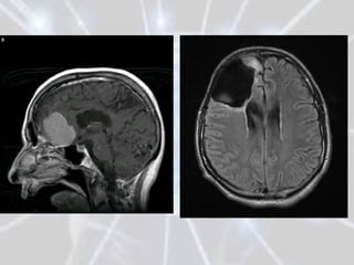
The document discusses different types of brain tumors, including gliomas, meningiomas, and medulloblastomas. It covers symptoms, risk factors, diagnosis methods like MRI and CT scans, and treatment options for brain tumors such as surgery, radiation therapy, chemotherapy, and managing increased intracranial pressure. The prognosis depends on the specific type of brain tumor, with some like glioblastoma multiforme having a very poor median survival rate.
you will learn about brain tumor, types of brain tumor, grading of brain tumor, risk factors for brain tumor, diagnosis for brain tumor, treatment for brain tumor, supportive care and rehabilitation for patients with brain tumor.
The slides give brief information about brain tumor ,its types and symptoms.
The presentation will help you to know more about a condition leading to neurological disturbances.
all about brain tumors. clinical presentation of brain tumors also CT scan MRI of different tumors available to interpret the tumors of brain and spinal cord.
Medulloblastoma- A primitive neuroectodermal tumors (PNETs) is the most common malignant brain tumor of childhood (WHO IV)
arising from the vermis in the inferior medullary velum.
It comprises up to 18% of all pediatric brain tumors.
WNT and Shh pathway plays major role in its pathogenesis.
c-erbB-2 (HER2/neu) oncogene expression has prognostic value. Norcantharidin, Vismodegib, Sonidegib are the future in medulloblastoma.
Spinal Tumors: approach and managementAmit Agrawal
The spinal cord consists of
Central canal surrounded by an H-shaped gray matter region containing neurons
Outer myelinated nerve tracts, termed white matter, surround the central gray matter
Central canal is lined with ependymal cells
Astrocytes support gray matter neurons and white matter axons
Gliomas are the commonest tumor of brain arising from the supportive cells of the brain with diverse form and presentation the treatment of which is surgical and demands adjuvant therapy for most of circumstances.
Dandy–Walker malformation (DWM) encompasses cystic dilatation of the fourth ventricle, complete or partial agenesis of cerebella vermis and enlarged posterior fossa while Dandy–Walker variant (DWV) comprises cystic posterior mass with variable hypoplasia of the cerebella vermis and no enlargement of the posterior fossa.
This PPT presentation talks about osteosarcoma from the clinical point of view, summarizing the recent guidelines in diagnosis and treatment of osteosarcoma.
Learn about the process of radiation therapy to treat soft tissue sarcoma, and how new radiation technology has improved treatment of the disease.
This presentation was given by Elizabeth H. Baldini, MD, MPH, radiation oncology director for the Center for Sarcoma and Bone Oncology at Dana-Farber Cancer Institute. It was originally presented as part of the "15 Years of GIST/Soft Tissue Sarcoma Symposium," held on Sept. 12, 2015 at Dana-Farber in Boston, Mass.
Conformal Radiotherapy in Head and neck cancers is essential in terms of improving quality of life and local control in this era. This presentation aimed at giving an overview of conformal radiotherapy and its role in HNC to a 'general audience'.
When most normal cells grow
old or get damaged, they die, and new cells take their place. Sometimes, this
process goes wrong. New cells form when the body doesn't need them, and old or
damaged cells don't die as they should. The buildup of extra cells often forms
a mass of tissue called a growth or tumor.
Primary brain tumors can be benign or malignant:
Benign brain tumors do not contain cancer cells:
Usually, benign tumors can be removed, and
they seldom grow back.
Benign brain tumors usually have an obvious
border or edge. Cells from benign tumors rarely invade tissues around them.
They don't spread to other parts of the body. However, benign tumors can press
on sensitive areas of the brain and cause serious health problems.
Unlike benign tumors in most other parts of
the body, benign brain tumors are sometimes life threatening.
Benign brain tumors may become malignant.
Malignant brain tumors (also called brain
cancer) contain cancer cells:
Malignant brain tumors are generally more
serious and often are a threat to life.
They are likely to grow rapidly and crowd
or invade the nearby healthy brain tissue.
Cancer cells may break away from malignant
brain tumors and spread to other parts of the brain or to the spinal cord. They
rarely spread to other parts of the body.
Tumor Grade
Doctors group brain tumors by grade.
The grade of a tumor refers to the way the cells look under a microscope:
Grade I: The tissue is benign. The cells
look nearly like normal brain cells, and they grow slowly.
Grade II: The tissue is malignant. The
cells look less like normal cells than do the cells in a Grade I tumor.
Grade III: The malignant tissue has cells
that look very different from normal cells. The abnormal cells are actively
growing (anaplastic).
Grade IV: The malignant tissue has cells
that look most abnormal and tend to grow quickly.
Cells from low-grade tumors (grades I and
II) look more normal and generally grow more slowly than cells from high-grade
tumors (grades III and IV).
Over time, a low-grade tumor may become a
highgrade tumor. However, the change to a high-grade tumor happens more often
among adults than children.
You may want to read the NCI fact sheet Tumor
Grade.
Types of Primary Brain
Tumors
There are many types of primary brain
tumors. Primary brain tumors are named according to the type of cells or the
part of the brain in which they begin. For example, most primary brain tumors
begin in glial cells. This type of tumor is called a glioma.
Among adults, the most common types are:
Astrocytoma:
The tumor arises from star-shaped glial cells called astrocytes.
It can be any grade. In adults, an astrocytoma most often arises in the
cerebrum.
Grade I or II astrocytoma: It may be called
a low-grade glioma.
Grade III astrocytoma: It's sometimes
called a high-grade or an anaplastic astrocytoma.
Grade IV astrocytoma: It may be called a glioblastoma or
malignant astrocytic glioma.
Meningioma:
The tumor arises i
Malignant Bone Tumours - A lecture for undergraduate students and demonstrators / Tutors featuring general aspects and three common malignant bone tumours viz. Osteosarcoma, Ewing's Sarcoma and Multiple Myeloma
Ozempic: Preoperative Management of Patients on GLP-1 Receptor Agonists Saeid Safari
Preoperative Management of Patients on GLP-1 Receptor Agonists like Ozempic and Semiglutide
ASA GUIDELINE
NYSORA Guideline
2 Case Reports of Gastric Ultrasound
Explore natural remedies for syphilis treatment in Singapore. Discover alternative therapies, herbal remedies, and lifestyle changes that may complement conventional treatments. Learn about holistic approaches to managing syphilis symptoms and supporting overall health.
ARTIFICIAL INTELLIGENCE IN HEALTHCARE.pdfAnujkumaranit
Artificial intelligence (AI) refers to the simulation of human intelligence processes by machines, especially computer systems. It encompasses tasks such as learning, reasoning, problem-solving, perception, and language understanding. AI technologies are revolutionizing various fields, from healthcare to finance, by enabling machines to perform tasks that typically require human intelligence.
micro teaching on communication m.sc nursing.pdfAnurag Sharma
Microteaching is a unique model of practice teaching. It is a viable instrument for the. desired change in the teaching behavior or the behavior potential which, in specified types of real. classroom situations, tends to facilitate the achievement of specified types of objectives.
New Directions in Targeted Therapeutic Approaches for Older Adults With Mantl...i3 Health
i3 Health is pleased to make the speaker slides from this activity available for use as a non-accredited self-study or teaching resource.
This slide deck presented by Dr. Kami Maddocks, Professor-Clinical in the Division of Hematology and
Associate Division Director for Ambulatory Operations
The Ohio State University Comprehensive Cancer Center, will provide insight into new directions in targeted therapeutic approaches for older adults with mantle cell lymphoma.
STATEMENT OF NEED
Mantle cell lymphoma (MCL) is a rare, aggressive B-cell non-Hodgkin lymphoma (NHL) accounting for 5% to 7% of all lymphomas. Its prognosis ranges from indolent disease that does not require treatment for years to very aggressive disease, which is associated with poor survival (Silkenstedt et al, 2021). Typically, MCL is diagnosed at advanced stage and in older patients who cannot tolerate intensive therapy (NCCN, 2022). Although recent advances have slightly increased remission rates, recurrence and relapse remain very common, leading to a median overall survival between 3 and 6 years (LLS, 2021). Though there are several effective options, progress is still needed towards establishing an accepted frontline approach for MCL (Castellino et al, 2022). Treatment selection and management of MCL are complicated by the heterogeneity of prognosis, advanced age and comorbidities of patients, and lack of an established standard approach for treatment, making it vital that clinicians be familiar with the latest research and advances in this area. In this activity chaired by Michael Wang, MD, Professor in the Department of Lymphoma & Myeloma at MD Anderson Cancer Center, expert faculty will discuss prognostic factors informing treatment, the promising results of recent trials in new therapeutic approaches, and the implications of treatment resistance in therapeutic selection for MCL.
Target Audience
Hematology/oncology fellows, attending faculty, and other health care professionals involved in the treatment of patients with mantle cell lymphoma (MCL).
Learning Objectives
1.) Identify clinical and biological prognostic factors that can guide treatment decision making for older adults with MCL
2.) Evaluate emerging data on targeted therapeutic approaches for treatment-naive and relapsed/refractory MCL and their applicability to older adults
3.) Assess mechanisms of resistance to targeted therapies for MCL and their implications for treatment selection
- Video recording of this lecture in English language: https://youtu.be/lK81BzxMqdo
- Video recording of this lecture in Arabic language: https://youtu.be/Ve4P0COk9OI
- Link to download the book free: https://nephrotube.blogspot.com/p/nephrotube-nephrology-books.html
- Link to NephroTube website: www.NephroTube.com
- Link to NephroTube social media accounts: https://nephrotube.blogspot.com/p/join-nephrotube-on-social-media.html
The prostate is an exocrine gland of the male mammalian reproductive system
It is a walnut-sized gland that forms part of the male reproductive system and is located in front of the rectum and just below the urinary bladder
Function is to store and secrete a clear, slightly alkaline fluid that constitutes 10-30% of the volume of the seminal fluid that along with the spermatozoa, constitutes semen
A healthy human prostate measures (4cm-vertical, by 3cm-horizontal, 2cm ant-post ).
It surrounds the urethra just below the urinary bladder. It has anterior, median, posterior and two lateral lobes
It’s work is regulated by androgens which are responsible for male sex characteristics
Generalised disease of the prostate due to hormonal derangement which leads to non malignant enlargement of the gland (increase in the number of epithelial cells and stromal tissue)to cause compression of the urethra leading to symptoms (LUTS
Title: Sense of Smell
Presenter: Dr. Faiza, Assistant Professor of Physiology
Qualifications:
MBBS (Best Graduate, AIMC Lahore)
FCPS Physiology
ICMT, CHPE, DHPE (STMU)
MPH (GC University, Faisalabad)
MBA (Virtual University of Pakistan)
Learning Objectives:
Describe the primary categories of smells and the concept of odor blindness.
Explain the structure and location of the olfactory membrane and mucosa, including the types and roles of cells involved in olfaction.
Describe the pathway and mechanisms of olfactory signal transmission from the olfactory receptors to the brain.
Illustrate the biochemical cascade triggered by odorant binding to olfactory receptors, including the role of G-proteins and second messengers in generating an action potential.
Identify different types of olfactory disorders such as anosmia, hyposmia, hyperosmia, and dysosmia, including their potential causes.
Key Topics:
Olfactory Genes:
3% of the human genome accounts for olfactory genes.
400 genes for odorant receptors.
Olfactory Membrane:
Located in the superior part of the nasal cavity.
Medially: Folds downward along the superior septum.
Laterally: Folds over the superior turbinate and upper surface of the middle turbinate.
Total surface area: 5-10 square centimeters.
Olfactory Mucosa:
Olfactory Cells: Bipolar nerve cells derived from the CNS (100 million), with 4-25 olfactory cilia per cell.
Sustentacular Cells: Produce mucus and maintain ionic and molecular environment.
Basal Cells: Replace worn-out olfactory cells with an average lifespan of 1-2 months.
Bowman’s Gland: Secretes mucus.
Stimulation of Olfactory Cells:
Odorant dissolves in mucus and attaches to receptors on olfactory cilia.
Involves a cascade effect through G-proteins and second messengers, leading to depolarization and action potential generation in the olfactory nerve.
Quality of a Good Odorant:
Small (3-20 Carbon atoms), volatile, water-soluble, and lipid-soluble.
Facilitated by odorant-binding proteins in mucus.
Membrane Potential and Action Potential:
Resting membrane potential: -55mV.
Action potential frequency in the olfactory nerve increases with odorant strength.
Adaptation Towards the Sense of Smell:
Rapid adaptation within the first second, with further slow adaptation.
Psychological adaptation greater than receptor adaptation, involving feedback inhibition from the central nervous system.
Primary Sensations of Smell:
Camphoraceous, Musky, Floral, Pepperminty, Ethereal, Pungent, Putrid.
Odor Detection Threshold:
Examples: Hydrogen sulfide (0.0005 ppm), Methyl-mercaptan (0.002 ppm).
Some toxic substances are odorless at lethal concentrations.
Characteristics of Smell:
Odor blindness for single substances due to lack of appropriate receptor protein.
Behavioral and emotional influences of smell.
Transmission of Olfactory Signals:
From olfactory cells to glomeruli in the olfactory bulb, involving lateral inhibition.
Primitive, less old, and new olfactory systems with different path
These lecture slides, by Dr Sidra Arshad, offer a quick overview of physiological basis of a normal electrocardiogram.
Learning objectives:
1. Define an electrocardiogram (ECG) and electrocardiography
2. Describe how dipoles generated by the heart produce the waveforms of the ECG
3. Describe the components of a normal electrocardiogram of a typical bipolar leads (limb II)
4. Differentiate between intervals and segments
5. Enlist some common indications for obtaining an ECG
Study Resources:
1. Chapter 11, Guyton and Hall Textbook of Medical Physiology, 14th edition
2. Chapter 9, Human Physiology - From Cells to Systems, Lauralee Sherwood, 9th edition
3. Chapter 29, Ganong’s Review of Medical Physiology, 26th edition
4. Electrocardiogram, StatPearls - https://www.ncbi.nlm.nih.gov/books/NBK549803/
5. ECG in Medical Practice by ABM Abdullah, 4th edition
6. ECG Basics, http://www.nataliescasebook.com/tag/e-c-g-basics
TEST BANK for Operations Management, 14th Edition by William J. Stevenson, Ve...kevinkariuki227
TEST BANK for Operations Management, 14th Edition by William J. Stevenson, Verified Chapters 1 - 19, Complete Newest Version.pdf
Lung Cancer: Artificial Intelligence, Synergetics, Complex System Analysis, S...Oleg Kshivets
RESULTS: Overall life span (LS) was 2252.1±1742.5 days and cumulative 5-year survival (5YS) reached 73.2%, 10 years – 64.8%, 20 years – 42.5%. 513 LCP lived more than 5 years (LS=3124.6±1525.6 days), 148 LCP – more than 10 years (LS=5054.4±1504.1 days).199 LCP died because of LC (LS=562.7±374.5 days). 5YS of LCP after bi/lobectomies was significantly superior in comparison with LCP after pneumonectomies (78.1% vs.63.7%, P=0.00001 by log-rank test). AT significantly improved 5YS (66.3% vs. 34.8%) (P=0.00000 by log-rank test) only for LCP with N1-2. Cox modeling displayed that 5YS of LCP significantly depended on: phase transition (PT) early-invasive LC in terms of synergetics, PT N0—N12, cell ratio factors (ratio between cancer cells- CC and blood cells subpopulations), G1-3, histology, glucose, AT, blood cell circuit, prothrombin index, heparin tolerance, recalcification time (P=0.000-0.038). Neural networks, genetic algorithm selection and bootstrap simulation revealed relationships between 5YS and PT early-invasive LC (rank=1), PT N0—N12 (rank=2), thrombocytes/CC (3), erythrocytes/CC (4), eosinophils/CC (5), healthy cells/CC (6), lymphocytes/CC (7), segmented neutrophils/CC (8), stick neutrophils/CC (9), monocytes/CC (10); leucocytes/CC (11). Correct prediction of 5YS was 100% by neural networks computing (area under ROC curve=1.0; error=0.0).
CONCLUSIONS: 5YS of LCP after radical procedures significantly depended on: 1) PT early-invasive cancer; 2) PT N0--N12; 3) cell ratio factors; 4) blood cell circuit; 5) biochemical factors; 6) hemostasis system; 7) AT; 8) LC characteristics; 9) LC cell dynamics; 10) surgery type: lobectomy/pneumonectomy; 11) anthropometric data. Optimal diagnosis and treatment strategies for LC are: 1) screening and early detection of LC; 2) availability of experienced thoracic surgeons because of complexity of radical procedures; 3) aggressive en block surgery and adequate lymph node dissection for completeness; 4) precise prediction; 5) adjuvant chemoimmunoradiotherapy for LCP with unfavorable prognosis.
10. 1- Consequences of intracranial
hypertension:
• are the most common symptom in brain
tumor patients.
• present at the initial
encounter with at least 40% of patients.
• must be distinguished from seizure.
•
13. 3-Irritation
• 25% to 50% of all patients with brain tumors experience
seizures at some point in their disease course.
•
•
•
14. Brain tumors can be benign or malignant: •
• Benign brain tumors do not contain cancer cells:
Usually, benign tumors can be removed, and they seldom grow back.
The border or edge of a benign brain tumor can be clearly seen. Cells
from benign tumors do not invade tissues around them or spread to
other parts of the body. However, benign tumors can press on
sensitive areas of the brain and cause serious health problems.
Unlike benign tumors in most other parts of the body, benign brain
tumors are sometimes life threatening.
15. Malignant brain tumors contain cancer cells:
Malignant brain tumors are generally more serious and often are life
threatening.
They are likely to grow rapidly and crowd or invade the surrounding
healthy brain tissue.
Very rarely, cancer cells may break away from a malignant brain
tumor and spread to other parts of the brain, to the spinal cord, or
even to other parts of the body. The spread of cancer is
called metastasis.
Sometimes, a malignant tumor does not extend into healthy tissue.
The tumor may be contained within a layer of tissue. Or the bones of
the skull or another structure in the head may confine it. This kind of
tumor is called encapsulated.
16. Grade I: The tissue is benign. The cells look nearly
like normal brain cells, and cell growth is slow.
Grade II: The tissue is malignant. The cells look less
like normal cells than do the cells in a grade I tumor.
Grade III: The malignant tissue has cells that look
very different from normal cells. The abnormal cells are
actively growing. These abnormal-appearing cells are
termed anaplastic.
Grade IV: The malignant tissue has cells that look
most abnormal and tend to grow very fast.
17. Tumor type :
1. Primary brain tumor:
Tumors that begin in brain tissue are known as primary
tumors of the brain.
Primary brain tumors are named according to the type of cells
or the part of the brain in which they begin.
The most common primary brain tumors are gliomas. They
begin in glial cells. There are many types of gliomas:
18. Astrocytoma - The tumor arises from star-shaped
glial cells called astrocytes. In adults, astrocytomas
most often arise in the cerebrum. In children, they
occur in the brain stem, the cerebrum and the
cerebellum. A grade III astrocytoma is sometimes
called an anaplastic astrocytoma. A grade IV
astrocytoma is usually called a glioblastoma
multiforme.
Brain stem glioma - The tumor occurs in the
lowest part of the brain. Brain stem gliomas most
often are diagnosed in young children and middle-
aged adults.
19. Ependymoma - The tumor arises from cells that
line the ventricles or the central canal of the
spinal cord. They are most commonly found in
children and young adults.
Oligodendroglioma - This rare tumor arises from
cells that make the fatty substance that covers
and protects nerves. These tumors usually occur
in the cerebrum. They grow slowly and usually do
not spread into surrounding brain tissue. They
are most common in middle-aged adults.
20. Some types of brain tumors do not begin in glial
cells. The most common of these are:
Medulloblastoma - This tumor usually arises in the
cerebellum. It is the most common brain tumor in children.
It is sometimes called aprimitive neuroectodermal tumor.
Meningioma - This tumor arises in the meninges. It usually
grows slowly.
Schwannoma - A tumor that arises from a Schwann cell.
These cells line the nerve that controls balance and
hearing. This nerve is in the inner ear. The tumor is also
called an acoustic neuroma. It occurs most often in adults.
21. Craniopharyngioma - The tumor grows at the base of the
brain, near the pituitary gland. This type of tumor most
often occurs in children.
Germ cell tumor of the brain - The tumor arises from
a germ cell. Most germ cell tumors that arise in the brain
occur in people younger than 30. The most common type
of germ cell tumor of the brain is agerminoma.
Pineal region tumor - This rare brain tumor arises in or
near the pineal gland. The pineal gland is located between
the cerebrum and the cerebellum.
22. 2. Secondary brain tumor (metastatic) :
Metastatic brain tumors are made of cancerous cells that
spread through the bloodstream from a tumor located
elsewhere in the body (e.g. lung, breast and kidney).
Metastatic brain tumors are the most common type of tumor
found in the brain and are much more common than primary
brain tumors.
Metastatic tumors are usually named after the type of tissue
from which the original cancer cells arose (for example,
metastatic lung or metastatic breast cancer).
23. No one knows the exact causes of brain tumors.
Research has shown that people with certain risk
factors are more likely than others to develop a brain
tumor.
The following risk factors are associated with an
increased chance of developing a primary brain
tumor:
24. I. Being male - In general, brain tumors are more common in
males than females. However, meningiomas are more
common in females.
II. Race - Brain tumors occur more often among white people
than among people of other races.
III. Age - Most brain tumors are detected in people who are 70
years old or older. However, brain tumors are the second
most common cancer in children. Brain tumors are more
common in children younger than 8 years old than in older
children.
IV. Family history - People with family members who have
gliomas may be more likely to develop this disease.
V. Being exposed to radiation or certain chemicals at work (e.g.
Formaldehyde, Vinyl chloride)
reference : http://www.medicinenet.com/brain_tumor/
25. • The Common Brain Tumor in Child :
1- Astrocytoma
2-neuroblastoma
3-Medulloblastoma
4-ependymoma
• Which one of this tumor is not a gliomas :
1- Ependymoma
2- Germ cell tumor
3- Astrocytoma
4-Brain stem
27. Increase Intracranial pressure
Neurological symptoms
History examination Investigation
meningitis vitals EEC
Space occupying fundoscope Spinal fluid analysis
lesion
Complete Radiological work up
neurological
examination
28. • computed tomography (CT)-scans
• magnetic resonance imaging (MRI).
• Neoplasms will often show as
differently colored masses (also
referred to as processes) in CT or MRI
results.
29. Benign brain tumors often
show up as hypodense (darker
than brain tissue) mass lesions
on cranial CT-scans.
On MRI, they appear either
hypo- (darker than brain
tissue) or isointense (same
intensity as brain tissue) on T1-
weighted scans, or
hyperintense (brighter than
brain tissue) on T2-weighted
MRI, although the appearance
is variable.
30.
31. • Contrast agent uptake, sometimes
in characteristic patterns, can be
demonstrated on either CT or MRI-
scans in most malignant primary
and metastatic brain tumors.
• Perifocal edema
32. • histological examination of tumor tissue samples
obtained either by means of brain biopsy or
open surgery.
• This examination, performed by a pathologist, typically
has three stages:
• interoperative examination of fresh tissue,
• preliminary microscopic examination of prepared
tissues,
• Follow up examination of prepared tissues after
immuno histochemical staining or genetic analysis.
33.
34. Overview
These various types of treatment are available depending
on neoplasm type and location and may be combined
to give the best chances of survival:
• surgery: complete or partial ressection of the tumor with
the objective of removing as many tumor cells as possible
• radiotherapy
• Chemotherapy: with the aim of killing as many as possible of
cancerous cells left behind after surgery and of putting
remaining tumor cells into a non dividing, sleeping state for
as long as possible
35. A)Surgery
• The primary and most desired course of action
described in medical literature is surgical removal
(resection) via craniotomy.
• The prime remediating objective of surgery is to
remove as many tumor cells as possible, with complete
removal being the best outcome
and cytoreduction ("debulking") of the tumor
otherwise.
• In some cases access to the tumor is impossible and
impedes or prohibits surgery.
36.
37. • Any person undergoing brain surgery may
suffer from epileptic seizures. Medication is
prescribed and administered to minimize or
eliminate the occurrence of seizures.
38. B) Radiation therapy
• The goal of radiation therapy is to selectively
kill tumor cells while leaving normal brain
tissue unharmed.
• Radiotherapy is the most common treatment
for secondary brain tumors.
• The amount of radiotherapy depends on the
size of the area of the brain affected by cancer.
39. • Conventional external beam
1-'whole brain radiotherapy treatment' (WBRT)
or 'whole brain irradiation‘
may be suggested if there is a risk that other
secondary tumors will develop in the future
2- Stereotactic radiotherapy is usually
recommended in cases involving fewer than
three small secondary brain tumors.
40. C) Chemotherapy
• Patients undergoing chemotherapy are administered drugs
designed to kill tumor cells. Although chemotherapy may improve
overall survival in patients with the most malignant primary brain
tumors, it does so in only about 20 percent of patients.
• Chemotherapy is often used in young children instead of
radiation, as radiation may have negative effects on the developing
brain.
• The decision to prescribe this treatment is based on a patient’s
overall health, type of tumor, and extent of the cancer. The toxicity
and many side effects of the drugs, and the uncertain outcome of
chemotherapy in brain tumors puts this treatment further down
the line of treatment options with surgery and radiation therapy
preferred.
41. Type Treatment prognosis
Glioma Surgical debulking 90% deis with treatment
radiation
meningioma Preoperative
embolisation & surgical
resection
Cerebellar astrocytoma Surgical resection 90% exceed 5 years
survival rate
Medulloblastoma Surgical removal 50% exceed 5 years
Radio survival rate
Chemotherapy
42. Other
• A shunt is used not as a cure but to relieve
symptoms by reducing hydrocephalus caused
by blockage of cerebrospinal fluid.
• Researchers are presently investigating a
number of promising new treatments
including gene therapy, highly focused
radiation therapy, immunotherapy and novel
chemotherapies.
43.
44. • The prognosis of brain cancer varies based on the type of cancer.
• Medulloblastoma
has a good prognosis with chemotherapy, radiotherapy, and surgical
resection while
• glioblastoma multiforme
has a median survival of only 12 months even with
aggressive chemoradiotherapy and surgery.
• Brainstem gliomas
have the poorest prognosis of any form of brain cancer, with most patients dying
within one year, even with therapy that typically consists of radiation to the
tumor along with corticosteroids.
However, one type of brainstem glioma, a focal seems open to exceptional
prognosis and long-term survival has frequently been reported.
Editor's Notes
or pressure-areas, hyperintense on T2-weighted MRI, they might indicate the presence a diffuse neoplasm (unclear outline)This is because these tumors disrupt the normal functioning of the blood-brain barrier and lead to an increase in its permeability.
Survival rates in primary brain tumors depend on the type of tumor, age, functional status of the patient, the extent of surgical tumor removal and other factors specific to each case.[9]
Several current research studies aim to improve the surgical removal of brain tumors by labeling tumor cells with 5-aminolevulinic acid that causes them to fluoresce. Postoperative radiotherapy and chemotherapy are integral parts of the therapeutic standard for malignant tumors. Radiotherapy may also be administered in cases of "low-grade" gliomas, when a significant tumor burden reduction could not be achieved surgically.
Multiple metastatic tumors are generally treated with radiotherapy and chemotherapy rather than surgery and the prognosis in such cases is determined by the primary tumor, but is generally poor.
Radiotherapy may be used following, or in some cases in place of, resection of the tumor. Forms of radiotherapy used for brain cancer include external beam radiation therapy, brachytherapy, and in more difficult cases, stereotactic radiosurgery, such as Gamma knife, Cyberknife or NovalisTx radiosurgery.[11]
In 2008 a study published by the University of Texas M. D. Anderson Cancer Center indicated that cancer patients who receive stereotactic radiosurgery (SRS) and whole brain radiation therapy (WBRT) for the treatment of metastatic brain tumors have more than twice the risk of developing learning and memory problems than those treated with SRS alone.[13][14]