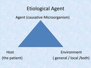
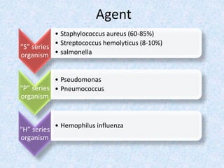
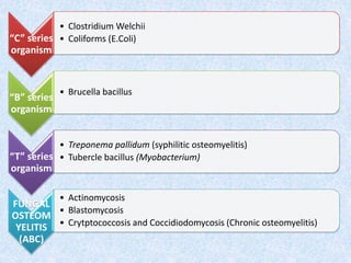
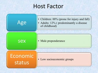
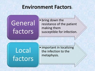
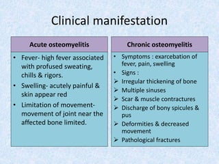
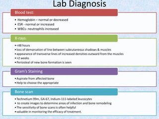
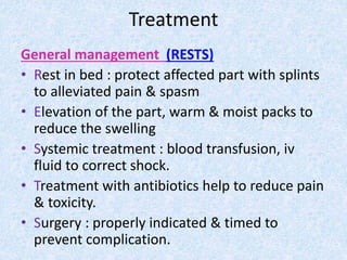
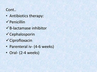
Bone and Skin Infection :
Osteomyelitis & Prosthetic Joint Infection
Name: Nur A’isyah Binti Idris
Matric No.: 082012100068
Serial No.: 61
Objectives
• We should able to know:
Definition
Etiological Agent
Clinical Manifestation
Lab diagnosis
Treatment
Osteomyelitis
• Definition: It is a suppurative process of bone caused
by:
 Pyogenic organisms;
 Pyogenic infection of the cancellous portion the
bone
• Causes : bacterias, fungi, virus, parasites
Etiological Agent
Agent (causative Microorganism)
Host Environment
(the patient) ( general / local /both)
“S” series
organism
• Staphylococcus aureus (60-85%)
• Streptococcus hemolyticus (8-10%)
• salmonella
“P” series
organism
• Pseudomonas
• Pneumococcus
“H” series
organism
• Hemophilus influenza
Agent
“C” series
organism
• Clostridium Welchii
• Coliforms (E.Coli)
“B” series
organism
• Brucella bacillus
“T” series
organism
• Treponema pallidum (syphilitic osteomyelitis)
• Tubercle bacillus (Myobacterium)
FUNGAL
OSTEOM
YELITIS
(ABC)
• Actinomycosis
• Blastomycosis
• Crytptococcosis and Coccidiodomycosis (Chronic osteomyelitis)
• Children: 88% (prone for injury and fall)
• Adults: 12% ( predominantly a disease
of childhood)
Age
• Male preponderancesex
• Low socioeconomic groups
Economic
status
Host Factor
• bring down the
resistance of the patient
making them
susceptible for infection.
General
factors
• important in localizing
the infection to the
metaphysis.
Local
factors
General Factor
• Anemia
• Debility
• Infection
• Poor nutrition
• Poor immune status
Local Factor
• Hairpin bend vessels
• Metaphyseal hemorrhage
• Defective phagocytosis
• Rapid growth at metaphysis
• Necrotic tissue
• Vasospasm
• Anoxia
Pathogenesis
Focus of infection
through blood reaches
metaphysis
Slow capillary flow
flavoring lodgement of
bacteria
Acute inflammatory
reaction, exudate, build
up pressure
Inflammation spread in
medulla, penetrate
endosteum
Through haversian
canal, form abscess
below periosteum
Abscess trek between
periosteum & cortical
surface of shaft –
rupturing of blood
supply to cortex
Form sequestrum
Pus rupture through
periosteum into
muscular & SC
compartment
Sinus tract formation
Clinical manifestation
Acute osteomyelitis
• Fever- high fever associated
with profused sweating,
chills & rigors.
• Swelling- acutely painful &
skin appear red
• Limitation of movement-
movement of joint near the
affected bone limited.
Chronic osteomyelitis
• Symptoms : exarcebation of
fever, pain, swelling
• Signs :
 Irregular thickening of bone
 Multiple sinuses
 Scar & muscle contractures
 Discharge of bony spicules &
pus
 Deformities & decreased
movement
 Pathological fractures
Lab Diagnosis
• Hemoglobin – normal or decreased
• ESR - normal or increased
• WBCs- neutrophils increased
Blood test:
•<48 hours
•loss of demarcation of line between subcutaneous shadows & muscles
•appearance of transverse lines of increased densities outward from the muscles
•>2 weeks
•Periosteal of new bone formation is seen
X-rays:
•Aspirate from affected bone
•Help to choose the appropriate
Gram’s Staining
•Technetium 99m, GA-67, Indium-111-labeled leucocytes
• to create images to determine areas of infection and bone remodeling
•The sensitivity of bone scans is often helpful
•valuable in monitoring the efficacy of treatment.
Bone scan
Treatment
General management (RESTS)
• Rest in bed : protect affected part with splints
to alleviated pain & spasm
• Elevation of the part, warm & moist packs to
reduce the swelling
• Systemic treatment : blood transfusion, iv
fluid to correct shock.
• Treatment with antibiotics help to reduce pain
& toxicity.
• Surgery : properly indicated & timed to
prevent complication.
Cont..
• Antibiotics therapy:
Penicillin
B-lactamase inhibitor
Cephalosporin
Ciprofloxacin
• Parenteral iv- (4-6 weeks)
• Oral- (2-4 weeks)
• Local management
The focus is on well-timed surgery
Nade’s indication for surgery:
Abscess formation
Severely ill and moribund child
Failure to respond to iv antibiotics for more
than 48 hours.
SURGICAL METHODS
• Aspiration  Helps in decompression and used to identify
organism and check for antibiotic sensitivity
• Incision and drainage  Drain the subcutaneous abscess
• Multiple drill holes  Drain abscess in subperiosteal by
making holes in the cortex
• Small bone window  If MDH failed, small window of
bone is removed from the cortex and
pus is evacuated
Prosthetic Joint Infection
• Definition : infection associated with joint
replacement.
• Etiological agents :
Staphylococci remains
coagulase-negative staphylococci
Gram-negative organisms (10%)
fungal species :Propionibacterium
acnes (prosthetic shoulder joint infections, 40% )
pathogenesis
• two main mechanisms of PJI:
1. direct inoculation at the time of surgery
2. haematogenous seeding of the prosthesis at
a later time.
pathogenesis
microorganisms
attach to the
prosthesis
they undergo a
phenotypic
change to
become the
sessile bacteria
form
These sessile
bacteria secrete
an extracellular
matrix
Cause infection
Clinical manifestation
• classification systems for PJI
• classified as :-
Early- (developing in the first 3 months after
surgery),
 delayed- (occurring 3–24 months after surgery)
late- (greater than 24 months)
Clinical presentation
• wound complications from close to
the time of their original joint
surgery.
Early PJI
• associated with history of slowly
increasing pain involving the
prosthetic joint
Delayed and late
presentations
• associated with a history of a joint
that was free of any problems for
several years before an acute
episode of sepsis suddenly occurs
Haematogenous PJIs-
• acute onset with swelling, erythema,
discharge, warmth, and tenderness seen in
the acute postoperative and hematogenous
settings
• chronic infections show pain and more subtle
symptoms
– function deteriorates over time
– pain worsens over time
Lab diagnosis
• Culture media
• Identify causative organisms
JOINT ASPIRATION
• Elevated WBC
• Elevated ESR
• Elevated C-Reactive Protein
LABORATORY TESTING
• X rays
• Bone scan
IMAGING STUDIES
• Gram StainingHistology
• polymerase chain reaction (PCR) of the
aspirate
• amplifies bacterial DNA to detect it
Molecular testing
treatment
• suppressive antibiotic therapy
– indications
• patients unfit for surgery and patients who refuse
surgery
– outcomes
• goal is to prevent systemic spread and maintain joint
motion with symptomatic relief
• one-stage replacement arthroplasty
– indications
• no sinus tract, healthy patient and soft tissue,
prolonged antibiotic use, no bone graft
• low-virulence organism with good antibiotic sensitivity
– technique
• use antibiotic-impregnated cement
• two-stage replacement arthroplasty
–indications
• gold standard for an infected joint >4 weeks
after arthroplasty
• must be medically fit for multiple surgeries
• requires adequate bone stock
–Techniques
• prosthesis removal, antibiotic spacer, IV
antibiotics for 4-6 weeks and delayed
reconstruction
• resection arthroplasty
–indications
• poor bone and soft tissue quality
• recurrent infections with multi-drug
resistant organisms
–technique
• remove all infected tissue and components
with no subsequent reimplantation
summary
• Osteomyelitis is inflammation of bone and
bone marrow.
• Prosthetic joint infection is infection
associated with joint replacement
• Causes : bacterias, fungi, virus,
References
TEXTBOOK OF ORTHOPAEDICS, 4th
EDITION, JOHN EBNEZAR
ATLAS OF PATHOLOGY, 2nd
EDITION, KLATT
• http://www.orthobullets.com/recon/5004/prosthetic-joint-infection
• http://www.uphs.upenn.edu/bugdrug/antibiotic_manual/idsaprostheticjoint201
3.pdf
• http://www.medscape.com/viewarticle/777837
Thank you 
osteomyelitis & prosthetic joint infection

osteomyelitis & prosthetic joint infection

  • 1.
    Bone and SkinInfection : Osteomyelitis & Prosthetic Joint Infection Name: Nur A’isyah Binti Idris Matric No.: 082012100068 Serial No.: 61
  • 2.
    Objectives • We shouldable to know: Definition Etiological Agent Clinical Manifestation Lab diagnosis Treatment
  • 3.
    Osteomyelitis • Definition: Itis a suppurative process of bone caused by:  Pyogenic organisms;  Pyogenic infection of the cancellous portion the bone • Causes : bacterias, fungi, virus, parasites
  • 4.
    Etiological Agent Agent (causativeMicroorganism) Host Environment (the patient) ( general / local /both)
  • 5.
    “S” series organism • Staphylococcusaureus (60-85%) • Streptococcus hemolyticus (8-10%) • salmonella “P” series organism • Pseudomonas • Pneumococcus “H” series organism • Hemophilus influenza Agent
  • 6.
    “C” series organism • ClostridiumWelchii • Coliforms (E.Coli) “B” series organism • Brucella bacillus “T” series organism • Treponema pallidum (syphilitic osteomyelitis) • Tubercle bacillus (Myobacterium) FUNGAL OSTEOM YELITIS (ABC) • Actinomycosis • Blastomycosis • Crytptococcosis and Coccidiodomycosis (Chronic osteomyelitis)
  • 7.
    • Children: 88%(prone for injury and fall) • Adults: 12% ( predominantly a disease of childhood) Age • Male preponderancesex • Low socioeconomic groups Economic status Host Factor
  • 8.
    • bring downthe resistance of the patient making them susceptible for infection. General factors • important in localizing the infection to the metaphysis. Local factors
  • 9.
    General Factor • Anemia •Debility • Infection • Poor nutrition • Poor immune status Local Factor • Hairpin bend vessels • Metaphyseal hemorrhage • Defective phagocytosis • Rapid growth at metaphysis • Necrotic tissue • Vasospasm • Anoxia
  • 10.
    Pathogenesis Focus of infection throughblood reaches metaphysis Slow capillary flow flavoring lodgement of bacteria Acute inflammatory reaction, exudate, build up pressure Inflammation spread in medulla, penetrate endosteum Through haversian canal, form abscess below periosteum Abscess trek between periosteum & cortical surface of shaft – rupturing of blood supply to cortex Form sequestrum Pus rupture through periosteum into muscular & SC compartment Sinus tract formation
  • 13.
    Clinical manifestation Acute osteomyelitis •Fever- high fever associated with profused sweating, chills & rigors. • Swelling- acutely painful & skin appear red • Limitation of movement- movement of joint near the affected bone limited. Chronic osteomyelitis • Symptoms : exarcebation of fever, pain, swelling • Signs :  Irregular thickening of bone  Multiple sinuses  Scar & muscle contractures  Discharge of bony spicules & pus  Deformities & decreased movement  Pathological fractures
  • 14.
    Lab Diagnosis • Hemoglobin– normal or decreased • ESR - normal or increased • WBCs- neutrophils increased Blood test: •<48 hours •loss of demarcation of line between subcutaneous shadows & muscles •appearance of transverse lines of increased densities outward from the muscles •>2 weeks •Periosteal of new bone formation is seen X-rays: •Aspirate from affected bone •Help to choose the appropriate Gram’s Staining •Technetium 99m, GA-67, Indium-111-labeled leucocytes • to create images to determine areas of infection and bone remodeling •The sensitivity of bone scans is often helpful •valuable in monitoring the efficacy of treatment. Bone scan
  • 15.
    Treatment General management (RESTS) •Rest in bed : protect affected part with splints to alleviated pain & spasm • Elevation of the part, warm & moist packs to reduce the swelling • Systemic treatment : blood transfusion, iv fluid to correct shock. • Treatment with antibiotics help to reduce pain & toxicity. • Surgery : properly indicated & timed to prevent complication.
  • 16.
    Cont.. • Antibiotics therapy: Penicillin B-lactamaseinhibitor Cephalosporin Ciprofloxacin • Parenteral iv- (4-6 weeks) • Oral- (2-4 weeks)
  • 17.
    • Local management Thefocus is on well-timed surgery Nade’s indication for surgery: Abscess formation Severely ill and moribund child Failure to respond to iv antibiotics for more than 48 hours.
  • 18.
    SURGICAL METHODS • Aspiration Helps in decompression and used to identify organism and check for antibiotic sensitivity • Incision and drainage  Drain the subcutaneous abscess • Multiple drill holes  Drain abscess in subperiosteal by making holes in the cortex • Small bone window  If MDH failed, small window of bone is removed from the cortex and pus is evacuated
  • 19.
  • 20.
    Prosthetic Joint Infection •Definition : infection associated with joint replacement. • Etiological agents : Staphylococci remains coagulase-negative staphylococci Gram-negative organisms (10%) fungal species :Propionibacterium acnes (prosthetic shoulder joint infections, 40% )
  • 21.
    pathogenesis • two mainmechanisms of PJI: 1. direct inoculation at the time of surgery 2. haematogenous seeding of the prosthesis at a later time.
  • 22.
    pathogenesis microorganisms attach to the prosthesis theyundergo a phenotypic change to become the sessile bacteria form These sessile bacteria secrete an extracellular matrix Cause infection
  • 23.
    Clinical manifestation • classificationsystems for PJI • classified as :- Early- (developing in the first 3 months after surgery),  delayed- (occurring 3–24 months after surgery) late- (greater than 24 months)
  • 24.
    Clinical presentation • woundcomplications from close to the time of their original joint surgery. Early PJI • associated with history of slowly increasing pain involving the prosthetic joint Delayed and late presentations • associated with a history of a joint that was free of any problems for several years before an acute episode of sepsis suddenly occurs Haematogenous PJIs-
  • 25.
    • acute onsetwith swelling, erythema, discharge, warmth, and tenderness seen in the acute postoperative and hematogenous settings • chronic infections show pain and more subtle symptoms – function deteriorates over time – pain worsens over time
  • 26.
    Lab diagnosis • Culturemedia • Identify causative organisms JOINT ASPIRATION • Elevated WBC • Elevated ESR • Elevated C-Reactive Protein LABORATORY TESTING • X rays • Bone scan IMAGING STUDIES • Gram StainingHistology • polymerase chain reaction (PCR) of the aspirate • amplifies bacterial DNA to detect it Molecular testing
  • 27.
    treatment • suppressive antibiotictherapy – indications • patients unfit for surgery and patients who refuse surgery – outcomes • goal is to prevent systemic spread and maintain joint motion with symptomatic relief • one-stage replacement arthroplasty – indications • no sinus tract, healthy patient and soft tissue, prolonged antibiotic use, no bone graft • low-virulence organism with good antibiotic sensitivity – technique • use antibiotic-impregnated cement
  • 28.
    • two-stage replacementarthroplasty –indications • gold standard for an infected joint >4 weeks after arthroplasty • must be medically fit for multiple surgeries • requires adequate bone stock –Techniques • prosthesis removal, antibiotic spacer, IV antibiotics for 4-6 weeks and delayed reconstruction
  • 29.
    • resection arthroplasty –indications •poor bone and soft tissue quality • recurrent infections with multi-drug resistant organisms –technique • remove all infected tissue and components with no subsequent reimplantation
  • 30.
    summary • Osteomyelitis isinflammation of bone and bone marrow. • Prosthetic joint infection is infection associated with joint replacement • Causes : bacterias, fungi, virus,
  • 31.
    References TEXTBOOK OF ORTHOPAEDICS,4th EDITION, JOHN EBNEZAR ATLAS OF PATHOLOGY, 2nd EDITION, KLATT • http://www.orthobullets.com/recon/5004/prosthetic-joint-infection • http://www.uphs.upenn.edu/bugdrug/antibiotic_manual/idsaprostheticjoint201 3.pdf • http://www.medscape.com/viewarticle/777837
  • 32.