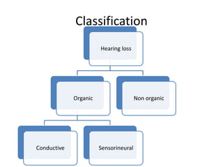
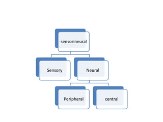
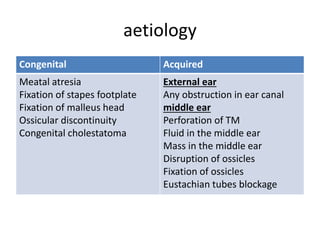
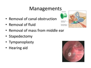
Hearing loss
Name: Nur Aisyah Binti Idris
ID. No.: 068
Classification
Hearing loss
Organic
Conductive Sensorineural
Non organic
sensorineural
Sensory Neural
Peripheral central
Conductive hearing loss & its
management
• Disease process which interfere with the
conduction of sound to reach cochlea
• Characteristics:
– Negative Rinne’s test
– Weber’s test-lateralised to poorer ear
– Normal ABC
– Low frequency affected more
– Audiometry-BC>AC with air bone gap
– Loss is not more than 60dB
– Speech discrimination is good
Average hearing loss seen in different
lesion of conductive apparatus
• Complete obstruction of ear canal-30dB
• Perforation of TM-10-40dB
• Ossicular interruption with intact drum-54dB
• Ossicular interruption with perforation-38dB
• Malleus fixation- 10-25dB
• Closure of oval window- 60dB
aetiology
Congenital Acquired
Meatal atresia
Fixation of stapes footplate
Fixation of malleus head
Ossicular discontinuity
Congenital cholestatoma
External ear
Any obstruction in ear canal
middle ear
Perforation of TM
Fluid in the middle ear
Mass in the middle ear
Disruption of ossicles
Fixation of ossicles
Eustachian tubes blockage
Managements
• Removal of canal obstruction
• Removal of fluid
• Removal of mass from middle ear
• Stapedectomy
• Tympanoplasty
• Hearing aid
Tympanoplasty
• To eradicate disease in middle ear
• To reconstruct hearing mechanism
• Type of middle ear reconstruction depends
damage present
– Repair only the TM (myringoplasty)
– To reconstruct ossicular chain ( ossiculoplasty)
– Both ( tympanoplasty)
Types
• Wullstein
Type 1 Perforation of TM which repaired with a graft
Type 2 Perforation of TM with erosion of malleus.
Type 3 Malleus & incus absent. Graft place directly on stapes
head (gastopedioplexy/ columella tympanoplasty)
Type 4 Only footplate of stapes is present. Graft place between
oval & round window.
Type 5 Stapes footplate is fixed but round window is
functioning.
Another window is created & covered with graft
(fenestration operation)
Myringoplasty
• Repair of TM
• Graft material: temporalis fascia,
perichondrium, dura, vein, fascia, cadever TM
• 2 technique: underlay, overlay
Ossicular reconstruction
• Used of autograft incus or cartilage,
homograft ossicles, prosthetic implants
• Common technique are incus transposition,
sculptured ossicle
• Most common ossicular fixation is stapes
footplate and head of malleus
• Prosthetic implants : TORP, PORP
Sensorineural hearing loss & its
management
• Result from lesion of cochlea, VIIIth nerve
• Can be congenital / acquired
• Characteristic
– Positive Rinne’s test
– Weber’s test-lateralized to better ear
– BC reduced on schwabach & ABC
– high frequency affected more
– Audiometry-no gap between air &bone conduction curve
– Loss is more than 60dB
– Speech discrimination is poor
– Difficult in hearing in the presence of noise
Aetiology
Congenital Acquired
Annomalies of inner ear
Damage to the hearing
apparatus
Genetic / non genetic
Manifest late & affect only the
hearing
Infection of labyrinth
Trauma to labyrinth / VIIIth nerve
Noise-induced hearing loss
Ototoxic drugs
Prebyscusis
Meniere’s disease
Acoustic neuroma
Sudden hearing loss
Familial progressive SNHL
Systemic disoreder
Diagnosis
• History- congenital/ acquired
• Severity of deafness- mild/ mod., mod.
Severe, severe, profound or total
• Type of audiogram- high frequency/low/ mid-
frequency/ flat
• Site of lesion- cochlea/ retrocochlea/ central
• Lab. Test
Management
• Early detection is important
• Syphilis of inner ear- penicillin & steroid
• Hypothyroidism- reversed with replacement
therapy
• Serous labyrinthitis-treat the infection
• SNHL due to perilymph fistula- surgical sealing
the fistula with fat
• Ototoxic drugs- discontinued the drugs
• Noise induced HL- removed from noisy
surroundings
Specific
forms of
hearing
loss
Inflammation
of labyrinth
Familial
progressive
SNHL
ototoxicity
Noise
trauma
Autoimmune
inner ear
disease
Sudden
hearing loss
presbycusis
Inflammation of labyrinth
• Viral, bacterial, syphilitic
• Viral labyrinth
– By blood stream affecting stria vascularis,
endolymph, organ of corti
– Measles, mumps, CMV, rubella, herpes zoster,
herpes simplex, epstein-barr, influenza
• Bacterial
– Through middle ear, CSF
– Invade along nerves, vessels, cochlear aqueduct
Syphilitic
• Congenital/acquired syphilis
• Congenital: early form (age 2), late form (age 8-20)
• Cause:
– sudden SNHL- uni/bilateral
– Mienere’s syndrome
– Henneber’s sign
– Tullio phenomenon
• Diagnosis:
– clinical evidence of late syphilis
– Lab test: VDRL, RPR, FTA-ABS
• Treatment
– Penicillin & steroid
Familial progressive SNHL
• Genetic disorder
• Prog. degenerative of cochlea
• Hearing loss - bilateral with flat audiogram
• Excellent speech discrimination
Ototoxicity
• Drugs & chemical that damage the inner ear
• SNHL, tinnitus, vertigo
Aminoglycoside antibiotic:
• streptomycin, gentamicin, tobramycin
(vestibulotoxic)
• Neomycin, kanamycin, amikacin, sisomycin,
dihydrostreptomycin (cochleotoxic)
Having impaired renal function
Elderly people age 65
Concomitantly receiving other ototoxic drugs
High dose of ototoxic drugs with high serum level of
drugs
Genetic susceptible to aminoglycoside
Diuretics
• Furosemide, bumetanide, ethacrynic acid
• Cause edema & cystic changes in stria
vascularis of cochlear duct
• Bilateral & symmetrical, sudden in onset
Salicylates
• Tinnitus, bilateral SNHL –high frequency
• Cochlear involvement
• Interfere at enzymatic level
• Quinine, chloroquine, hydroxychloroquine
• Cytotoxic drugs
Topical ear drops
• Damage to cochlea by absorption through OW
& RW
• Deafness occur with the use of chlorhexidine,
containing aminoglycoside
• Polymycin B, propylene glycol & antifungal
agents
Noise trauma
• Hearing loss A/W exposure to noise
• 2 groups:
• Acoustic trauma
• Permanent damage to hearing due to single brief
exposure (impulse noise)
• E.g.: explosion, gun fire, powerful cracker
• Noise induced hearing loss
• Hearing loss due to chronic exposure to less intense
sounds
• Damage hair cells starting from basal turn of cochlea.
Outer hair cells affected before inner hair cells
Factors affecting damage by noise trauma
 Frequency of noise
 Intensity and duration of noise
 Continuous noise
 Susceptibility of the individual
 Pre-existing ear disease
Autoimmune inner ear disease
• Prog. bilateral SNHL
• Between 40-50 years, equal incidence of sexes
• Vestibular symptoms: disequilibrium, motion
intolerance, positional/ episodic vertigo
• Investigation: audiogram, speech audiogram,
evoked response audiometry, contrast
enhanced MRI, blood test, western blot assay
for ant-Hsp 70
Bilateral SNHL =/> 30dB at any frequency & evidence of
progression in atleast one ear on 2 serial audiogram that
are done at = /< 3 months apart
Treatment
• Prednisolone 1mg/kg/day upto 60mg/day (4weeks)
No response, tapered off in 12 days
Continue in maintenance does of 10-20mg every alternate day
(6month)
• Methotraxate 15mg/week (6-8weeks)
If respond, Continue (6month)
If not response, discontinued
• Other: cyclophosphomide, intratympanic steroid inj.,
systemic IgG inj., plasmapheresis
Sudden hearing loss
• 30dB or > of SNHL over at least 3 contiguous
frequency occurring within a period of 3 days
or less
• Unilateral, tinnitus, temporary spell of vertigo
• Aetiology: idiopahtic , viral, vascular, ruptured
of cochlear membrane
Need to exclude:
Infection In
trauma The
vascular Very
ear Ear
toxic Too
Neoplastic No
Miscellaneous Major
Psychogenic Pathology
Management:
• Investigation: audiometry, vestibular test, imaging
studies of temporal bones, ESR, test for syphilis,
diabetes, hypothyroidism, blood disorder, lipid profile
• Exploratory tympanotomy required perilymph fistula
• Empirical treatment: bed rest, steroid therapy,
inhalation of carbogen, Vasodilator drugs, LMW
dextran, hyperbaric oxygen therapy, low-salt diet &
diuretics, intratympanic steroid therapy
Prognostic factors in sudden SNHL
Good prognosis Bad prognosis
Mild loss
Low & medium frequency loss
Recovery starting in 2 weeks
No history of vertigo
Young patients
Early treatment
Severe loss
High frequency loss
Recovery does not start in 2
weeks
history of vertigo
older patients
Late treatment
Presbycusis
• SNHL A/W physiological aging process in the
ear
• Manifest age 65 years
• Hereditary predisposition, chronic noise
exposure, generalized vascular disease
• Difficulty in hearing in the presence of
background noise, all sounds become
intolerable in raised volume
• tinnitus
Types• 4 types:
• Sensory-degeneration of organ of corti, basal coilapex, high
frequency affected, speech discrimination good
• Neural-degeneration of cells of spiral ganglion, basal
coilapex, neurons may also affected, high tone loss, speech
discrimination poor & out of proportion to pure tone loss
• Strial or metabolic-atrophy of stria vascularis, physical &
chemical process of energy production affected, runs in
families, audiogram flat, speech discrimination is good
• Cochlear conductive-stiffening of basilar membrane,
audiogram sloping types
Treatments
• Hearing aid
• Speech reading through visual cues
• Curtailment of smoking & stimulants
decrease tinnitus
Nonorganic hearing loss
• Hearing loss due to no organic lesion
• Malingering/psychogenic
• Patients may present:
– Total hearing loss in both ears
– Total loss only one ear
– Exaggerated loss in one / both ears
Diagnose
• Inconsistent result on repeat pure tone &
speech audiometry test
• Absence of shadow curve
• Inconsistency in PTA and SRT
• Strenger test
• Acoustic reflex threshold
• Electric response audiometry
References
• Disease of ear, nose and throat,6th edition,
shruti dhingra
Thank you 

Hearing loss

  • 1.
    Hearing loss Name: NurAisyah Binti Idris ID. No.: 068
  • 2.
  • 3.
  • 4.
    Conductive hearing loss& its management • Disease process which interfere with the conduction of sound to reach cochlea • Characteristics: – Negative Rinne’s test – Weber’s test-lateralised to poorer ear – Normal ABC – Low frequency affected more – Audiometry-BC>AC with air bone gap – Loss is not more than 60dB – Speech discrimination is good
  • 5.
    Average hearing lossseen in different lesion of conductive apparatus • Complete obstruction of ear canal-30dB • Perforation of TM-10-40dB • Ossicular interruption with intact drum-54dB • Ossicular interruption with perforation-38dB • Malleus fixation- 10-25dB • Closure of oval window- 60dB
  • 6.
    aetiology Congenital Acquired Meatal atresia Fixationof stapes footplate Fixation of malleus head Ossicular discontinuity Congenital cholestatoma External ear Any obstruction in ear canal middle ear Perforation of TM Fluid in the middle ear Mass in the middle ear Disruption of ossicles Fixation of ossicles Eustachian tubes blockage
  • 7.
    Managements • Removal ofcanal obstruction • Removal of fluid • Removal of mass from middle ear • Stapedectomy • Tympanoplasty • Hearing aid
  • 8.
    Tympanoplasty • To eradicatedisease in middle ear • To reconstruct hearing mechanism • Type of middle ear reconstruction depends damage present – Repair only the TM (myringoplasty) – To reconstruct ossicular chain ( ossiculoplasty) – Both ( tympanoplasty)
  • 9.
    Types • Wullstein Type 1Perforation of TM which repaired with a graft Type 2 Perforation of TM with erosion of malleus. Type 3 Malleus & incus absent. Graft place directly on stapes head (gastopedioplexy/ columella tympanoplasty) Type 4 Only footplate of stapes is present. Graft place between oval & round window. Type 5 Stapes footplate is fixed but round window is functioning. Another window is created & covered with graft (fenestration operation)
  • 11.
    Myringoplasty • Repair ofTM • Graft material: temporalis fascia, perichondrium, dura, vein, fascia, cadever TM • 2 technique: underlay, overlay
  • 12.
    Ossicular reconstruction • Usedof autograft incus or cartilage, homograft ossicles, prosthetic implants • Common technique are incus transposition, sculptured ossicle • Most common ossicular fixation is stapes footplate and head of malleus • Prosthetic implants : TORP, PORP
  • 14.
    Sensorineural hearing loss& its management • Result from lesion of cochlea, VIIIth nerve • Can be congenital / acquired • Characteristic – Positive Rinne’s test – Weber’s test-lateralized to better ear – BC reduced on schwabach & ABC – high frequency affected more – Audiometry-no gap between air &bone conduction curve – Loss is more than 60dB – Speech discrimination is poor – Difficult in hearing in the presence of noise
  • 15.
    Aetiology Congenital Acquired Annomalies ofinner ear Damage to the hearing apparatus Genetic / non genetic Manifest late & affect only the hearing Infection of labyrinth Trauma to labyrinth / VIIIth nerve Noise-induced hearing loss Ototoxic drugs Prebyscusis Meniere’s disease Acoustic neuroma Sudden hearing loss Familial progressive SNHL Systemic disoreder
  • 16.
    Diagnosis • History- congenital/acquired • Severity of deafness- mild/ mod., mod. Severe, severe, profound or total • Type of audiogram- high frequency/low/ mid- frequency/ flat • Site of lesion- cochlea/ retrocochlea/ central • Lab. Test
  • 17.
    Management • Early detectionis important • Syphilis of inner ear- penicillin & steroid • Hypothyroidism- reversed with replacement therapy • Serous labyrinthitis-treat the infection • SNHL due to perilymph fistula- surgical sealing the fistula with fat • Ototoxic drugs- discontinued the drugs • Noise induced HL- removed from noisy surroundings
  • 18.
  • 19.
    Inflammation of labyrinth •Viral, bacterial, syphilitic • Viral labyrinth – By blood stream affecting stria vascularis, endolymph, organ of corti – Measles, mumps, CMV, rubella, herpes zoster, herpes simplex, epstein-barr, influenza • Bacterial – Through middle ear, CSF – Invade along nerves, vessels, cochlear aqueduct
  • 20.
    Syphilitic • Congenital/acquired syphilis •Congenital: early form (age 2), late form (age 8-20) • Cause: – sudden SNHL- uni/bilateral – Mienere’s syndrome – Henneber’s sign – Tullio phenomenon • Diagnosis: – clinical evidence of late syphilis – Lab test: VDRL, RPR, FTA-ABS • Treatment – Penicillin & steroid
  • 21.
    Familial progressive SNHL •Genetic disorder • Prog. degenerative of cochlea • Hearing loss - bilateral with flat audiogram • Excellent speech discrimination
  • 22.
    Ototoxicity • Drugs &chemical that damage the inner ear • SNHL, tinnitus, vertigo Aminoglycoside antibiotic: • streptomycin, gentamicin, tobramycin (vestibulotoxic) • Neomycin, kanamycin, amikacin, sisomycin, dihydrostreptomycin (cochleotoxic) Having impaired renal function Elderly people age 65 Concomitantly receiving other ototoxic drugs High dose of ototoxic drugs with high serum level of drugs Genetic susceptible to aminoglycoside
  • 23.
    Diuretics • Furosemide, bumetanide,ethacrynic acid • Cause edema & cystic changes in stria vascularis of cochlear duct • Bilateral & symmetrical, sudden in onset Salicylates • Tinnitus, bilateral SNHL –high frequency • Cochlear involvement • Interfere at enzymatic level
  • 24.
    • Quinine, chloroquine,hydroxychloroquine • Cytotoxic drugs Topical ear drops • Damage to cochlea by absorption through OW & RW • Deafness occur with the use of chlorhexidine, containing aminoglycoside • Polymycin B, propylene glycol & antifungal agents
  • 25.
    Noise trauma • Hearingloss A/W exposure to noise • 2 groups: • Acoustic trauma • Permanent damage to hearing due to single brief exposure (impulse noise) • E.g.: explosion, gun fire, powerful cracker • Noise induced hearing loss • Hearing loss due to chronic exposure to less intense sounds • Damage hair cells starting from basal turn of cochlea. Outer hair cells affected before inner hair cells Factors affecting damage by noise trauma  Frequency of noise  Intensity and duration of noise  Continuous noise  Susceptibility of the individual  Pre-existing ear disease
  • 26.
    Autoimmune inner eardisease • Prog. bilateral SNHL • Between 40-50 years, equal incidence of sexes • Vestibular symptoms: disequilibrium, motion intolerance, positional/ episodic vertigo • Investigation: audiogram, speech audiogram, evoked response audiometry, contrast enhanced MRI, blood test, western blot assay for ant-Hsp 70 Bilateral SNHL =/> 30dB at any frequency & evidence of progression in atleast one ear on 2 serial audiogram that are done at = /< 3 months apart Treatment • Prednisolone 1mg/kg/day upto 60mg/day (4weeks) No response, tapered off in 12 days Continue in maintenance does of 10-20mg every alternate day (6month) • Methotraxate 15mg/week (6-8weeks) If respond, Continue (6month) If not response, discontinued • Other: cyclophosphomide, intratympanic steroid inj., systemic IgG inj., plasmapheresis
  • 27.
    Sudden hearing loss •30dB or > of SNHL over at least 3 contiguous frequency occurring within a period of 3 days or less • Unilateral, tinnitus, temporary spell of vertigo • Aetiology: idiopahtic , viral, vascular, ruptured of cochlear membrane Need to exclude: Infection In trauma The vascular Very ear Ear toxic Too Neoplastic No Miscellaneous Major Psychogenic Pathology
  • 28.
    Management: • Investigation: audiometry,vestibular test, imaging studies of temporal bones, ESR, test for syphilis, diabetes, hypothyroidism, blood disorder, lipid profile • Exploratory tympanotomy required perilymph fistula • Empirical treatment: bed rest, steroid therapy, inhalation of carbogen, Vasodilator drugs, LMW dextran, hyperbaric oxygen therapy, low-salt diet & diuretics, intratympanic steroid therapy
  • 29.
    Prognostic factors insudden SNHL Good prognosis Bad prognosis Mild loss Low & medium frequency loss Recovery starting in 2 weeks No history of vertigo Young patients Early treatment Severe loss High frequency loss Recovery does not start in 2 weeks history of vertigo older patients Late treatment
  • 30.
    Presbycusis • SNHL A/Wphysiological aging process in the ear • Manifest age 65 years • Hereditary predisposition, chronic noise exposure, generalized vascular disease • Difficulty in hearing in the presence of background noise, all sounds become intolerable in raised volume • tinnitus
  • 31.
    Types• 4 types: •Sensory-degeneration of organ of corti, basal coilapex, high frequency affected, speech discrimination good • Neural-degeneration of cells of spiral ganglion, basal coilapex, neurons may also affected, high tone loss, speech discrimination poor & out of proportion to pure tone loss • Strial or metabolic-atrophy of stria vascularis, physical & chemical process of energy production affected, runs in families, audiogram flat, speech discrimination is good • Cochlear conductive-stiffening of basilar membrane, audiogram sloping types
  • 32.
    Treatments • Hearing aid •Speech reading through visual cues • Curtailment of smoking & stimulants decrease tinnitus
  • 33.
    Nonorganic hearing loss •Hearing loss due to no organic lesion • Malingering/psychogenic • Patients may present: – Total hearing loss in both ears – Total loss only one ear – Exaggerated loss in one / both ears
  • 34.
    Diagnose • Inconsistent resulton repeat pure tone & speech audiometry test • Absence of shadow curve • Inconsistency in PTA and SRT • Strenger test • Acoustic reflex threshold • Electric response audiometry
  • 35.
    References • Disease ofear, nose and throat,6th edition, shruti dhingra Thank you 