
Recommended
Recommended
More Related Content
What's hot
What's hot (20)
Similar to Osteoarthritis of the Foot and Ankle
Similar to Osteoarthritis of the Foot and Ankle (20)
More from OARSI
More from OARSI (20)
Recently uploaded
Recently uploaded (20)
Osteoarthritis of the Foot and Ankle
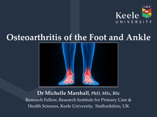
- 1. Osteoarthritis of the Foot and Ankle Dr Michelle Marshall, PhD, MSc, BSc Research Fellow, Research Institute for Primary Care & Health Sciences, Keele University, Staffordshire, UK
- 2. I have no financial relationships with commercial interests to disclose Disclosure Information
- 3. Overview Foot & ankle OA • Definitions • Prevalence & incidence • Burden • Phenotypes • Risk Factors • Diagnosis • Long-term course • Management
- 4. Background Number of annual research study publications in PubMed related to OA by joint site 0 500 1000 1500 2000 2500 3000 Numberofpublications Year Knee OA Hip OA Hand OA Foot OA Ankle OA
- 5. Atlas Knee Hip Hand Foot Ankle Kellgren & Lawrence (1963) Lane et al. (1993) Spector et al. (1994) OARSI atlas - Altman et al. (1995 & 2007) La Trobe Atlas - Menz et al. (2007) Kraus et al. (2015) La Trobe Atlas extension - Murray et al. (2018) Radiographic OA atlases
- 6. 1st MTPJ: first metatarsophalangeal joint 1st CMJ: first cuneo-metatarsal joint 2nd CMJ: second cuneo-metatarsal joint NCJ: navicular-first cuneiform joint TNJ: talo-navicular joint Foot: La Trobe Radiographic Atlas (Menz et al. 2007 OAC)
- 7. 1st MTPJ: first metatarsophalangeal joint 1st CMJ: first cuneo-metatarsal joint 2nd CMJ: second cuneo-metatarsal joint NCJ: navicular-first cuneiform joint TNJ: talo-navicular joint Foot: La Trobe Radiographic Atlas (Menz et al. 2007 OAC) • Views: weight-bearing DP & lateral • Features: osteophytes & JSN graded 0-3 • Reliability: Intra-rater k=0.52-0.95 Inter-rater k=0.13-0.87
- 8. Ankle: radiographic atlases Kraus et al. 2015 OAC Murray et al. 2018 PLOS ONE Joints: Tibiotalar, Talofibular & Subtalar Tibiotalar & Talofibular Views: Weight-bearing AP (Mortise) & Lateral Weight-bearing AP & Lateral Features: Osteophytes & JSN, Graded 0-3 Osteophytes & JSN, Graded 0-3 Reliability: Intra-rater k=0.15-0.94 Inter-rater k=0.09-0.72 Intra-rater k=0.87 Inter-rater k=0.30
- 10. How prevalent is foot & ankle OA?
- 11. Definitions foot & ankle OA • Symptoms e.g. pain, aching, stiffness
- 12. Definitions foot & ankle OA • Symptoms e.g. pain, aching, stiffness Population Prevalence Foot Pain 24% Population Prevalence Ankle Pain 15% (Thomas et al. 2011 Pain)
- 13. Definitions foot & ankle OA • Symptoms e.g. pain, aching, stiffness • Structural changes e.g. radiographic
- 14. Systematic reviews: prevalence of foot & ankle OA Trivedi et al. 2010 OAC Murray et al. 2018 PLoS One Region Foot Ankle No. studies 27 18 ≥2 x-ray views 22% 33% Weight bearing x-rays 22% 39% Assessment of radiographic OA 74% KL 7 other methods 14 different methods Joints 1st MTPJ = 74% Ankle Population prevalence of radiographic OA 6-39% 1st MTPJ in middle–older adults None
- 15. Definitions foot & ankle OA • Symptoms e.g. pain, aching, stiffness • Structural changes e.g. radiographic • Symptomatic (radiographic & symptoms)
- 16. Systematic reviews: prevalence of foot & ankle OA Trivedi et al. 2010 OAC Murray et al. 2018 PLoS One Region Foot Ankle No. studies 27 18 Two x-ray views 22% 33% Weight bearing x-rays 22% 39% Assessment of radiographic OA 74% KL 7 other methods 14 different methods Joints 1st MTPJ = 74% Ankle Population prevalence of radiographic OA 6-39% 1st MTPJ in middle–older adults None Population prevalence of symptomatic OA None None
- 18. Clinical Assessment Study of the Foot (CASF) • Postal survey to all adults aged 50 years & over • 4 general practices in North Staffordshire, UK • Adjusted response rate 56% (n=5109) • 1635 reported pain in an around the foot ≤12 months • 560 attended clinic where x-rays were obtained (Roddy et al. 2011 JFAR) X X
- 19. In adults aged 50 years and over: (Roddy et al. 2015 ARD; Murray et al. 2018 PLoS One) Foot Ankle Symptomatic OA (radiographic & pain in last month) 16.7% 3.4% Population prevalence of OA
- 20. 1st MTPJ 7.8% 1st CMJ 3.9% 2nd CMJ 6.8% NCJ 5.2% TNJ 5.8% (Roddy et al. 2015 ARD) Prevalence: symptomatic radiographic OA Midfoot 12.0%
- 21. Incidence Clearwater OA Study • N=1592 adults aged 40-91 free of 1st MTPJ OA • Mean follow-up 7 years • Incident radiographic OA (K&L ≥2) 25% in left 1st MTPJ 27% in right 1st MTPJ (Mahiquez et al. 2006 Foot Ankle Int) Chingford Study • Follow-up 19 years • Incident radiographic OA (La Trobe ≥2) 7% in left 1st MTPJ 17% in right 1st MTPJ (OARSI 2019 Poster 367)
- 22. What is the burden of foot & ankle OA?
- 23. Burden of foot & ankle OA • 69% experience disabling pain (Roddy et al. 2015 ARD) • Functional limitation & impairment in balance, strength & locomotor ability (Menz et al. 2001 J Am Podiatr Med Assoc) • Disabling foot pain is a risk factor for falls (Menz et al. 2006 J Gerontol A Biol Sci Med Sci) • Poorer physical & social health (Bergin et al. 2012 AC&R) • Reduced ability to work (OR=1.9) (Sayre et al. 2010 PLoS One) • Common cause for GP consultation; 8% MSK consultations (Menz et al. 2010 Rheumatol)
- 24. Are there any identifiable patterns of joint involvement?
- 25. Across both feet: • 42% multiple (≥2) joints affected • OA clustered across both feet • OA was highly symmetrical 0-20.7 1st MTPJ OR = 10.5 1st CMJ OR = 11.5 2nd CMJ OR = 10.0 NCJ OR = 20.7 TNJ OR = 10.3 (Rathod et al. 2016 AC&R) Patterns of foot OA
- 26. Within a foot: (Rathod et al. 2016 AC&R) Patterns of foot OA
- 27. No/minimal foot OA Isolated 1st MTPJ OA Polyarticular OA class size - n (%) 339 (64%) 112 (22%) 82 (15%) (Rathod et al. 2016 AC&R)
- 28. No/minimal foot OA Isolated 1st MTPJ OA Polyarticular OA class size - n (%) 339 (64%) 112 (22%) 82 (15%) % Foot pain on most / all days in last month 50% 51% 69% Mean foot pain severity (NRS 0-10)† 5.2 4.9 6.0 MFPDI Function score (-2 to 2) † -0.7 -0.9 0.0 † Higher scores indicate greater pain and functional difficulties
- 29. No/minimal foot OA Isolated 1st MTPJ OA Polyarticular OA class size - n (%) 339 (64%) 112 (22%) 82 (15%) % Female Sex 52% 54% 77% Mean Age (years) 63.9 66.1 67.3 Mean BMI (kg/m2) 29.9 30.1 32.5 % Nodal OA 21% 22% 34%
- 30. Risk factors for foot & ankle OA
- 31. Clinical Assessment Study of the Foot (CASF) (Roddy et al. 2015 ARD; Thomas et al. 2015 OAC; Murray et al. 2018 PLoS One) Foot 1st MTPJ Midfoot Ankle Gender: Men Women 14.3% 18.9% 6.7% 8.8% 10.3% 13.7% 2.9% 3.9% Age: 50-64 65-74 ≥75 15.9% 17.0% 18.5% 6.9% 8.7% 9.0% 11.8% 11.1% 14.4% 3.6% 3.2% 3.1% Socio-economic class: Managerial & professional Intermediate occupations Routine & manual 10.2% 17.9% 18.2% 4.8% 8.7% 8.3% 6.9% 12.6% 13.3% 2.4% 3.0% 4.1% Evidence of possible risk factors
- 32. 1st MTPJ • Bone shape & size differences (Zammit et al. 2009 J Orthop Sports Phys Ther) • Increasing 1st MTPJ OA severity was associated with hallux valgus & greater foot pronation (Menz et al. 2015 OAC) • Individuals with >5o foot pronation 23% more likely to develop 1st MTPJ OA than normal alignment (Mahiquez et al. 2006 Foot & Ankle Int) Evidence of possible risk factors Arch Index
- 33. Midfoot Case control study midfoot OA associated with: • Increase pronation • Increased midfoot pressures (Menz et al. 2010 OAC) CASF Study - Symptomatic midfoot OA associated with: • Pain in other lower limb joints = aOR 8.5 • Obesity (BMI ≥30 vs <30) = aOR 2.0 • Diabetes = aOR 1.9 • Previous injury/trauma = aOR 1.6 (Thomas et al. 2015 AR&T) Evidence of possible risk factors
- 34. Ankle Tertiary care patients with KL grades 3-4 • 70-78% Ankle OA is post-traumatic – Malalignment – Instability – Incongruity • 13-23% Secondary OA • 7-9% Primary OA (Saltzman et al. 2005 Iowa Orthop J; Valderranano et al. 2009 Clin Orthop Relat Res; Nelson et al. 2017 JFAR) Evidence of possible risk factors
- 35. Evidence of possible risk factors Foot 1st MTPJ Midfoot Ankle Female sex Older age ? Lower socioeconomic status Bone shape & size Foot posture, alignment or deformity Injury/trauma Obesity Diabetes
- 36. Diagnosis of foot & ankle OA
- 37. Symptoms • Pain • Stiffness Clinical signs • Swelling • Pain on palpation • Limited range of motion • Crepitus • Dorsal exotosis Diagnosis foot & ankle OA
- 38. • N=181 people with 1st MTPJ pain • 77% radiographic OA • Diagnostic variables – pain > 25 months – dorsal exostosis of 1st MTPJ – hard-end feel – Crepitus – <64 degrees of dorsiflexion • ≥3 of these observations – sensitivity 88%, specificity 71%, AUC 0.87 (Zammit et al. 2011 OAC) Diagnosis 1st MTPJ OA
- 39. • N=274 people with midfoot pain • 43% symptomatic radiographic OA • Diagnostic variables – Older age – Female sex – Increased BMI – Increased arch index (flatter foot) • Poor model fit – sensitivity 30%, specificity 88%, AUC 0.64 (Thomas et al. 2015 OAC) Diagnosis midfoot OA
- 40. Course of foot & ankle OA
- 41. Radiographic course • Clearwater OA Study • 36-42 months • Progression of 1st MTPJ = 21-29% & 1st CMJ = 3-7% (Wilder et al. 2005 J Am Podiatr Med Assoc) • Chingford Study • 19 years • Progression of 1st MTPJ = 29% in left & 35% in right (OARSI 2019 Poster 367) Course of foot OA Symptomatic course • CASF study • 18 months • Few differences seen in pain and function (Downes et al. 2018 AC&R)
- 42. Management of foot & ankle OA (non-surgical)
- 43. • Physical therapy – 1st MPTPJ OA – Package physical interventions vs package plus strengthening exercises, gait training & sesamoid mobilisation, 12 sessions in 4wks (n=20) (Shamus et al. 2004 J Orthop Sports Phys Ther) • Footwear & foot orthoses – 1st MTPJ OA – RCT Rocker-soled footwear vs foot orthoses (n=88) (Menz et al. 2016 AC&R) – Midfoot OA - feasibility pilot RCT functional foot orthoses vs sham orthoses (n=33) (Halstead et al. 2016 Clin Rheumatol) Management foot & ankle OA
- 44. Analgesia • No evidence use of paracetamol (acetaminophen); topical NSAIDs & capsaicin Oral NSAIDs • 2 studies in foot OA similarly effective – 1000mg naproxen vs 20mg piroxicam for 8 wks (Jennings 1994 J Am Podiatr Med Assoc) – 1000mg naproxen vs 800mg etodolac for 5wks (Jennings 1997 Lower Extremity) Management foot & ankle OA
- 45. Intra-articular Injections • Systematic review use in ankle OA – 27 studies inc 7 RCTs evaluated Hyaluronic acid – Pooled results 3 studies (n=109) Hyaluronic acid vs placebo (saline) significantly improved pain, function & stiffness over 6m (Vannabouathong et al. 2018 Foot & Ankle Int) • 2 RCTs 1st MTPJ – Hyaluronic acid vs corticosteroid over 3m (n=37) (Pons et al. 2007 Foot & Ankle Int) – Hyaluronic acid vs saline over 6m (n=151) (Munteanu et al. 2011 ARD) Management foot & ankle OA
- 46. Summary & future directions • Foot & ankle OA have been relatively neglected • Symptomatic radiographic foot OA is a common problem; ankle OA less common. • Foot & ankle OA are disabling, & adversely affects physical & social functioning, & ability to work • Ankle OA accepted post traumatic form; isolated 1st MTPJ OA & polyarticular OA may be different clinical entities • Longitudinal studies are needed to investigate risk factors, & the incidence, progression & prognosis of foot and ankle OA • Limited evidence for the conservative management of foot and ankle OA; further RCTs are needed.
- 47. International Foot and Ankle OA Consortium Sat 4th May 6.30pm In Chesnut East
- 48. Acknowledgments Keele University • Prof George Peat • Prof Hylton Menz • Dr Edward Roddy • Dr Martin Thomas • Trishna Rathod • Dr Milisa Bucknall-Blagojevic • Dr Charlotte Murray • Dr Thomas Downes • Dr Bansari Trivedi Funding for the CASF study: • Arthritis Research UK (now VERSUS ARTHRITIS) • Service support costs through West Midlands North CLRN
- 50. Any Questions
Editor's Notes
- I would like to thank the organisers for inviting me to talk at OARSI. This afternoon, I am going to provide you with an overview of foot and ankle OA, an area that has been relatively neglected, but which I am glad to say has a growing interest.
- I have no conflicts of interest to declare.
- In this presentation, I am going to describe the definitions that can be used, the estimates of prevalence & incidence that have been obtained for foot & ankle OA, describe what is known about the disease burden, the possible different phenotypes that might exist, and the potential risk factors that have been investigated. I will also examine how OA at these sites might be diagnosed, & present evidence on the course & possible management options for foot & ankle OA.
- Since the 1950s, research has been published annually in small numbers at the different joint sites, while we have seen a rise in publications for knee & hip OA since the 1990s, there has only been a small increase in the number of annual publications for hand, foot & ankle OA. While the foot and ankle have been relatively neglected, we should note that not all research needs to be replicated at each joint site, and insights from other joints can be helpful.
- A significant barrier to the study of foot & ankle OA has been the lack of specific grading systems to define structural changes. The studies that were undertaken used the generic Kellgren & Lawrence system, which although it has allowed different joint sites to be compared, it has been criticised for its over reliance on the presence of an osteophyte & the assumed chronological development of features. The rise in number of studies examining foot & ankle OA is in part due to recent publication of specific radiographic atlases for the foot & ankle, which up to this point were notably absent.
- The La Trobe radiographic foot atlas scores five joints: the 1st metatarsophalangeal joint, the 1st & 2nd cuneo-metatarsal joints, the navicular 1st cuneiform joint & the talo-navicular joint, on weight bearing, dorso-plantar & lateral views.
- It scores osteophytes & joint space narrowing in each joint on a 0-3 scale. A joint is defined as having OA if it has grade ≥2 for either feature on either view. The atlas was found to have moderate to excellent intra-rater reliability, the inter-rater reliability was lower but was not too dissimilar with other reliability studies that have been undertaken at the hip & knee.
- Two radiographic atlases have been developed for use at the ankle. They are largely similar, in that they both use weight-bearing AP & lateral views to grade osteophytes & joint space narrowing on 0-3 scales. However, the Kraus atlas does additionally allow the subtalar joint to be scored. The atlas published by Murray was developed to be an extension of the La Trobe foot atlas, so that the foot & ankle can be examined as a complex.
- As we have seen at the knee and other sites, we know that the direction of travel is to use 3D imaging that allows visualization of the different tissues affected by OA, and recently a CT atlas has been developed for ankle OA, by Cohen & colleagues in Miami, & an MRI atlas has been developed for foot OA, by Jill Halstead & the team in Leeds, in the UK. There has also been study looking at the use of ultrasound in foot OA by an OMERACT task force.
- So how prevalent is foot & ankle OA?
- OA can be defined in different ways, & prevalence estimates will be sensitive to the definitions used, as well as the populations examined. The first set of estimates obtained was based on symptoms.
- A systematic review with meta-analysis of representative populations, using comparable definitions of frequent pain on most days, has estimated that the population prevalence in adults aged 45 years & over for foot pain was 24% & ankle pain was 15%. These estimates are consistent with subsequently completed studies by the Foot Pain Consortium, lead by Lucy Gates which found estimates between 13-36% for foot pain, & in work undertaken by Murray where estimates of 12% were obtained for ankle pain.
- However, we know that symptoms alone may overestimate OA, so definitions of OA can be based on structural changes.
- To date, prevalence estimates for structural change at the foot & ankle have been limited to those determined radiographically. Systematic reviews have examined how radiographic foot & ankle OA has been examined, defined & what prevalence estimates have been obtained. At both sites, there was infrequent use of multiple views & weight-bearing films. In the foot, the joint most commonly examined was the 1st MTP joint & estimates of between 6-39% were found for this joint in middle–older aged adults (≥35 years). In the ankle, in fact no true population prevalence estimates were found, estimates were only reported for selected sporting or medical populations.
- OA can also been defined using a combination of structural change & symptoms commonly referred to as symptomatic OA.
- The systematic reviews found, that up to the point each was carried out, there were no estimates of the prevalence of symptomatic radiographic OA for either the foot or the ankle.
- Based on this, my colleagues & I secured funding & undertook a study at Keele University, in Staffordshire, in the UK.
- In the Clinical Assessment Study of the Foot, CASF, we sent a general health survey to over 9000 adults aged 50 years & over, at 4 general practices in the area. In the UK, 96% of the population is registered with a GP & so they make a convenient sampling frame for the general population. We received responses from over 5000 individuals, a third of whom reported having experienced pain in & around the foot in the last 12 months. These individuals were invited to attend a research clinic where an interview, clinical assessments & x-rays were obtained.
- Using multiple imputation & weighted logistic regression we were able to derive population prevalence estimates for symptomatic radiographic OA in adults aged 50 years and over. Estimates of 17% were obtained for foot OA & 3% for ankle OA. In comparison, meta-analysis completed by Pereira in 2011 obtained estimates of 18% for sym rad OA at the knee, 15% at the hand OA & 6% at the hip.
- We also obtained population prevalence estimates for the foot joints assessed by the radiographic atlas in adults aged 50 years and over. The highest prevalence of 8% was found for the symptomatic radiographic OA in either 1st MTP joint & the lowest was 4% for OA in either 1st CMJ. An estimate of 12% was also obtained for OA in any 1 or more midfoot joints.
- Up till this conference, there had only been 1 study that has examined the incidence of OA. This was in the Clearwater OA Study, a longitudinal cohort study. In the 1592 adults aged 40-91, who were free of 1st MTP OA at baseline, the cumulative incident radiographic OA (K&L ≥2) was 25% in the left & 27% in the right 1st MTP joints over a mean follow-up of 7 years. At this conference, poster 367, work is being presented on the incidence of 1st MTP OA in the Chingford population cohort of women. The cumulative incidence over 19 years was found to be 7% in the left & 17% in the right 1st MTP joints.
- What is the burden of foot & Ankle OA?
- Foot & ankle OA has a significant impact on individuals. 69% people with symptomatic radiographic foot OA have reported experiencing disabling pain (Roddy et al. 2015 ARD) Pain has been shown to result in functional limitation & significant impairment in balance, strength & locomotor ability (Menz et al. 2001 J Am Podiatr Med Assoc) Disabling foot pain is also a significant & independent risk factor for falls (Menz et al. 2006 J Gerontol A Biol Sci Med Sci) Foot & ankle OA leads to poorer physical & social health (Bergin et al. 2012 AC&R) We don’t know much about the economic impact but people with foot OA have been found to have a reduced ability to work (Sayre et al. 2010 PLoS One) And foot & ankle problems are a common cause for consulting a GP, accounting for 8% of musculoskeletal consultations (Menz et al. 2010 Rheumatol)
- The feet are similar to the hands, with numerous small bones & joints. In the hands patterns have been identified & a number of subgroups are recognised. But what about the feet, can we identify any patterns of joint involvement? Better understanding of patterns of involvement across the different joints in the feet & their associated risk factor profiles has the potential to provide new insights into aetiology.
- In our CASF study, we examined the patterning of radiographic OA. Across both feet we found that 42% of the study population had radiographic OA in 2 or more foot joints. We found OA affected joints in both feet, significantly more than was expected by chance. We explored this further & found this might be due to a strong symmetrical patterning, as the odds of having OA in the same joint in both feet were between 10 & 20 fold. This mirrors the findings at the hand and implies the involvement of systemic determinants.
- Within a foot, we found that OA in the 1st MTP joint occurred in 61% of feet in isolation, without OA being presented in other joints in the same foot. In comparison, OA in the NCJ occurred frequently with OA in other joints in the same foot, as was also the case for the 1st & 2nd CM joints.
- Further to this we undertook latent class analysis to examine whether people can be grouped into distinct presentations based on radiographic joint involvement. The model with the best fit was a 3-class solution. The 3 classes were those with no or minimal foot OA – which was the largest group with 64% of the study population, a smaller group of 22% that had isolated 1st MTP OA & a group consisting of 15% had both the midfoot & 1st MTP joints affected which we called polyarticular OA.
- We found these three groups differed in terms of their symptoms. The polyarticular OA group had after adjustment for age & sex had more persistent & severe foot pain & greater functional limitation, as indicated by the higher scores, compared to the no or minimal foot OA & isolated 1st MTP OA groups.
- We also found that after adjustment for age, the polyarticular group had the highest proportion of females. After adjustment for sex, they were also the oldest. After adjustment for both age & sex the polyarticular group also had a higher BMI & more nodal OA than the no or minimal foot OA & the isolated 1st MTP groups. The differences we’ve seen in the symptomatic & risk profiles does suggest a possible distinction between the isolated 1st MTP & Polyarticular forms of foot OA.
- We have seen that sex, age, BMI & nodal OA might be risk factors for foot OA but what other evidence is there of possible risk factors for foot & ankle OA?
- Well, stratification of the prevalence estimates we obtained in our CASF study, indicates that similar to other joint sites, there are trends for higher estimates in women, older ages & individuals of lower socio economic class. The only exception to this was for ankle OA where an increase with age was not seen, however the estimates had wide confidence intervals due to the small numbers with ankle OA.
- Exploring possible risk factors, firstly in the 1st MTP joint. A systematic review found that those with 1st MTP OA had a number of differences in bone size and shape, including longer & wider phalanges, than controls that were free of radiographic disease. In the CASF study, we found that increasing OA severity in the 1st MTP joint was associated with hallux valgus & greater foot pronation that was consistent across 3 different measures of foot posture (foot posture index, arch index & navicular height) In the Clearwater OA study, individuals with >5o ankle pronation were 23% more likely to subsequently develop 1st MTP OA than individuals with normal alignment
- In the midfoot, a case control study (cases n=35, controls n=170) found that increase pronation (Arch index, Radiographic arch measurements) & increased midfoot pressures were associated with midfoot OA. In the CASF study, we found that symptomatic midfoot OA was associated with having pain in other lower limb joints, obesity, diabetes and reporting a previous foot injury or trauma, although the injury was across either foot or ankle, & not specific to the side with OA. The strong association seen between symptomatic midfoot OA and pain in other joints could be indicative of OA at other joints, and a more generalised OA presentation, but as this association was specific to weight-bearing joints in the lower limb this could also suggest that more localised biomechanical factors are at play.
- At the ankle, post-traumatic OA is a recognised form of ankle OA in its own right. Two studies of tertiary care patients with moderate to severe radiographic OA have found that in this setting: The majority of ankle OA is post-traumatic from sprains & intra-articular fractures. This is thought to occur through contributory factors such as chronically altered joint mechanics due to malalignment, instability & incon-gru-ity. Between 13-23% was due to secondary OA related to the presence of other conditions such as RA, hemochromatosis, haemophilia & gout & only 7-9% of ankle OA was thought to be primary OA. Half of individuals with this form were found to have foot deformities such as flat feet or feet with a very high arch. Work by Amanda Nelson on the Johnston County OA project has shown that joint shape differs in people with a history of ankle injury, but whether this predisposes someone to injury or whether it is a consequence of injury, has yet to be determined.
- While there is some indication that the possible person-level & localised risk factors for foot & ankle OA, are similar to those at other joint sites, the majority of this evidence is from single, cross-sectional studies & further longitudinal studies are needed to sort out which are truly risk factors.
- Diagnosing foot & ankle OA.
- The signs & symptoms of foot & ankle OA are similar to those at other joint sites, but evidence from medical record and qualitative studies is that foot and ankle OA is underdiagnosed. Currently there are no accepted, clinical diagnostic criteria, for foot or ankle OA, so imaging remains common, but is it needed to diagnose OA?
- There have been a couple of attempts to develop diagnostic models, one for 1st MTP OA & one for midfoot OA. In a study of 181 people with 1st MTP pain, the presence of three of more features from having, foot pain longer than 25 months, dorsal exostosis (bony enlargement) of the joint, a hard-end feel when the joint was dorsiflexed, crepitus & less than 64 degrees dorsiflexion had a sensitivity of 88%, a specificity of 71% & an area under the curve of 0.87. The model performed well and this would suggest that radiographic imaging is not essential to make a diagnosis of 1st MTP OA.
- In our CASF study, a diagnostic model for the midfoot was investigated. While several foot assessments were associated with the presence of symptomatic radiographic midfoot OA, only an increased arch index made the final model along with demographic & anthropometric characteristics. This model did not perform well, with a sensitivity of 30%, a specificity 88% & an area under the curve of 0.64. So in contrast, to the 1st MTP joint clinical signs and symptoms do not give a good indication of what was found on the radiographs.
- So what do we know about the course of foot & ankle OA over time? Well there have been very few longitudinal observational studies that have examined this.
- Up till this conference, there were only two published studies, one which examined change in symptoms & one which examined radiographic progression. Symptomatic progression of foot OA was examined in the CASF study, but over a very short 18-month period, and few symptomatic differences were seen in pain and function. Radiographic progression of foot OA was examined in the Clearwater OA Study over a longer period of 36-42 months. Progression was found to occur in 1st MTP joint in about 1 in 4 individuals (21-29%) & in the 1st CMJ in about 1 in 20 individuals (3-7%). Work presented in poster 367 at this conference from the Chingford study has found that 29% of left and 35% of right 1st MTP joints progressed radiographically over 19 years.
- Clinically, management of foot & ankle OA generally commences with conservative treatments including pharmaceutical options, but if ineffective then surgery may be considered. There are clinical guidelines for the treatment of 1st MTPJ OA published by the American College of Foot & Ankle Surgeons, but they were published in 2003 & so are quite dated. However, since then, there have only been a small number of RCTs for the conservative treatments in the foot & ankle, which I will now describe.
- Looking first at physical therapy. A package of physical therapy interventions were compared to an enhanced package which additionally included sesamoid mobilisation, gait training, & strengthening exercises, for 1st MTP OA, the treatments were delivered in 12 sessions over 4 wks, the arm with the enhanced package showed significant improvements in strength & function over this time period compared to other arm, although the sample sizes was very small, with only 10 people in each arm (Shamus et al. 2004 J Orthop Sports Phys Ther) There have been two studies looking at use of specialised Footwear & Foot orthoses. - One RCT compared Rocker-soled footwear vs foot orthoses in individuals with 1st MTPJ OA. Rocker soled shoes are thought reduce the amount of dorsi flexion that is needed in the 1st MTP joint. Over 12 weeks both had clinically meaningful reductions in pain but no significant differences were seen between groups (Menz et al. 2016 AC&R). - A further pilot RCT has investigated the use of a semi-rigid contoured foot orthoses to a sham orthoses over a 12-week period & found the semi-rigid contoured orthoses had significant improvements in pain & function compared to the sham orthoses. So there is evidence of some conservative non-pharmaceutical interventions being beneficial to patients with foot OA, however, a study by Kade Paterson found that currently in a primary care setting, Australian GPs predominately manage patients with foot OA pharmaceutically.
- - With regard to pharmacological management, there are no clinical trials of paracetamol, topical NSAIDs & cap-say-cin in foot & ankle OA, but there is no reason to believe that the effects of these drugs would differ from other joints sites. - There have been 2 RCTs comparing the effectiveness of different oral NSAIDs. One over 8 weeks comparing naproxen to pir-ox-icam which found both drugs to be equally effective at reducing pain. Similar results were obtained in the other study which compared naproxen to etod-olac over 5 weeks.
- Intra-articular injections – in comparison to the other treatments the ankle as a site was frequently investigated. - A systematic review of intra-articular injections in ankle OA found that there were 27 studies that examined the use of hyaluronic acid, corticosteroids, platelet-rich plasma & mesen-chymal stem cells, although only 7 were RCTS. All 7 RCTs examined hyaluronic acid. It was possible to pool data from 3 of these studies & meta-analysis found that hyaluronic acid in comparison to a saline placebo led significant improvements in pain, function & stiffness over 6 months but these beneficial effects were not over shorter time periods. - There have been 2 RCTs investigating the use of intra-articular injections for the 1st MTP joint. One compared hyaluronic acid to corticosteroid over 3 months (Pons et al. 2007 Foot & Ankle Int) & the other compared hyaluronic acid to saline over 6 months (Munteanu et al. 2011 ARD). In both trials, all arms had clinically meaningful reductions in pain but no differences were found between the arms in each trial.
- In summary, - Foot and ankle OA until recently has been relatively neglected and so the evidence base lags behind that of other joints - Symptomatic radiographic foot OA is very common, with prevalence estimates that are similar to those for the hand and knee, but ankle OA is less common with estimates that are slightly lower than for hip OA. - They are disabling conditions causing pain, & adversely affecting physical & social functioning, & an individual’s ability to work While there are generally accepted post traumatic, secondary and primary forms of ankle OA, there is emerging evidence that isolated 1st MTP OA and polyarticular OA may be different clinical entities in the foot, that have different risk profiles. However, this needs further investigation in longitudinal studies, along with further research on the potential risk factors, & the incidence and progression of foot and ankle OA. Better understanding of patterns of involvement, risk factor profiles and prognosis, has the potential to provide new insights into the relative contribution of systemic & localised risk factors, which could help guide the devt of new interventions for foot & ankle OA and indicate who could be targeted for treatments. While there is guidance in a number of clinical guidelines for treating a person with OA, there is a limited evidence for the conservative management of foot and ankle OA. Further RCTs of existing and novel interventions are needed to improve treatment options for people affected with these painful and disabling conditions.
- There is an International Foot and Ankle OA Consortium meeting tomorrow at 6:30 PM. If you are interested please come along.
- The CASF study was undertaken by a team of researchers & students at Keele University, so I must acknowledge their contributions as well as their support in the preparation of this presentation. Funding for the CASF study was from a programme grant from Arthritis Research UK, now known as Versus Arthritis.
- Thank you for listening