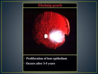
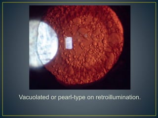
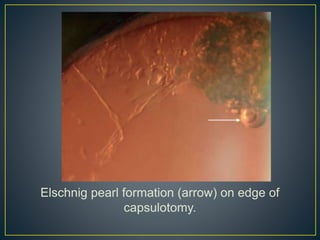
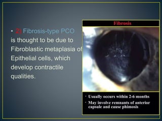
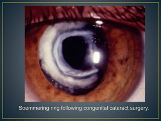
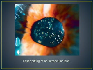
• Visually significant PCO, also known as 'after
cataract', is the most common late complication
of uncomplicated cataract surgery, occurring in
20-25% of patients.
• It is caused by the proliferation of lens epithelial
cells that have remained within the capsular bag
following cataract extraction. The incidence of
PCO is reduced when the capsulorhexis opening
is in complete contact with the anterior surface of
the IOL.
• PMMA “ polymethylmethacrylate “ (and probably
to a lesser extent hydro-gel) IOLs are particularly
prone to PCO. Implant design is more important
than material.
• Symptoms: include persistent slowly worsening
blurring, glare and sometimes monocular
diplopia.
• Visual Acuity is variably reduced, though
dysfunction may be more marked on contrast
sensitivity testing.
• Typically include more than one pattern of opacification.
• 1) Vacuolated (pearl-type) PCO consists of proliferating
swollen lens epithelial cells, similar to the bladder (Wedl)
cells seen in posterior subcapsular cataract . They are
commonly termed ‘Elschnig pearls’.
• particularly when grouped into clusters at the edge of a
capsulotomy, though strictly Hirschberg-Elschnig pearls
refers to globular or grape-like collections of swollen cells
seen following traumatic or surgical anterior capsular
rupture.
Vacuolated or pearl-type on retroillumination.
Elschnig pearl formation (arrow) on edge of
capsulotomy.
• 2) Fibrosis-type PCO
is thought to be due to
Fibroblastic metaplasia of
Epithelial cells, which
develop contractile
qualities.
Slit-lamp appearance of fibrotic capsule.
• 3) A Soemmering ring is a whitish annular or
doughnut-shaped proliferation of residual cells
that classically formed almost in the periphery of
the capsular bag following older methods of
cataract surgery, but is uncommon now. It may
form at the edge of a capsulorhexis or
capsulotomy.
Soemmering ring following congenital cataract surgery.
• Treatment involves the creation of an opening in
the posterior capsule using a Nd:YAG laser
(termed a posterior capsulotomy).
TiP From Kanski
Posterior capsular thickening is the
commonest cause of late visual deterioration
after small incisional cataract surgery and is
easily treated with a Nd:YAG laser.
Appearance following laser capsulotomy.
• Indications: The presence of visual symptoms is the main
indication (reduced VA and glare). Less commonly, capsulotomy
is performed to improve an inadequate fundus view impairing
assessment and treatment of posterior segment pathology.
• Technique: Safe and successful laser capsulotomy involves
accurate focusing and use of the minimum energy required. Laser
power is initially set at 1 mJ/pulse and may be increased if
necessary. A series of punctures is applied in a cruciate or
circumferential pattern using single-pulse shots. The opening
should equate approximately to the size of the physiologically
dilated pupil under scotopic conditions - this should average
around 4-5 mm in the pseudophakic eye. A larger capsulotomy
may be necessary if glare persists, or for retinal examination or
treatment, but the capsulotomy should not extend beyond the
edge of the optic in case vitreous prolapses around its edge. It
may be prudent to adopt a higher threshold for treatment, and
minimizing its extent, in eyes at risk of retinal detachment (e.g.
high myopia), CMO “ Cystoid Macular Oedema “ (e.g. history of
uveitis) or lens displacement (e.g. pseudoexfoliation). Some
research suggests that the total energy applied should be less
than 80 mJ in order to reduce the risk of a significant IOP spike.
• Complications: include pitting of the IOL that is
usually visually inconsequential. The IOP may
rise, particularly in patients with glaucoma, but is
typically mild and transient. A retinal tear or
detachment may follow the treatment and myopic
individuals should be warned to return if they
develop symptoms compatible with a posterior
vitreous detachment (PVD). CMO may occur, but
is less common when the capsulotomy is
delayed for 6 months or more after cataract
surgery. IOL subluxation or dislocation is rare.
Laser pitting of an intraocular lens.
PCO.pptx

PCO.pptx

  • 2.
    • Visually significantPCO, also known as 'after cataract', is the most common late complication of uncomplicated cataract surgery, occurring in 20-25% of patients. • It is caused by the proliferation of lens epithelial cells that have remained within the capsular bag following cataract extraction. The incidence of PCO is reduced when the capsulorhexis opening is in complete contact with the anterior surface of the IOL.
  • 3.
    • PMMA “polymethylmethacrylate “ (and probably to a lesser extent hydro-gel) IOLs are particularly prone to PCO. Implant design is more important than material.
  • 4.
    • Symptoms: includepersistent slowly worsening blurring, glare and sometimes monocular diplopia. • Visual Acuity is variably reduced, though dysfunction may be more marked on contrast sensitivity testing.
  • 5.
    • Typically includemore than one pattern of opacification. • 1) Vacuolated (pearl-type) PCO consists of proliferating swollen lens epithelial cells, similar to the bladder (Wedl) cells seen in posterior subcapsular cataract . They are commonly termed ‘Elschnig pearls’. • particularly when grouped into clusters at the edge of a capsulotomy, though strictly Hirschberg-Elschnig pearls refers to globular or grape-like collections of swollen cells seen following traumatic or surgical anterior capsular rupture.
  • 7.
    Vacuolated or pearl-typeon retroillumination.
  • 8.
    Elschnig pearl formation(arrow) on edge of capsulotomy.
  • 9.
    • 2) Fibrosis-typePCO is thought to be due to Fibroblastic metaplasia of Epithelial cells, which develop contractile qualities.
  • 10.
    Slit-lamp appearance offibrotic capsule.
  • 11.
    • 3) ASoemmering ring is a whitish annular or doughnut-shaped proliferation of residual cells that classically formed almost in the periphery of the capsular bag following older methods of cataract surgery, but is uncommon now. It may form at the edge of a capsulorhexis or capsulotomy.
  • 12.
    Soemmering ring followingcongenital cataract surgery.
  • 13.
    • Treatment involvesthe creation of an opening in the posterior capsule using a Nd:YAG laser (termed a posterior capsulotomy). TiP From Kanski Posterior capsular thickening is the commonest cause of late visual deterioration after small incisional cataract surgery and is easily treated with a Nd:YAG laser.
  • 14.
  • 15.
    • Indications: Thepresence of visual symptoms is the main indication (reduced VA and glare). Less commonly, capsulotomy is performed to improve an inadequate fundus view impairing assessment and treatment of posterior segment pathology. • Technique: Safe and successful laser capsulotomy involves accurate focusing and use of the minimum energy required. Laser power is initially set at 1 mJ/pulse and may be increased if necessary. A series of punctures is applied in a cruciate or circumferential pattern using single-pulse shots. The opening should equate approximately to the size of the physiologically dilated pupil under scotopic conditions - this should average around 4-5 mm in the pseudophakic eye. A larger capsulotomy may be necessary if glare persists, or for retinal examination or treatment, but the capsulotomy should not extend beyond the edge of the optic in case vitreous prolapses around its edge. It may be prudent to adopt a higher threshold for treatment, and minimizing its extent, in eyes at risk of retinal detachment (e.g. high myopia), CMO “ Cystoid Macular Oedema “ (e.g. history of uveitis) or lens displacement (e.g. pseudoexfoliation). Some research suggests that the total energy applied should be less than 80 mJ in order to reduce the risk of a significant IOP spike.
  • 16.
    • Complications: includepitting of the IOL that is usually visually inconsequential. The IOP may rise, particularly in patients with glaucoma, but is typically mild and transient. A retinal tear or detachment may follow the treatment and myopic individuals should be warned to return if they develop symptoms compatible with a posterior vitreous detachment (PVD). CMO may occur, but is less common when the capsulotomy is delayed for 6 months or more after cataract surgery. IOL subluxation or dislocation is rare.
  • 17.
    Laser pitting ofan intraocular lens.