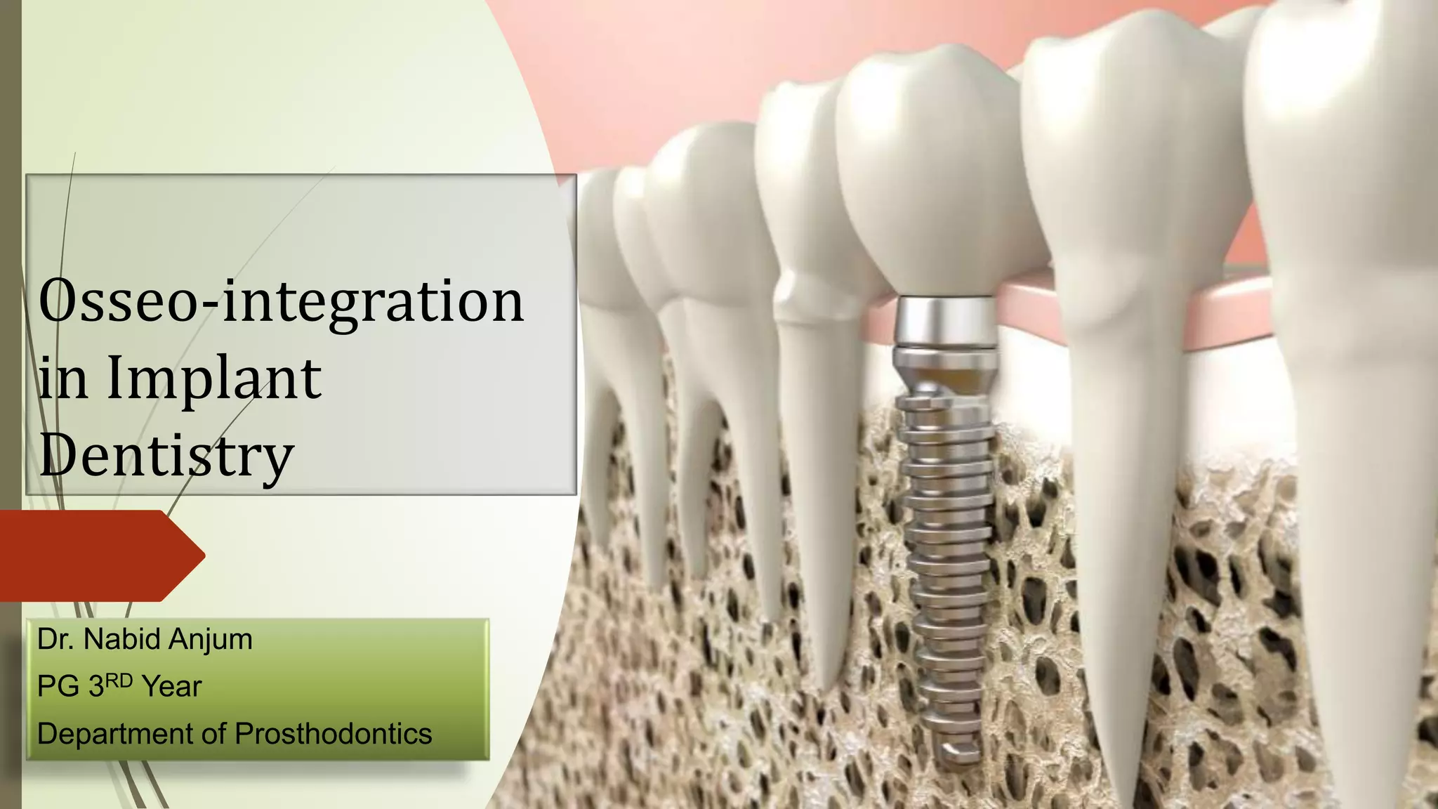
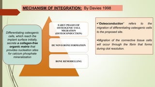
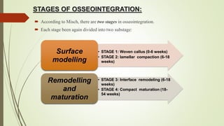
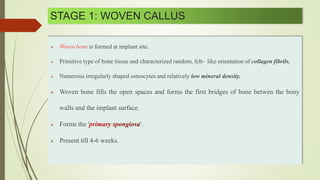
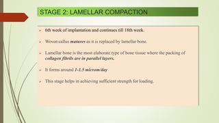
Osseo-integration
in Implant
Dentistry
Dr. Nabid Anjum
PG 3RD Year
Department of Prosthodontics
Contents:
 Introduction
 Development of concept of
Osseointegration
 Definitions
 Bone to Implant Interface
 Mechanism of Osseointegration
 Key Factors responsible for
successful Osseointegration
 Methods of evaluation of
Osseointegration
 Success criteria of Implants
 Conclusion
 References
Introduction:
 A successful replacement of missing natural tooth by dental implant supported
prosthesis is a major clinical advance in dental science. The successful outcome of the
treatment depends upon the degree of Osseointegration.
 Osseointegration in clinical dentistry depends on an understanding of the healing and
reparative capacities of hard and soft tissues. Its objective is a predictable tissue response
to placement of tooth root analogue.
 It is the most investigated area in implantology in recent times.
 Evidence based data reveals that Osseo integrated implants are predictable and highly
successful. This process is relatively complex and is influenced by various factors in
formation of bone neighboring implant surface.
 Word “osseointegration” has been stamped indelibly in implant nomenclature and has been
used at various times to describe lack of clinical mobility, lack of a perimplant space
radiographically and lack of a connective tissue interface at the implant/bone junction.
OSTEON INTEGRARE
OSSEO INTEGRATION
Development of concept of Osseointegration:
The concept of Osseo integration based on research that
began by Branemark in 1952.
Dr. Per-Ingvar Branemark
Orthopedic surgeon
Professor, University of Goteborg,
Sweden.
He wanted to observe the microcirculation of both soft
and hard tissues under various phases of injuries of a
rabbit bone.
He implanted titanium optic chamber in to rabbits fibula
and carried out the investigation with a vital microscopic
(essentially made of titanium) and when he tried to remove
the titanium chamber he found that bone was normally
adhered to the metal.
Coined the term ‘osseointegration’
“Father of Modern Implantology”
 Branemark and his teams designed titanium screws and inserted them into the jaws of
beagle dogs.
 By 1965, he felt it to be ready for use in humans and placed 4 implants in a Swede named
Mr. Gosta Larsson {severe chin and jaw deformities}.
 Branemark began training the first Swedish dental experts his techniques in October 1977.
However , it was
the 1983 Toronto
conference when
Branemark’s
work finally was
universally
accepted.
In Sweden ,
osseo-
integrated
implants
became
acceptable by
1977
Definitions :
 American Academy of Implant Dentistry :
 GPT 9:
 Structurally oriented definition:
Contact established without interposition of non-bone tissue
between normal remodeled bone and an implant entailing a
sustained transfer and distribution of load from the implant to
and within the bone tissue.
The apparent direct attachment or connection of osseous tissue to an inert,
alloplastic material without intervening connective tissue.
Direct structural and functional connection between the ordered, living bone and the
surface of a load carrying implants.
- Branemark and associates {1977}
 Clinically:
 Histologically:
 Biologically oriented definition:
• Ankylosis of the implant bone interface.
-Schroeder and colleagues {1976}
- “Functional ankylosis”
• It is a process where by clinically asymptomatic rigid fixation of alloplastic material is
achieved and maintained in bone during functional loading.
- Zarb and T Albrektson {1991}
Direct anchorage of an implant by the formation of bone directly on the surface of an
implant without any intervening layer of fibrous tissue.
- Albrektsson and Johnson (2001)
Attachment resistant to shear as well as tensile forces.
Steinmann et al (1986)
 Prosthetic rehabilitation of missing teeth
 Anchorage for the maxillofacial prosthesis
 For rehabilitation of congenital and developmental defects
 Complex maxillofacial defect rehabilitation
 Distraction osteogenesis {new bone formation}
 Orthodontic anchorage
Scope of Osseointegration in Dentistry:
Bone Physiology:
Bone can be classified as:
MATURE BONE :
Compact Bone
Spongy Bone
IMMATURE BONE:
Woven Bone
BONE TO IMPLANT INTERFACE:
 There are two basic theories regarding the bone implant interface:
Fibro osseous integration Osseo integration
Concept of soft tissue anchorage
(Linkow 1970, James 1975 and Weiss
1986)
Concept of Bony Anchorage
Branemark (1982), supported by
Zarb and Albrektsson (1985)
 Fibro-osseous integration:
(pseudo-ligament, peri-implant ligament, peri-implant membrane.)
 This theory was proposed by Weiss and was supported by Linkow and James.
 AAID (1986) defined fibrous integration as “tissue to implant contact with interposition of
healthy dense collagenous tissue between the implant and bone”.
Inhibition of bone remodeling
Failure of Fibro osseous theory:
 According the theory , pseudo-peri implant fibrous membrane gave a cushion effect and
acted as similar as periodontal membrane in natural dentition. However:
 No real evidence
 Forces are not transmitted through the fibers - remodeling was not expected
 Forces applied resulted in widening fibrous encapsulation, inflammatory reactions, and
gradual bone resorption there by leading to failure.
 Hence, a failure in today’s standard.
Smuzkler-Moncler et al. 1998 - micromotion >150 microns at the bone implant interface
results in fibrous encapsulation instead of osseointegration
 Osseo integration:
 Direct bone to implant interface without any intervening layer of fibrous tissue.
 ‘direct bone deposition on the implant surface’ (Branemark et al. 1997), and also
'functional ankylosis’ (Schroederet al. 1981 ).
 If osseointegration does not occur or osseointegration is lost for some reason, a fibrous
connective tissue forms around the implant.
Meffert et al in 1987 subdivided osseointegration:
MECHANISM OF OSSEO INTEGRATION:
 Healing process may be primary bone healing or
secondary bone healing.
 In primary bone healing, there is well organized
bone formation with minimal granulation tissue
formation – ideal
 Secondary bone healing may have granulation
tissue formation and infection at the site, prolonging
healing period. Fibrocartilage is sometimes formed
instead of bone - undesirable
Healing around endosteal dental implant:
Stage 1
• Seen at 3- 7 days following implantation
• Earliest angiogenesis and osteogenesis phase
Stage 1
• Angiogenesis seen at the broken ends of blood vessel in prepared osteotomy site
• There is sprouting or budding extension of the pre-existing blood vessel
Stage 1
• Pluripotent cells activated,more active in threaded grooves or acute angles of
interface geometry.
• After 1st week , these are rapidly filled with fine collagen fibers and fibroblasts
VASCULARSPROUTINGSTAGE
 The damage caused during the surgical procedure and the interlocking of the
implant to the hard and soft tissues initiate the process of healing.
Stage 2
• Two weeks after implantation
• Ridge-like bone with sinusoidal capillaries fill the grooves on implant surface
Stage 2
• Discontinous bone segments at the base adhere with the basket-like capillary
network and develop into continous new bone.
EARLYBONEFORMATIONSTAG
Stage 3
• 4 weeks following implantation
• Primary spongiosa transforms into secondary spongiosa and proliferates to form
new alveolar bone
Stage 3
• Bone trabeculae arising from the osteotomy over the peri-implant space
perpendicular to the interface form a bone plate
BONEGROWTHSTAGE
Stage 4
• 6-8 weeks following implantation
• Formation of bone around implant is nearly completed.
Stage 4
• Capillary plexus evident between the original bone bordering the osteotomy and new
bone bordering the implant.
• Bone within the threading and grooves begins to fill in.
Stage 4
• At the implant socket base, thick plates of trabeculae appear.
BONEMATURATIONSTAGE
The three main phases of bone healing necessary for osseointegration are –
BIOLOGICAL PROCESS OF OSSEOINTEGRATION: (BRANEMARK)
OSTEOPHYLIC PHASE
OSTEOCONDUTIVE PHASE
OSTEOADAPTIVE PHASE
OSTEOPHYLIC PHASE:
 Blood clot formation
 Inflammatory cells infiltration
 Neovascularisation (3rd day)
 Ossification begins during first week
 This phase lasts about 1 month
OSTEOCONDUCTIVE PHASE:
 Woven bone - Foot plate
 lamellar bone formation
 Lasts for 4 months
OSTEOADAPTIVE PHASE:
 A balanced remodelling occured
 The footplate/ woven bone thickened in response to load
transmitted through the implant
 Some reorientation of vascular pattern may be seen
BONE TISSUE RESPONSE:
MECHANISM OF INTEGRATION: By Osborn and Newesley – 1980
Proposed 2 different phenomenon by which bone can become juxtaposed to an implant
surface.
•Distance Osteogenesis:
 A gradual process of bone healing inward from
the edge of the osteotomy toward the implant.
Bone does not grow directly on the implant
surface.
 Impossible to achieve a phenomenon called
bone bonding as implant surface-obscured by
interveneing cells and connective tissue extra-
cellular matrix.
•Contact Osteogenesis :
 The direct migration of bone-
building cells through the clot matrix
to the implant surface.
 Bone is quickly formed directly on
the implant surface.
 bone bonding will occur provided
appropriate surface topography of
implant
MECHANISM OF INTEGRATION: By Davies 1998
EARLY PHASES OF
OSTEOGENIC CELL
MIGRATION
(OSTEOCONDUCTION)
DE NOVO BONE FORMATION
BONE REMODELLING
“Osteoconduction” refers to the
migration of differentiating osteogenic cells
to the proposed site.
Migration of the connective tissue cells
will occur through the fibrin that forms
during clot resolution.
Differentiating osteogenic
cells, which reach the
implant surface initially,
secrete a collagen-free
organic matrix that
provides nucleation sites
for calcium phosphate
mineralization
STAGES OF OSSEOINTEGRATION:
 According to Misch, there are two stages in osseointegration.
 Each stage been again divided into two substage:
• STAGE 1: Woven callus (0-6 weeks)
• STAGE 2: lamellar compaction (6-18
weeks)
Surface
modelling
• STAGE 3: Interface remodeling (6-18
weeks)
• STAGE 4: Compact maturation (18-
54 weeks)
Remodelling
and
maturation
STAGE 1: WOVEN CALLUS
 is formed at implant site.
 Primitive type of bone tissue and characterized random, felt- like orientation of collagen fibrils.
 Numerous irregularly shaped osteocytes and relatively low mineral density.
 Woven bone fills the open spaces and forms the first bridges of bone betwen the bony
walls and the implant surface.
 Forms the ' '.
 Present till 4-6 weeks.
STAGE 2: LAMELLAR COMPACTION
 6th week of implantation and continues till 18th week.
 Woven callus matures as it is replaced by lamellar bone.
 Lamellar bone is the most elaborate type of bone tissue where the packing of
collagen fibrils are in parallel layers.
 It forms around 1-1.5 microm/day
 This stage helps in achieving sufficient strength for loading.
STAGE 3: INTERFACE REMODELLING
 This stage begins at the same time when woven callus is completing lamellar
compaction.
 During this stage callus starts to resorb, and remodeling of devitalized interface
begins.
 The helps in establishing a viable interface between the
implant and original bone.
 It helps in adaptation of bone structure to load.
STAGE 4: COMPACT BONE MATURATION
 This is the of osseointegration.
 This occurs form 18th week of implantation and continues till the 54th week.
 During this stage compact bone matures by series of modeling and remodeling
processes.
 The callus volume is further decreased and interface remodeling continues.
KEY FACTORS RESPONSIBLE FOR SUCCESSFUL OSSEO-
INTEGRATION:
 Six different factors known to be important for the establishment of a reliable, long
term osseous anchorage of an implanted device :
DESIGN
CHARACTERISTICS
SURFACE
CHARACTERISTICS
STATE OF THE
HOST BED
SURGICAL
FACTORS
LOADING
CONDITIONS
IMPLANT
BIOCOMPATABILITY
 IMPLANT BIOCOMPATABILITY:
 Commercially pure titanium (CP Ti) 99.75% is widely used as an
implant material as:
 highly biocompatible
 good resistance to corrosion, and
 no toxicity on macrophages or fibroblasts,
 lack of inflammatory response in peri-implant tissues and
 it’s composed of an oxide layer and has the ability to repair itself by
reoxidation when damaged. (biologically inert) --
 Another material used for implants,
Titanium -6 Aluminum-4 Vanadium (TI-6AL-4 V) alloy exhibits soft tissue
reactions very similar to those reported to Cp Ti and showed inferior bone
attachment.
Grouping of hard tissue replacement materials according to their
compatibility to bony tissue:
Degree of
Compatibility
Characteristics of Reactions of
Bony Tissue
Materials
Biotolerant Implants separated from adjacent
bone by a soft tissue layer along
most of the interface: distance
osteogenesis
Stainless steels: CoCrMo and
CoCrMoNi alloys
Bioinert Direct contact to bony tissue
contact osteogenesis
Alumina ceramics, zirconia
ceramics, titanium, niobium,
carbon.
Bioactive Bonding to bony tissue:
bonding osteogenesis
HA, Calcium phosphate-
containing glasses, glass-
ceramics, ceramics,
titanium
 IMPLANT DESIGN:
 Implant design can be broadly classified as and
 Root form implants : Cylindrical and Screw shaped.
Cylindrical Screw shaped implants
Non threaded implants Threaded implants
The design of the thread:
 Functional surface area per unit length of implant may be modified by the three thread
geometry parameters:
Thread shape
Thread pitch
Thread depth
THREAD SHAPE:
The shear force on the face of a V thread is about 10 times greater than the shear force
on a square thread.
THREAD PITCH:
The number of thread per unit length.
Pitch distance is inversely related to the
number of threads in the unit area.
The greater the number of threads, the lesser
the pitch, hence the more will be surface area.
THREAD DEPTH
It is the distance between the minor and
major diameterplant.
The deeper the thread depth, the greater
the surface area of the implant.
 IMPLANT SURFACE CHARACTERISTICS:
Topographic properties
Implant surface texture &
roughness
Physiochemical properties
Implant surface chemistry
Physical properties
Surface energy and charge
 The First generation titanium implants were machined with a smooth surface texture.
 Implant surfaces have been recognized to play an important role in molecular
interactions, cellular response and Osseo integration.
 The Second generation implants with surface modification can accelerate and
improve implant osseointegration.
 Implants underwent mechanical blasting, acid etching, bioactive coatings, more recently
, laser modified surfaces.
 The main objective for the development of implant surface modifications is to promote
Osseo integration, with faster and stronger bone formation.
Surface topography
 The surface topography relates to the degree of roughness of the surface and the orientation
of the surface irregularities.
 The chemical composition of the implant interface on the implant surface was shown to
affect initial cell attachment
 Macro and micro surface irregularity can influence the response of cells and tissues.
 This has stimulated a great interest on implant surface modification as a way to
NEED FOR SURFACE MODIFICATION:
Toincrease the surface area.
Toremove surface contaminants
Tobring better bonding
Toincrease surface roughness of metal
Toincrease corrosion resistance of metal.
 Commercially available implants have been categorized acc to Roughness Value (Sa) into
4 groups by into:
Smooth ( Sa <0.5 um)
Minimally rough ( Sa= 0.5-1.0 um)
Moderately rough (Sa= 1.0-2.0um) - most commonly used
Rough (Sa >2.0um)
TECHNIQUES FOR SURFACE MODIFICATION OF IMPLANTS:
 The implant modifications can be achieved either by additive or subtractive methods.
Impregnation
implies that
the
material/chemi
cal agent is
fully
integrated into
the titanium
core, such as
calcium
phosphate
crystals
within TiO2
layer or
incorporation
Titanium plasma spraying (TPS), plasma
sprayed hydroxyapatite (HA) coating,
alumina coating, and using biomimetic
materials
Additive methods Subtractive methods
The subtractive techniques are the
procedure to either remove the layer
of core material or plastically deform
the superficial surface and thus
roughen the surface of core material.
The common subtractive techniques are
Large-grit sands or ceramic particle
blasts, Acid etch and Anodization
PLASMA SPRAYED SURFACE:
This is the process of spraying molten metal on the titanium base which results in
surface irregularity like valley, pores.
The growth of bone to this irregularity will create a mechanical interlock and surface
irregularity will increase surface area which aid in initial fixation of implant, especially in
soft bone
Titanium plasma sprayed (TPS) or Hydroxyapatite coatings (HA)
Hydroxyapatite bonds well with bone and accelerates new bone formation in initial
healing period with formation of osteophylic surface.
In order to increase bone formation in initial stages in cases like immediate implant
placement and poor bone quality, HA surface is a good choice.
ACID ETCHING
The most commonly used solutions for
acid pickling of titanium and titanium
alloy are,
or
Dual acid etching:
This is done by immersing the titanium
implant in a
and heat above 100°C for several
minutes.
Dual acid etched surfaces accelerate
the osteoconductive process by
attachment of fibrin and osteogenic
cells, leading to bone formation straight
on the implant.
Etching the implant surface with strong acids aids in cleaning the surface and attaining
homogenous roughening.
GRIT BLASTING / SAND BLASTING
Different ceramic particles have been used, like
glass, silica, alumina and titanium oxide particles.
Blasting with biocompatible material is always
advised.
TiO2-blasted implants were suggested as a certain
long term support for fixed prostheses in both the
maxilla and the mandible
SAND BLASTED AND ACID ETCHED (SLA)
 The surface modification can be made by
combination of sandblasting and acid etching.
 Blasting is done with various particles like Al2O3
and TiO2., which is followed by etching with HCl
and H2SO4.
 This will create a micro and macro structure
modification.
 It has got more Osseo conductive properties
and higher ability to induce cell proliferation.
LASER PEENING
 An advantage of lasers in surface modification is that laser has the
 In addition, laser processing is contactless and the thermal, mechanical deformation of
the substrate is generally low.
 and are used.
DRUG INCORPORATED
Surface treatment of implant with antibacterial coating serves the possible way to prevent
surgical site from infection.
Gentamicin can be used along with HA coating.
is also an efficient method for decontamination and
detoxification of implant surface.
BIOMIMETIC DEPOSITION
 Biomimetic surface treatment is still a
developing topic of research in
implantology.
 Desirable properties of biomimetic agents
are :
1) it should be able to bring about
differentiation of cell for bone formation;
2) itshould not delaminate
3) easy to manufacture
4) affordable
5) chemically stable
6) non-immunogenic.
 Bioceramics
• Hydroxyapatite(HA)
•Calcium phosphate phases.
 Bioactive proteins
• Bone morphogenic proteins (BMP)
• Type1collagen
• RGD peptide sequence.
 Ions
• Fluoride.
 Polymers
• Chitosan
Physical characteristic
 Physical characteristic refers to the factors such as surface energy and
charge.
 Hypothesis : A surface with high energy →high affinity for adsorption → show
stronger osseointegration.
 Baier RE (1986) – results in high surface energy as
well as the implant sterilization, being conductive to tissue integration.
 Charge affects the hydrophilic and hydrophobic characteristic of the surface.
 A hydrophilic / easily wettable implant surface : Increases a initial phase of wound
healing.
 Fact : Increase surface energy would disappear immediately after implant placement.
Implant surface chemistry:
• Chemical alteration  increases bioactivity  increase implant bone anchorage.
 Chemical surfaces :
• Ceramic coated – hydroxyapatite (HA), Calcium phosphate
• Oxidized/anodized surfaces with electrolytes containing phosphorous, sulfur, calcium,
magnesium and flouride.
• Ionization, implantation of calcium ion, floride ions
• Doped surfaces with the BONE STIMULATING FACTORS / GROWTH FACTORS.
 BONE FACTOR:
Bone quality  bone with well formed cortex and densely trabaculated
medullary spaces
Bone quantity  Refers to the dimension of available bone in reference to
length, width and depth.
LIKHOM AND ZARB CLASSIFICATION 1985
Class I : Jaw consist
almost exclusively of
of homogeneous
compact bone
Class II : Thick compact
bone surrounds highly
trabecular core
Class III : Thin cortical
bone surrounds highly
trabecular core
Class IV : Thin cortical bone
surrounds loose, spongy core
MISCH CLASSIFICATION 1988
According to Branemark and Misch
D1 and D2 bone  initial stability / better osseointegration
osseointegration
D3 and D4  poor prognosis
D1 bone – least risk
D4 bone - most at risk
D1 D2 D3 D4
}loss of osseointegration
Selection of implant
D1 and D2 – conventional
threaded implants
D3 and D4 – HA coated or
Titanium plasma coated
implants
Branemark system (5 year
documentation)
Mandible – 95% success
Maxilla – 85-90% success
 STATE OF THE IMPLANTATION OR HOST BED:
 Healthy implant host site
- Vascularity
Cellularity (osteogenic potential)
Factors influencing osseointegration:
• Patient Considerations : Age
• History of proposed host bed : Previous irradiation
Infection
History of smoking
Advanced ridge resorption
Osteoporosis or osteoporotic like
bone lesion
Age :
 The fact that the implant behaves as an ankylosed unit restricts its use to individuals who
have completed their jaw growth.
 Possible complications of placing implants too early in life
Submerging of implant into the jaw
Relocation of the implant
Interference with the normal growth
Smoking and osseointegration :
 History of smoking affect the healing response in osseointegration.
 Lower success rates with oral implants
 Mean failure rates in smokers is about twice than in non smoker.
 Radiation therapy and osseointegration :
• Jacobsson (1985): Previous irradiation – for implant placement.
• Expected success rate 10-15% lower than the non irrradiated patients.
• The main complication associated with placing implants implants in irradiated bone is
'osteoradionecrosis.'
 Granstrom G (1998): HBO (Hyperbaric oxygen therapy) can counteract some of the
negative effect from irradiation and act as a stimulator for osseointegration.
 Compromised oral hygiene
 Vitamin C deficiency
 Uncontrolled Diabetes.
 Available bone
 SURGICAL FACTORS:
 Minimal tissue violence at surgery is
essential for osseointegration.
 This objective depends on continuous
and careful cooling while surgical
drilling is performed at low speed.
 The critical time/temperature
relationship for bone tissue necrosis is
around 47 ºC applied for 1 min.
 Use of sharp drills with a drill speed of
less than 2000rpm is desired.
 56ºC is the critical temperature to
prevent bone overheating .
 Use of torque wrench with moderate
torque of 45 N/cm is ideal.
 LOADING CONDITIONS:
 Objective : “No loading while healing”  successful
osseointegration.
 Movement of the implant within the bone – fibrous tissue
rather than osseointegration. ( )
Premature loading
leads to implant
movement
The end result “Soft
tissue interface”
“Bony interface”
 branemark, albrektsson
 Two stage implant insertion.
 First stage – Installation of fixture into bone
Maxilla: 6 months
Mandible: 3 months
 Second stage – Connection of abutment to the fixtures
 Misch –
 Progressive / Gradual loading
 Suggested in
Softer bone
less number of implants to be used
METHODS FOR EVALUATION OF
OSSEOINTEGRATION:
 Implant stability plays a critical role for successful osseointegration. Successful
osseointegration is a prerequisite for functional dental implants.
• Primary stability
• Secondary stability
 Primary stability is a function of the Bone quality, Implant design and Surgical
technique.
 During the osseointegration healing and maturation process , the initial stability
changes with increases in bone- to –implant contact and osseous remodeling.
 Two terms osseointegration and rigid fixation {absence of observed
clinical mobility} are interchangeably used.
 A healthy implant moves less than 73 microns – appears as zero clinical
mobility .
 There are different methods to assess implant stability:
 They can be grouped as invasive/destructive methods and noninvasive/nondestructive
methods
Following methods were included:
Histologic/histomorphologic analysis
Tensional test
Push-out/pull-out test and
Removal torque analysis.
These include the following:
The surgeon's perception
Radiographical analysis
Cutting torque resistance (for primary
stability)
Insertion torque measurement
Reverse torque
Percussion test
Pulsed oscillation waveform (POWF)
Periotest
Resonance frequency analysis (RFA)
Magnetic technology.
INVASIVE METHODS:
NON INVASIVE METHODS:
The surgeon’s perception
Radiographical analysis/imaging techniques
Insertion torque measurements
Reverse torque test
Cutting torque resistance Analysis
Percussion test
Resonance frequency analysis
• It was suggested by Meredith in 1998.
• It is a noninvasive diagnostic method that measures implant stability and bone density at
various time points using vibration and a principle of structural analysis.
• A bending load is applied which mimics the clinical load and direction and provides information
about the stiffness of the implant–bone junction.
1st generation
Ostell transducer3rd generation
4th generation Ostell Mentor
Newer methods under research an development :
 Implatest conventional impulse testing
 Electro-mechanical impedance method
 Micro motion detecting device
 Highly nonlinear solitary waves method
Swami V, Vijayaraghavan V, Swami V. Current trends to measure implant stability. J Indian
Prosthodont Soc 2016;16:124-30.
 Harvard success criteria:
 The dental implant must provide functional service for 5 years in 75% of cases
Objective criteria:
Bone loss no greater than 33% of vertical length of
implant
Good occlusal balance and vertical dimension
Gingival inflammation amenable to treatment
Mobility of less than 1mm in any direction
Absence of symptoms of infection
Absence of damage to surrounding structure
Healthy connective tissues
Subjective criteria:
Adequate function
Absence of discomfort
Improved aesthetics
Improved emotional and psychological
wellbeing
SCHUITMAN AND SCHULMAN {1979}
The mobility of the implant must be less than 1mm when
tested clinically.
There must be no evidence of radiolucency
Bone loss should be less than 1/3rd of the height of the
implant.
There should be an absence of infection, damage to
structure or violation of body cavity, inflammation present
must be amneable to treatment.
The success rate must be 75% or more after 5 years of
functional service.
ALBREKTSON AND ZARB G {1980}
The individual unattached implant should be immobile
when tested clinically.
The radiographic evaluation should not show any
evidence of radiolucency.
The vertical bone loss around the fixtures should be
less than 0.2mm per year after first year of implant
loading
The implant should not show any signs of pain,
infection, neuropathies, paresthesia, violation of
mandible canals and sinus drainage.
The success rate of 85% at the end of 5 year and 80%
at the end
CONCLUSION:
 Osseointegration has revolutionized the treatment
options open to prosthodontist. For edentulous
patients, restoration of oral health, function and
appearance have now become predictable and
reliable.
 Osseo integrated implants provide scope for
diversifying and enhancing treatment planning for
both partially edentulous arches and maxillofacial
patients.
 In implant dentistry , an undisturbed healing period
is always required to ensure osseointegration.
Provided that the implant has primary stability ,
studies have shown that the survival of loaded
implants can be analogous to the unloaded protocol.
REFERENCES:
 Misch CE. Contemporary implant dentistry, 3rd edition, Mosby Elsevier publication, St Louis, 2008,
pp:27, 70, 621
 Brånemark PI. Osseointegration and its experimental studies. J Prosthet Dent 1983;50:399-410.
 Smith DE, Zarb GA. Criteria for success of Osseo integrated endosseous implants, J Prosthet Dent
1989;62:567-72.
 Macha D et al. Osseointegration in Implants: A Review. J Res Adv Dent 2014; 3:3:67-72.
 Davies JE. Understanding Peri-Implant Endosseous Healing. J Dent Edu 2005;67(8):932-949
 Albrektsson T and Jansson T. Osseo integrated dental implants. Dent clin North Am, 1986,30:151
 Swami V, Vijayaraghavan V, Swami V. Current trends to measure implant stability. J Indian
Prosthodont Soc 2016;16:124-30.

Osseointegration in Dental Implants

  • 1.
    Osseo-integration in Implant Dentistry Dr. NabidAnjum PG 3RD Year Department of Prosthodontics
  • 2.
    Contents:  Introduction  Developmentof concept of Osseointegration  Definitions  Bone to Implant Interface  Mechanism of Osseointegration  Key Factors responsible for successful Osseointegration  Methods of evaluation of Osseointegration  Success criteria of Implants  Conclusion  References
  • 3.
    Introduction:  A successfulreplacement of missing natural tooth by dental implant supported prosthesis is a major clinical advance in dental science. The successful outcome of the treatment depends upon the degree of Osseointegration.  Osseointegration in clinical dentistry depends on an understanding of the healing and reparative capacities of hard and soft tissues. Its objective is a predictable tissue response to placement of tooth root analogue.  It is the most investigated area in implantology in recent times.  Evidence based data reveals that Osseo integrated implants are predictable and highly successful. This process is relatively complex and is influenced by various factors in formation of bone neighboring implant surface.
  • 4.
     Word “osseointegration”has been stamped indelibly in implant nomenclature and has been used at various times to describe lack of clinical mobility, lack of a perimplant space radiographically and lack of a connective tissue interface at the implant/bone junction. OSTEON INTEGRARE OSSEO INTEGRATION
  • 5.
    Development of conceptof Osseointegration: The concept of Osseo integration based on research that began by Branemark in 1952. Dr. Per-Ingvar Branemark Orthopedic surgeon Professor, University of Goteborg, Sweden. He wanted to observe the microcirculation of both soft and hard tissues under various phases of injuries of a rabbit bone. He implanted titanium optic chamber in to rabbits fibula and carried out the investigation with a vital microscopic (essentially made of titanium) and when he tried to remove the titanium chamber he found that bone was normally adhered to the metal. Coined the term ‘osseointegration’ “Father of Modern Implantology”
  • 6.
     Branemark andhis teams designed titanium screws and inserted them into the jaws of beagle dogs.  By 1965, he felt it to be ready for use in humans and placed 4 implants in a Swede named Mr. Gosta Larsson {severe chin and jaw deformities}.  Branemark began training the first Swedish dental experts his techniques in October 1977. However , it was the 1983 Toronto conference when Branemark’s work finally was universally accepted. In Sweden , osseo- integrated implants became acceptable by 1977
  • 7.
    Definitions :  AmericanAcademy of Implant Dentistry :  GPT 9:  Structurally oriented definition: Contact established without interposition of non-bone tissue between normal remodeled bone and an implant entailing a sustained transfer and distribution of load from the implant to and within the bone tissue. The apparent direct attachment or connection of osseous tissue to an inert, alloplastic material without intervening connective tissue. Direct structural and functional connection between the ordered, living bone and the surface of a load carrying implants. - Branemark and associates {1977}
  • 8.
     Clinically:  Histologically: Biologically oriented definition: • Ankylosis of the implant bone interface. -Schroeder and colleagues {1976} - “Functional ankylosis” • It is a process where by clinically asymptomatic rigid fixation of alloplastic material is achieved and maintained in bone during functional loading. - Zarb and T Albrektson {1991} Direct anchorage of an implant by the formation of bone directly on the surface of an implant without any intervening layer of fibrous tissue. - Albrektsson and Johnson (2001) Attachment resistant to shear as well as tensile forces. Steinmann et al (1986)
  • 9.
     Prosthetic rehabilitationof missing teeth  Anchorage for the maxillofacial prosthesis  For rehabilitation of congenital and developmental defects  Complex maxillofacial defect rehabilitation  Distraction osteogenesis {new bone formation}  Orthodontic anchorage Scope of Osseointegration in Dentistry:
  • 10.
    Bone Physiology: Bone canbe classified as: MATURE BONE : Compact Bone Spongy Bone IMMATURE BONE: Woven Bone
  • 11.
    BONE TO IMPLANTINTERFACE:  There are two basic theories regarding the bone implant interface: Fibro osseous integration Osseo integration Concept of soft tissue anchorage (Linkow 1970, James 1975 and Weiss 1986) Concept of Bony Anchorage Branemark (1982), supported by Zarb and Albrektsson (1985)
  • 12.
     Fibro-osseous integration: (pseudo-ligament,peri-implant ligament, peri-implant membrane.)  This theory was proposed by Weiss and was supported by Linkow and James.  AAID (1986) defined fibrous integration as “tissue to implant contact with interposition of healthy dense collagenous tissue between the implant and bone”. Inhibition of bone remodeling
  • 13.
    Failure of Fibroosseous theory:  According the theory , pseudo-peri implant fibrous membrane gave a cushion effect and acted as similar as periodontal membrane in natural dentition. However:  No real evidence  Forces are not transmitted through the fibers - remodeling was not expected  Forces applied resulted in widening fibrous encapsulation, inflammatory reactions, and gradual bone resorption there by leading to failure.  Hence, a failure in today’s standard. Smuzkler-Moncler et al. 1998 - micromotion >150 microns at the bone implant interface results in fibrous encapsulation instead of osseointegration
  • 14.
     Osseo integration: Direct bone to implant interface without any intervening layer of fibrous tissue.  ‘direct bone deposition on the implant surface’ (Branemark et al. 1997), and also 'functional ankylosis’ (Schroederet al. 1981 ).  If osseointegration does not occur or osseointegration is lost for some reason, a fibrous connective tissue forms around the implant. Meffert et al in 1987 subdivided osseointegration:
  • 15.
    MECHANISM OF OSSEOINTEGRATION:  Healing process may be primary bone healing or secondary bone healing.  In primary bone healing, there is well organized bone formation with minimal granulation tissue formation – ideal  Secondary bone healing may have granulation tissue formation and infection at the site, prolonging healing period. Fibrocartilage is sometimes formed instead of bone - undesirable
  • 16.
    Healing around endostealdental implant: Stage 1 • Seen at 3- 7 days following implantation • Earliest angiogenesis and osteogenesis phase Stage 1 • Angiogenesis seen at the broken ends of blood vessel in prepared osteotomy site • There is sprouting or budding extension of the pre-existing blood vessel Stage 1 • Pluripotent cells activated,more active in threaded grooves or acute angles of interface geometry. • After 1st week , these are rapidly filled with fine collagen fibers and fibroblasts VASCULARSPROUTINGSTAGE  The damage caused during the surgical procedure and the interlocking of the implant to the hard and soft tissues initiate the process of healing.
  • 17.
    Stage 2 • Twoweeks after implantation • Ridge-like bone with sinusoidal capillaries fill the grooves on implant surface Stage 2 • Discontinous bone segments at the base adhere with the basket-like capillary network and develop into continous new bone. EARLYBONEFORMATIONSTAG Stage 3 • 4 weeks following implantation • Primary spongiosa transforms into secondary spongiosa and proliferates to form new alveolar bone Stage 3 • Bone trabeculae arising from the osteotomy over the peri-implant space perpendicular to the interface form a bone plate BONEGROWTHSTAGE
  • 18.
    Stage 4 • 6-8weeks following implantation • Formation of bone around implant is nearly completed. Stage 4 • Capillary plexus evident between the original bone bordering the osteotomy and new bone bordering the implant. • Bone within the threading and grooves begins to fill in. Stage 4 • At the implant socket base, thick plates of trabeculae appear. BONEMATURATIONSTAGE
  • 19.
    The three mainphases of bone healing necessary for osseointegration are –
  • 20.
    BIOLOGICAL PROCESS OFOSSEOINTEGRATION: (BRANEMARK) OSTEOPHYLIC PHASE OSTEOCONDUTIVE PHASE OSTEOADAPTIVE PHASE
  • 21.
    OSTEOPHYLIC PHASE:  Bloodclot formation  Inflammatory cells infiltration  Neovascularisation (3rd day)  Ossification begins during first week  This phase lasts about 1 month
  • 22.
    OSTEOCONDUCTIVE PHASE:  Wovenbone - Foot plate  lamellar bone formation  Lasts for 4 months OSTEOADAPTIVE PHASE:  A balanced remodelling occured  The footplate/ woven bone thickened in response to load transmitted through the implant  Some reorientation of vascular pattern may be seen
  • 23.
    BONE TISSUE RESPONSE: MECHANISMOF INTEGRATION: By Osborn and Newesley – 1980 Proposed 2 different phenomenon by which bone can become juxtaposed to an implant surface. •Distance Osteogenesis:  A gradual process of bone healing inward from the edge of the osteotomy toward the implant. Bone does not grow directly on the implant surface.  Impossible to achieve a phenomenon called bone bonding as implant surface-obscured by interveneing cells and connective tissue extra- cellular matrix. •Contact Osteogenesis :  The direct migration of bone- building cells through the clot matrix to the implant surface.  Bone is quickly formed directly on the implant surface.  bone bonding will occur provided appropriate surface topography of implant
  • 25.
    MECHANISM OF INTEGRATION:By Davies 1998 EARLY PHASES OF OSTEOGENIC CELL MIGRATION (OSTEOCONDUCTION) DE NOVO BONE FORMATION BONE REMODELLING “Osteoconduction” refers to the migration of differentiating osteogenic cells to the proposed site. Migration of the connective tissue cells will occur through the fibrin that forms during clot resolution. Differentiating osteogenic cells, which reach the implant surface initially, secrete a collagen-free organic matrix that provides nucleation sites for calcium phosphate mineralization
  • 26.
    STAGES OF OSSEOINTEGRATION: According to Misch, there are two stages in osseointegration.  Each stage been again divided into two substage: • STAGE 1: Woven callus (0-6 weeks) • STAGE 2: lamellar compaction (6-18 weeks) Surface modelling • STAGE 3: Interface remodeling (6-18 weeks) • STAGE 4: Compact maturation (18- 54 weeks) Remodelling and maturation
  • 27.
    STAGE 1: WOVENCALLUS  is formed at implant site.  Primitive type of bone tissue and characterized random, felt- like orientation of collagen fibrils.  Numerous irregularly shaped osteocytes and relatively low mineral density.  Woven bone fills the open spaces and forms the first bridges of bone betwen the bony walls and the implant surface.  Forms the ' '.  Present till 4-6 weeks.
  • 28.
    STAGE 2: LAMELLARCOMPACTION  6th week of implantation and continues till 18th week.  Woven callus matures as it is replaced by lamellar bone.  Lamellar bone is the most elaborate type of bone tissue where the packing of collagen fibrils are in parallel layers.  It forms around 1-1.5 microm/day  This stage helps in achieving sufficient strength for loading.
  • 29.
    STAGE 3: INTERFACEREMODELLING  This stage begins at the same time when woven callus is completing lamellar compaction.  During this stage callus starts to resorb, and remodeling of devitalized interface begins.  The helps in establishing a viable interface between the implant and original bone.  It helps in adaptation of bone structure to load.
  • 30.
    STAGE 4: COMPACTBONE MATURATION  This is the of osseointegration.  This occurs form 18th week of implantation and continues till the 54th week.  During this stage compact bone matures by series of modeling and remodeling processes.  The callus volume is further decreased and interface remodeling continues.
  • 31.
    KEY FACTORS RESPONSIBLEFOR SUCCESSFUL OSSEO- INTEGRATION:  Six different factors known to be important for the establishment of a reliable, long term osseous anchorage of an implanted device : DESIGN CHARACTERISTICS SURFACE CHARACTERISTICS STATE OF THE HOST BED SURGICAL FACTORS LOADING CONDITIONS IMPLANT BIOCOMPATABILITY
  • 32.
     IMPLANT BIOCOMPATABILITY: Commercially pure titanium (CP Ti) 99.75% is widely used as an implant material as:  highly biocompatible  good resistance to corrosion, and  no toxicity on macrophages or fibroblasts,  lack of inflammatory response in peri-implant tissues and  it’s composed of an oxide layer and has the ability to repair itself by reoxidation when damaged. (biologically inert) --  Another material used for implants, Titanium -6 Aluminum-4 Vanadium (TI-6AL-4 V) alloy exhibits soft tissue reactions very similar to those reported to Cp Ti and showed inferior bone attachment.
  • 33.
    Grouping of hardtissue replacement materials according to their compatibility to bony tissue: Degree of Compatibility Characteristics of Reactions of Bony Tissue Materials Biotolerant Implants separated from adjacent bone by a soft tissue layer along most of the interface: distance osteogenesis Stainless steels: CoCrMo and CoCrMoNi alloys Bioinert Direct contact to bony tissue contact osteogenesis Alumina ceramics, zirconia ceramics, titanium, niobium, carbon. Bioactive Bonding to bony tissue: bonding osteogenesis HA, Calcium phosphate- containing glasses, glass- ceramics, ceramics, titanium
  • 34.
     IMPLANT DESIGN: Implant design can be broadly classified as and  Root form implants : Cylindrical and Screw shaped. Cylindrical Screw shaped implants Non threaded implants Threaded implants
  • 35.
    The design ofthe thread:  Functional surface area per unit length of implant may be modified by the three thread geometry parameters: Thread shape Thread pitch Thread depth THREAD SHAPE: The shear force on the face of a V thread is about 10 times greater than the shear force on a square thread.
  • 36.
    THREAD PITCH: The numberof thread per unit length. Pitch distance is inversely related to the number of threads in the unit area. The greater the number of threads, the lesser the pitch, hence the more will be surface area. THREAD DEPTH It is the distance between the minor and major diameterplant. The deeper the thread depth, the greater the surface area of the implant.
  • 37.
     IMPLANT SURFACECHARACTERISTICS: Topographic properties Implant surface texture & roughness Physiochemical properties Implant surface chemistry Physical properties Surface energy and charge
  • 38.
     The Firstgeneration titanium implants were machined with a smooth surface texture.  Implant surfaces have been recognized to play an important role in molecular interactions, cellular response and Osseo integration.  The Second generation implants with surface modification can accelerate and improve implant osseointegration.  Implants underwent mechanical blasting, acid etching, bioactive coatings, more recently , laser modified surfaces.  The main objective for the development of implant surface modifications is to promote Osseo integration, with faster and stronger bone formation.
  • 39.
    Surface topography  Thesurface topography relates to the degree of roughness of the surface and the orientation of the surface irregularities.  The chemical composition of the implant interface on the implant surface was shown to affect initial cell attachment  Macro and micro surface irregularity can influence the response of cells and tissues.  This has stimulated a great interest on implant surface modification as a way to NEED FOR SURFACE MODIFICATION: Toincrease the surface area. Toremove surface contaminants Tobring better bonding Toincrease surface roughness of metal Toincrease corrosion resistance of metal.
  • 40.
     Commercially availableimplants have been categorized acc to Roughness Value (Sa) into 4 groups by into: Smooth ( Sa <0.5 um) Minimally rough ( Sa= 0.5-1.0 um) Moderately rough (Sa= 1.0-2.0um) - most commonly used Rough (Sa >2.0um)
  • 41.
    TECHNIQUES FOR SURFACEMODIFICATION OF IMPLANTS:  The implant modifications can be achieved either by additive or subtractive methods. Impregnation implies that the material/chemi cal agent is fully integrated into the titanium core, such as calcium phosphate crystals within TiO2 layer or incorporation Titanium plasma spraying (TPS), plasma sprayed hydroxyapatite (HA) coating, alumina coating, and using biomimetic materials Additive methods Subtractive methods The subtractive techniques are the procedure to either remove the layer of core material or plastically deform the superficial surface and thus roughen the surface of core material. The common subtractive techniques are Large-grit sands or ceramic particle blasts, Acid etch and Anodization
  • 42.
    PLASMA SPRAYED SURFACE: Thisis the process of spraying molten metal on the titanium base which results in surface irregularity like valley, pores. The growth of bone to this irregularity will create a mechanical interlock and surface irregularity will increase surface area which aid in initial fixation of implant, especially in soft bone Titanium plasma sprayed (TPS) or Hydroxyapatite coatings (HA) Hydroxyapatite bonds well with bone and accelerates new bone formation in initial healing period with formation of osteophylic surface. In order to increase bone formation in initial stages in cases like immediate implant placement and poor bone quality, HA surface is a good choice.
  • 43.
    ACID ETCHING The mostcommonly used solutions for acid pickling of titanium and titanium alloy are, or Dual acid etching: This is done by immersing the titanium implant in a and heat above 100°C for several minutes. Dual acid etched surfaces accelerate the osteoconductive process by attachment of fibrin and osteogenic cells, leading to bone formation straight on the implant. Etching the implant surface with strong acids aids in cleaning the surface and attaining homogenous roughening.
  • 44.
    GRIT BLASTING /SAND BLASTING Different ceramic particles have been used, like glass, silica, alumina and titanium oxide particles. Blasting with biocompatible material is always advised. TiO2-blasted implants were suggested as a certain long term support for fixed prostheses in both the maxilla and the mandible
  • 45.
    SAND BLASTED ANDACID ETCHED (SLA)  The surface modification can be made by combination of sandblasting and acid etching.  Blasting is done with various particles like Al2O3 and TiO2., which is followed by etching with HCl and H2SO4.  This will create a micro and macro structure modification.  It has got more Osseo conductive properties and higher ability to induce cell proliferation.
  • 46.
    LASER PEENING  Anadvantage of lasers in surface modification is that laser has the  In addition, laser processing is contactless and the thermal, mechanical deformation of the substrate is generally low.  and are used. DRUG INCORPORATED Surface treatment of implant with antibacterial coating serves the possible way to prevent surgical site from infection. Gentamicin can be used along with HA coating. is also an efficient method for decontamination and detoxification of implant surface.
  • 47.
    BIOMIMETIC DEPOSITION  Biomimeticsurface treatment is still a developing topic of research in implantology.  Desirable properties of biomimetic agents are : 1) it should be able to bring about differentiation of cell for bone formation; 2) itshould not delaminate 3) easy to manufacture 4) affordable 5) chemically stable 6) non-immunogenic.  Bioceramics • Hydroxyapatite(HA) •Calcium phosphate phases.  Bioactive proteins • Bone morphogenic proteins (BMP) • Type1collagen • RGD peptide sequence.  Ions • Fluoride.  Polymers • Chitosan
  • 49.
    Physical characteristic  Physicalcharacteristic refers to the factors such as surface energy and charge.  Hypothesis : A surface with high energy →high affinity for adsorption → show stronger osseointegration.  Baier RE (1986) – results in high surface energy as well as the implant sterilization, being conductive to tissue integration.  Charge affects the hydrophilic and hydrophobic characteristic of the surface.  A hydrophilic / easily wettable implant surface : Increases a initial phase of wound healing.  Fact : Increase surface energy would disappear immediately after implant placement.
  • 50.
    Implant surface chemistry: •Chemical alteration  increases bioactivity  increase implant bone anchorage.  Chemical surfaces : • Ceramic coated – hydroxyapatite (HA), Calcium phosphate • Oxidized/anodized surfaces with electrolytes containing phosphorous, sulfur, calcium, magnesium and flouride. • Ionization, implantation of calcium ion, floride ions • Doped surfaces with the BONE STIMULATING FACTORS / GROWTH FACTORS.
  • 51.
     BONE FACTOR: Bonequality  bone with well formed cortex and densely trabaculated medullary spaces Bone quantity  Refers to the dimension of available bone in reference to length, width and depth. LIKHOM AND ZARB CLASSIFICATION 1985 Class I : Jaw consist almost exclusively of of homogeneous compact bone Class II : Thick compact bone surrounds highly trabecular core Class III : Thin cortical bone surrounds highly trabecular core Class IV : Thin cortical bone surrounds loose, spongy core
  • 52.
    MISCH CLASSIFICATION 1988 Accordingto Branemark and Misch D1 and D2 bone  initial stability / better osseointegration osseointegration D3 and D4  poor prognosis D1 bone – least risk D4 bone - most at risk D1 D2 D3 D4 }loss of osseointegration Selection of implant D1 and D2 – conventional threaded implants D3 and D4 – HA coated or Titanium plasma coated implants Branemark system (5 year documentation) Mandible – 95% success Maxilla – 85-90% success
  • 53.
     STATE OFTHE IMPLANTATION OR HOST BED:  Healthy implant host site - Vascularity Cellularity (osteogenic potential) Factors influencing osseointegration: • Patient Considerations : Age • History of proposed host bed : Previous irradiation Infection History of smoking Advanced ridge resorption Osteoporosis or osteoporotic like bone lesion
  • 54.
    Age :  Thefact that the implant behaves as an ankylosed unit restricts its use to individuals who have completed their jaw growth.  Possible complications of placing implants too early in life Submerging of implant into the jaw Relocation of the implant Interference with the normal growth Smoking and osseointegration :  History of smoking affect the healing response in osseointegration.  Lower success rates with oral implants  Mean failure rates in smokers is about twice than in non smoker.
  • 55.
     Radiation therapyand osseointegration : • Jacobsson (1985): Previous irradiation – for implant placement. • Expected success rate 10-15% lower than the non irrradiated patients. • The main complication associated with placing implants implants in irradiated bone is 'osteoradionecrosis.'  Granstrom G (1998): HBO (Hyperbaric oxygen therapy) can counteract some of the negative effect from irradiation and act as a stimulator for osseointegration.  Compromised oral hygiene  Vitamin C deficiency  Uncontrolled Diabetes.  Available bone
  • 56.
     SURGICAL FACTORS: Minimal tissue violence at surgery is essential for osseointegration.  This objective depends on continuous and careful cooling while surgical drilling is performed at low speed.  The critical time/temperature relationship for bone tissue necrosis is around 47 ºC applied for 1 min.  Use of sharp drills with a drill speed of less than 2000rpm is desired.  56ºC is the critical temperature to prevent bone overheating .  Use of torque wrench with moderate torque of 45 N/cm is ideal.
  • 57.
     LOADING CONDITIONS: Objective : “No loading while healing”  successful osseointegration.  Movement of the implant within the bone – fibrous tissue rather than osseointegration. ( ) Premature loading leads to implant movement The end result “Soft tissue interface” “Bony interface”
  • 58.
     branemark, albrektsson Two stage implant insertion.  First stage – Installation of fixture into bone Maxilla: 6 months Mandible: 3 months  Second stage – Connection of abutment to the fixtures  Misch –  Progressive / Gradual loading  Suggested in Softer bone less number of implants to be used
  • 59.
    METHODS FOR EVALUATIONOF OSSEOINTEGRATION:  Implant stability plays a critical role for successful osseointegration. Successful osseointegration is a prerequisite for functional dental implants. • Primary stability • Secondary stability  Primary stability is a function of the Bone quality, Implant design and Surgical technique.  During the osseointegration healing and maturation process , the initial stability changes with increases in bone- to –implant contact and osseous remodeling.  Two terms osseointegration and rigid fixation {absence of observed clinical mobility} are interchangeably used.  A healthy implant moves less than 73 microns – appears as zero clinical mobility .
  • 60.
     There aredifferent methods to assess implant stability:  They can be grouped as invasive/destructive methods and noninvasive/nondestructive methods Following methods were included: Histologic/histomorphologic analysis Tensional test Push-out/pull-out test and Removal torque analysis. These include the following: The surgeon's perception Radiographical analysis Cutting torque resistance (for primary stability) Insertion torque measurement Reverse torque Percussion test Pulsed oscillation waveform (POWF) Periotest Resonance frequency analysis (RFA) Magnetic technology.
  • 61.
  • 62.
    NON INVASIVE METHODS: Thesurgeon’s perception Radiographical analysis/imaging techniques Insertion torque measurements Reverse torque test Cutting torque resistance Analysis Percussion test
  • 63.
    Resonance frequency analysis •It was suggested by Meredith in 1998. • It is a noninvasive diagnostic method that measures implant stability and bone density at various time points using vibration and a principle of structural analysis. • A bending load is applied which mimics the clinical load and direction and provides information about the stiffness of the implant–bone junction. 1st generation
  • 64.
    Ostell transducer3rd generation 4thgeneration Ostell Mentor
  • 65.
    Newer methods underresearch an development :  Implatest conventional impulse testing  Electro-mechanical impedance method  Micro motion detecting device  Highly nonlinear solitary waves method Swami V, Vijayaraghavan V, Swami V. Current trends to measure implant stability. J Indian Prosthodont Soc 2016;16:124-30.
  • 66.
     Harvard successcriteria:  The dental implant must provide functional service for 5 years in 75% of cases Objective criteria: Bone loss no greater than 33% of vertical length of implant Good occlusal balance and vertical dimension Gingival inflammation amenable to treatment Mobility of less than 1mm in any direction Absence of symptoms of infection Absence of damage to surrounding structure Healthy connective tissues Subjective criteria: Adequate function Absence of discomfort Improved aesthetics Improved emotional and psychological wellbeing
  • 67.
    SCHUITMAN AND SCHULMAN{1979} The mobility of the implant must be less than 1mm when tested clinically. There must be no evidence of radiolucency Bone loss should be less than 1/3rd of the height of the implant. There should be an absence of infection, damage to structure or violation of body cavity, inflammation present must be amneable to treatment. The success rate must be 75% or more after 5 years of functional service.
  • 68.
    ALBREKTSON AND ZARBG {1980} The individual unattached implant should be immobile when tested clinically. The radiographic evaluation should not show any evidence of radiolucency. The vertical bone loss around the fixtures should be less than 0.2mm per year after first year of implant loading The implant should not show any signs of pain, infection, neuropathies, paresthesia, violation of mandible canals and sinus drainage. The success rate of 85% at the end of 5 year and 80% at the end
  • 69.
    CONCLUSION:  Osseointegration hasrevolutionized the treatment options open to prosthodontist. For edentulous patients, restoration of oral health, function and appearance have now become predictable and reliable.  Osseo integrated implants provide scope for diversifying and enhancing treatment planning for both partially edentulous arches and maxillofacial patients.  In implant dentistry , an undisturbed healing period is always required to ensure osseointegration. Provided that the implant has primary stability , studies have shown that the survival of loaded implants can be analogous to the unloaded protocol.
  • 70.
    REFERENCES:  Misch CE.Contemporary implant dentistry, 3rd edition, Mosby Elsevier publication, St Louis, 2008, pp:27, 70, 621  Brånemark PI. Osseointegration and its experimental studies. J Prosthet Dent 1983;50:399-410.  Smith DE, Zarb GA. Criteria for success of Osseo integrated endosseous implants, J Prosthet Dent 1989;62:567-72.  Macha D et al. Osseointegration in Implants: A Review. J Res Adv Dent 2014; 3:3:67-72.  Davies JE. Understanding Peri-Implant Endosseous Healing. J Dent Edu 2005;67(8):932-949  Albrektsson T and Jansson T. Osseo integrated dental implants. Dent clin North Am, 1986,30:151  Swami V, Vijayaraghavan V, Swami V. Current trends to measure implant stability. J Indian Prosthodont Soc 2016;16:124-30.