Epithelial down growth can compromise osseointegration by preventing direct bone-to-implant contact. Modern implant designs and surgical techniques aim to prevent this.
…
ϒ Bone toimplant interface
ϒ Mechanism of osseointegration
ϒ Ultrastructure in osseointegration
ϒ Destruction of osseointegration
ϒ Soft tissue implant interface
ϒ Peri-implant membrane
ϒ Disease activity in peri-implant tissue
ϒ Neuromuscular system as it relates to the implant
5.
….
Factors influencingosseointegration
Osseointegration vs biointegration
Success criteria for osseointegrated implants
Clinical applications of osseointegration
Future of osseointegration
Summary & Conclusion
References
6.
Introduction
The ideal goalof modern dentistry is to restore the
patient to normal contour, function………..
Implant dentistry is unique because of its ability to
achieve this goal regardless of the stomatognathic
system.
7.
…..
Theprimary function of an implant is to act as an
abutment for prosthetic device.
The present surge in the use of implants was
initiated by Branemark (1952)………..
Described the relationship between titanium and
bone for which they coined the term
osseointegration.
8.
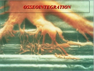
Definition
The word osseointegrationconsists of “OS”
the Latin word for bone and “integration”
derived from Latin word meaning the state of
being combined into a complete whole.
Osseointegration is defined as a direct bone
anchorage to an implant body which can
provide a foundation to support a prosthesis.
ϒ “Direct structural and functional connection between
ordered, living bone and surface of a load carrying implant ”.
9.
…..
American Academyof Implant Dentistry
defined it as “contact established without
interposition of non bone tissue between
normal remodeled bone and on implant
entailing a sustained transfer and
distribution of load from the implant to and
within bone tissue”.
10.
Historical Review
ϒ Theconcept of osseointegration was developed and the term was
coined by Dr. Per-Ingvar Branemark,
ϒ Professor at the institute for Applied Biotechnology, University of
Goteborg, Sweden.
11.
Initial concept ofosseointegration stemmed
from vital microscopic studies of
microcirculation in bone repair mechanisms.
Titanium chamber was surgically inserted
into the tibia of of a rabbit.
It was considered the best material for
artificial tooth root replacement.
12.
…..
Many studiesfollowed involving titanium
implants being placed into jaws of dogs.
Direct bone anchorage has been shown to
be very strong. A force of over 100kg was
applied to dislodge an implant.
Based on such a consequence the
foundation for Osseo integration and the
Branemark implant system was established
in 1952.
13.
Studies on humanswere
conducted by means of an
implant optical titanium chamber
in a twin pedicle skin tube on the
inside of the left upper arm of
volunteers.
Tissue reactions were studied in
long term experiments.
All this lead to the treatment of
first edentulous patient in 1965.
14.
History of Branemarksystem categorized in
three stages
ϒ Early stage (1965-1968)
ϒ Developmental stage (1968-1971)
ϒ Production stage (1971 – present)
Biological Considerations for
Osseointegration
Bone implant interface
ϒ When compared to compact bone
spongy bone has less density and
hardness is not a stable base for
primary fixture fixation.
ϒ In the mandible the spongy bone
is more dense than maxilla.
ϒ With primary fixation in
compact bone, osseointegration
in the maxilla require a longer
healing period.
17.
Bone remodeling
Osseointegration requires new bone formation
around the fixture. A process resulting from
remodeling within bone tissue.
Osteoblastic and osteoclastic activity helps
maintain blood calcium without change in
quantity of bone.
18.
…..
To maintaina constant level of bone
remodeling there should be proper local
stimulation, crucial levels of thyroid
hormone, calcitonin and vitamin D.
Occlusion or occlusal force stimulus are
both important to optimal bone remodeling.
19.
Foreign body reaction
Organization or an antigen antibody
reaction occurs when a foreign body is
present in the body.
This reaction occurs in the presence of a
protein but with implant materials devoid
of proteins no antigen antibody reaction.
20.
…..
When titaniumis used no foreign body reaction are
seen.
The implant material is an important factor for
Osseo integration to occur.
21.
Bone to implantinterface
Two basic theories
ϒ Fibro-osseous integration by Linkow, James & Weis
ϒ Osseointegration by Branemark
ϒ Meffert divided osseointegration
Adaptive osseointegration Biointegration
22.
American Academy ofimplant dentistry defined
fibrous integration as tissue to implant contact with
healthy dense collagenous tissue between the
implant and bone.
23.
…..
The fibersare arranged irregularly, parallel to the
implant body, when forces are applied they are not
transmitted through the fibers.
So no bone remodeling expected in fibro-
integration.
24.
Ichida & Caputo(1986) used photo-elastic analysis to
study the stress concentration along the implant
threads and sharp edges when a connective tissue like
structure was included in the analysis.
Even stress distribution was seen when there was
direct contact with a bone like structure.
They concluded that implants with fibro-osseous
integration had a tendency of increased mobility.
25.
A direct boneimplant interface occurs when an
implant is allowed to heal in bone undisturbed.
Main factors affecting osseointegration include
ϒ Implant oxide layer contamination.
ϒ Poor temperature control during drilling.
26.
……
A minimum of3 month healing in mandible and 6
months in maxilla is necessary before load is
applied.
If osseointegration does not occur or a fibrous
connective tissue forms around the implant
organization process continues.
27.
Biological process ofimplant osseointegration
The healing process of implant
system is similar to primary
bone healing.
Titanium dental implants show
three stages of healing.
28.
…..
OSTEOPHYLLIC STAGE
ϒ When a implant is placed into the cancellous marrow
space blood is initially present between implant and
bone.
ϒ Only a small amount of bone is in contact with the
implant surface; the rest is exposed to extracellular
fluids.
ϒ Generalized inflammatory response to the surgical
insult.
29.
…..
ϒ By theend of first week, inflammatory cells are
responding to foreign antigens.
ϒ Vascular ingrowth from the surrounding vital tissues
begins by third day.
ϒ A mature vascular network forms by 3 weeks.
ϒ Ossification also begins during the first week and the
initial response observed in the migration of osteoblasts
from the trabacular bone which can be due to the
release of BMP’s.
ϒ The osteophyllic phase lasts about 1 month.
30.
OSTEOCONDUCTIVE PHASE
ϒOnce theyreach the implant, the bone cells spread
along the metal surface laying down osteoid.
ϒInitially this is an immature connective tissue
matrix and bone deposited is a thin layer of woven
bone called foot plate.
31.
…..
ϒ Fibro-cartilaginouscallus is eventually
remodeled into bone callus.
ϒ This process occurs during the next 3 months
ϒ Four months after implant placement the
maximum surface area is covered by bone.
32.
OSTEOADAPTIVE PHASE
ϒ Thefinal phase begins approximately 4 months after
implant placement.
ϒ Once loaded implants do not gain or loose bone contact but
the foot plates thicken in response and some reorientation of
the vascular pattern may be seen.
33.
…..
ϒ Graftedbone integrates to a higher degree than the
natural host bone to the implant.
ϒ To achieve optimal results an osseointegration period
of 4 months is recommended for implants in graft
bone and 4 to 8 months for implant placed in normal
bone.
34.
Ultrastructure in osseointegration
ϒOsseointegrated fixtures
under occlusal loads are
surrounded by cortical and
spongy bone.
ϒ The cortical bone to fixture
surface interface has
canaliculi participating in
electrolyte transportation
near oxide layer.
35.
….
Osseointegration in spongybone occurs as
bone trabaculae approaches the fixture and
come into intimate contact with oxide layer.
Ground substance forms and fills spaces
between bone trabeculae this fuses with
oxide layer.
36.
Destruction of Osseointegration
Themain contributing factor to bone resorption are
local inflammation from plaque and trauma from
occlusion
ϒ Direct action of plaque products induces formation of
osteoclasts.
ϒ Plaque products at directly on bone destroying it
through a non cellular mechanism.
ϒ Stimulate gingival cells, which release mediators for
osteoclast formation.
37.
ϒ Plaque causesgingival cells to release agents which act as
cofactors in bone resorption and which destroy bone by
direct chemical action without osteoclasts.
Bone resorption can be caused by premature
loading.
12 months following fixture insertion vertical
bone loss is observed due to traumatic
surgical procedure.
ϒ Vertical bone loss approximately 1 to 1.5 mm in first year
ϒ Marginal bone loss is 0.05 to 0.1 mm in first year
ϒ These measurements can be used a reference and in a
bone loss condition should be evaluated to minimize
failure.
38.
Peri-implant membrane
ϒ Withthe osseointegrated implant
the abutment to fixture junction
corresponds to cementoenamel
junction present in natural
dentition.
ϒ Peri-implant membrane is similar
to that present in natural
dentition, consisting of peri-
implant free gingiva.
39.
….
The sulcular epitheliumforms the peri-
implant gingival crevice and junctional
epithelium attaches to the abutment forming
a cuff.
With a tight cuff and filamentous attachment
a membrane is sealed tightly and
functionally to the abutment surface.
40.
Disease activity inperi-implant tissue
The fibrous connective tissue capsule
formed around an implant generally has low
differentiating capabilities such that it also
has less resistance against bacterial bi-
products and does not respond well to
occlusal stimulation.
An osseointegrated implant has periosteum
directly covering the neck of the fixture.
Which may act as a barrier against
inflammation.
41.
….
Although the abutmentto junctional epithelium
attachment is not strong, a connective tissue band
is tightly attached to the abutment surface and acts
as resistant barrier.
42.
The neuromuscular systemas it relates to the
osseointegrated implant
A fixture site does not have periodontal ligament but
has nerve endings located near the fixture, sensing
pain and temperature.
Patients with osseointegrated implants have a high
threshold and low sensitivity for discriminating
thickness.
43.
…..
As theperiodontal ligament is lost the
fixture remains with reduce amount of
receptors.
Impulses from the fixture sites are
transmitted through motor nucleus of
trigeminal nerve.
Mechanical retention
metallic substratesystem such as titanium
or titanium alloy.
The retention is based on undercut forms
such as vents, slots, dimples, screws etc.,
Direct contact between the dioxide layer on
the titanium and bone with no chemical
bonding.
46.
Bioactive retention
Bioactivity
ϒ characteristicof an implant material that allows attachment to
living tissues, whereas a non bioactive material would form a
loosely adherent layer of fibrous tissue at the implant interface
Bioactive retention is achieved with
bioactive materials such as hydroxyapatite
(HA), which bond directly to bone
47.
Plasma spraying &ion sputter coating
Two techniques used to coat metallic implants
with HA.
48.
Plasma spraying
Involves heatingthe HA by a plasma flame
at a temperature of approximately 15,000° C
to 20,000°C.
The HA is then propelled onto the implant
body in an inert environment like argon, to a
thickness of 50 to 100 μm.
49.
Ion-sputter coating
Process bywhich a thin, dense layer of HA can be
coated onto an implant substrate.
Directing an ion beam at a solid-phase HA block,
Vaporising it to create a plasma and then
recondensing this plasma on the implant.
Bone formation and maturation occurs at a faster
rate in the initial phases on HA coated implants
than on non-coated implants
50.
Advantages of increasedsurface roughness of Cp Ti
implant
Increased surface area of the implant adjacent to
bone.
Improved cell attachment to the implant surface.
Increased bone present at the implant surface.
Increased biomechanical interactions of the implant
with bone.
Promoted inflammation of the periimplant area.
51.
Clinical advantages ofTPS or HA coatings
Increased surface area
Increased roughness for initial stability
Stronger bone-to- implant interface
Additional advantages of HA over TPS include the
following
ϒ Faster healing bone interface
ϒ Increased gap healing between bone and HA
ϒ Stronger interface than TPS
52.
Disadvantages of Coatings
Flaking,cracking, or scaling upon insertion
Increased plaque retention when placed above the
bone.
Increased bacteria adhesion and acts as a nidus for
infection
Complications of treating the failing implants
Increased cost
53.
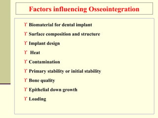
Factors influencing Osseointegration
ϒBiomaterial for dental implant
ϒ Surface composition and structure
ϒ Implant design
ϒ Heat
ϒ Contamination
ϒ Primary stability or initial stability
ϒ Bone quality
ϒ Epithelial down growth
ϒ Loading
54.
1.Biomaterial for dentalimplant
Implants must not induce a host immune
response Titanium and certain calcium-
phosphate ceramics are biocompatible
and do not stimulate a foreign body
rejection reaction.
55.
2. Surface composition and structure
ϒ It is thought that cp Ti owes its ability to form an
osseointegrated interface to the tough and relatively
inert oxide layer, which forms very rapidly on its
surface.
ϒ This surface has been described as osseoconductive,
that is, conducive to bone formation
ϒ Other substrates also have this property and may also
stimulate bone formation, a property known as
osseoinduction
56.
3. Implant Design
Thevast majority of commercially available
implants claiming osseointegration status
are cylindrical in shape.
Their design may be threaded or else lack
similar microscopic retentive/stabilization
aspects
57.
4. Heat
Heating ofbone to a temperature in
excess of 47°C during implant surgery can
result in cell death and denaturation of
collagen.
As a result, osseointegration may not
occur, instead the implant becomes
surrounded by a fibrous capsule and the
shear strength of the implant-host
interface is significantly reduced.
58.
5. Contamination
ϒ Contaminationof the implant site by organic and inorganic
debris can prejudice the achievement of osseointegration.
ϒ Material such as necrotic tissue, bacteria, chemical reagents
and debris from drills can all be harmful in this respect.
59.
6. Primary stabilityor Initial stability
ϒ It is known that where an implant fits tightly into its
osteotomy site then osseointegration is more likely to
occur.
ϒ This is often referred to as primary stability, and where
an implant body has this attribute when first placed
failure is less probable.
ϒ This property is related to the quality of fit of the
implant, its shape, and bone morphology and density.
60.
7. Bone quality
Itis a function of bone density, anatomy and volume, and
has been described using a number of indices.
ϒ The classifications of Lekholm & Zarb and of Cawood &
Howell are widely used to describe bone quality and
quantity.
ϒ The former relates to the thickness and density of cortical
and Cancellous bone,
ϒ and the latter to the amount of bone resorption.
ϒ Bone volume does not by itself influence osseointegration,
but is an important determinant of implant placement
61.
8. Epithelial down growth
ϒ Early implant designs were often associated with down
growth of oral epithelium, which eventually exteriorized
the device.
ϒ When the newer generation of cp Ti devices was
introduced great care was taken to prevent this by
initially covering the implant body with oral mucosa
while osseointegration occurred.
ϒ The implant body was then exposed and a superstructure
added, since it was known that the osseointegrated
interface was resistant to epithelial down growth.
62.
9. Loading schemes
ϒDelayed loading: The prosthesis is attached at the
second procedure after a conventional healing
period of 3 to 6 months 8, 23.
ϒ Early loading: The prosthesis is attached during a
second procedure, earlier than the conventional
healing period of 3 to 6 months. Time of loading
should be stated in days to weeks 8, 23.
ϒ Immediate / Direct loading: The prosthesis is
attached to the implants the same day the implants
are placed.
63.
Success criteria forOsseo integrated
Implants
ϒ Durability
ϒ Bone loss
ϒ Gingival health
ϒ Pocket depth
ϒ Effect of adjacent teeth
ϒ Functions
ϒ Esthetics
ϒ Presence of infection
ϒ Intrusion on the mandibular canal
ϒ Patient emotional and psychological attitude
64.
Revised criteria forimplant success
ϒ Individual unattached implant is immobile when
tested clinically.
ϒ No evidence of peri implant radiolucency is present as
assessed on an undistorted radiograph.
ϒ Mean vertical bone loss is less than 0.2 mm after 1st
year of service.
ϒ No persistent pain, discomfort or infection.
ϒ A success rate of 85% at the end of a 5-year
observation period and 80% at the end of a 10-year
period are minimum levels of success.
Futuristic concepts ofOsseointegration
OSSEOPERCEPTION
ϒ The interaction between
the osseointegrated
fixture bone tissue,
receptor systems and
nervous system has to be
studied.
“Owing to the nature of osseointegration it is not easy to
dissect the system of anchorage from the clinical level down
to the molecular level or even the real interface which is still
largely a mystery”
Mechanism of Osseointegration
Blood clot (between fixture & bone)
Clot transformed by phagocytic cell
(1st to 3rd day)
Procallus formation
(containing fibroblasts & phagocytes)
Procallus becomes dense connective tissue
(Differentiation of osteoblasts & fibroblasts)
Callus (Osteoblasts on the fixture)
Fibro cartilagenous callus (between fixture & bone)
Bone callus (Penetrates & matures)
Prosthesis attached to the fixtures stimulating bone remodeling
71.
It is becauseof the attention to training, research & clinical
studies that osseointegration has now become an accepted part of
the treatment regime in many countries world wide and no longer
regarded as the last resort when all else has failed but often as a
treatment of choice
72.
References
ϒ Hobo, Ichida,Garcia “Osseointegration and occlusal
rehabilitation” Quintessence Publishing.
ϒ Jan Lindhe “Clinical periodontology and implant
dentistry” 4th edition, Blackwell Publishing.
ϒ Elaine McClarence “Branemark and the development of
osseointegration” Quintessence publication
ϒ Carl E. Misch “Implant dentistry” 2nd edition, Mosby.
ϒ Hubertus Spiekermann “Color atlas of dental medicine
implantology” Theime Publishers.
73.
…..
ϒ CharlesM.Weis “Principles and practice of implant
dentistry” Mosby.
ϒ Charles Babbush “Dental implants the art and
science” W.B. Saunders.
ϒ Per Ingvar Branemark “Osseointegration and its
experimental background” JPD 1983 Vol. 50, 399-410.
ϒ Hanson, Alberktson “Structural aspects of the
interface between tissue and titanium implants” JPD
1983 vol. 50, 108-113.
74.
….
ϒ T. Alberktson“Osseointegrated dental implants” DCNA Vol.
30, Jan 1986, 151-189.
ϒ Richard Palmer “Introduction to dental implants” BDJ, Vol.
187, 1999, 127-132.
ϒ Geroge A. Zarb “Osseointegrated dental implants:
Preliminary report on a replication study”. JPD 1983, Vol
50, 271-276.
ϒ Bergman “Evaluation of the results of treatment with
osseointegrated implants by the Swedish National Board of
Health and Welfare”. JPD 1983, vol. 50, 114-116.