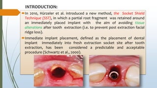
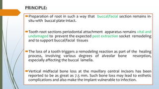
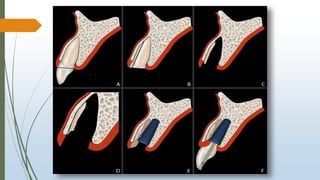
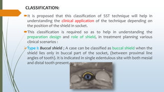
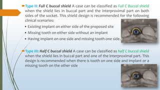
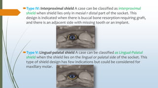
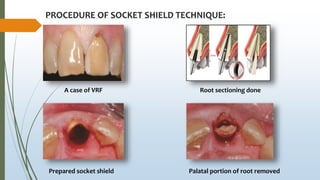
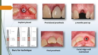
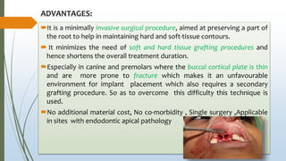
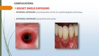
The socket-shield technique at molar sites: A proof-of-
principle technique report.
Schwimer W C, Gluckman H, Salama M, Nagy K and Toit J , J Prost Dent 2019
;121(2): 229-33
Presented by:
DR. NABID ANJUM
PG 2ND YEAR
DEPARTMENT OF PROSTHODONTICS
CONTENTS:
INTRODUCTION
PRINCIPLE OF TECHNIQUE
CLASSIFICATION OF SOCKET SHIELD
PROCEDURE
ADVANTAGES
LIMITATIONS
COMPLICATIONS
MASTER ARTICLE
DISCUSSION
CRITICAL ANALYSIS
CONCLUSION
REFERENCES
INTRODUCTION:
In 2010, Hürzeler et al. introduced a new method, the Socket Shield
Technique (SST), in which a partial root fragment was retained around
an immediately placed implant with the aim of avoiding tissue
alterations after tooth extraction (i.e. to prevent post extraction facial
ridge loss).
Immediate implant placement, defined as the placement of dental
implant immediately into fresh extraction socket site after tooth
extraction, has been considered a predictable and acceptable
procedure (Schwartz et al., 2000).
PRINCIPLE:
Preparation of root in such a way that buccal/facial section remains in-
situ with buccal plate intact.
Tooth root sections periodontal attachment apparatus remains vital and
undamaged to prevent the expected post extraction socket remodeling
and to support buccal/facial tissues
The loss of a tooth triggers a remodeling reaction as part of the healing
process, involving various degrees of alveolar bone resorption,
especially affecting the buccal lamella.
Vertical midfacial bone loss at the maxillary central incisors has been
reported to be as great as 7.5 mm. Such bone loss may lead to esthetic
complications and also make the implant vulnerable to infection.
INDICATIONS FOR SOCKET SHIELD TECHNIQUE:
The ideal extraction site for immediate implant placement:
Little or no periodontal bone loss on the tooth that is to be extracted
such as tooth being extracted due to
1) Endodontic involvement 2) Root fracture/Root resorption
3) Periapical pathology 4) Root perforation 5) Unfavorable crown to
root ratio 6)Residual deciduous tooth
A minimum of 4-5mm bone width at the alveolar crest and at least
10mm bone length from the alveolar crest to a safe distance above
closest anatomical structures are recommended (WORTHINGTON
2004)
CONTRAINDICATIONS OF SOCKET SHIELD TECHNIQUE:
Presence of pus.
Lack of bone beyond the apex.
Close proximity to anatomical vital structures.
Clinical conditions preventing primary closure.
CLASSIFICATION:
It is proposed that this classification of SST technique will help in
understanding the clinical application of the technique depending on
the position of the shield in socket.
This classification is required so as to help in understanding the
preparation design and role of shield, in treatment planning various
clinical scenarios :
Type I: Buccal shield ; A case can be classified as buccal shield when the
shield lies only in buccal part of the socket, (between proximal line
angles of tooth). It is indicated in single edentulous site with both mesial
and distal tooth present.
Type II: Full C buccal shield A case can be classified as Full C Buccal shield
when the shield lies in buccal part and the interproximal part on both
sides of the socket. This shield design is recommended for the following
clinical scenarios:
• Existing implant on either side of the proposed site
• Missing tooth on either side without an implant
• Having implant on one side and missing tooth one side.
Type III: Half C buccal shield A case can be classified as half C buccal shield
when the shield lies in buccal part and one of the interproximal part. This
design is recommended when there is tooth on one side and implant or a
missing tooth on the other side
Type IV: Interproximal shield A case can be classified as interproximal
shield when shield lies only in mesial r distal part of the socket. This
design is indicated when there is buccal bone resorption requiring graft,
and there is an adjacent side with missing tooth or an implant.
Type V: Lingual-palatal shield A case can be classified as Lingual-Palatal
shield when the shield lies on the lingual or palatal side of the socket. This
type of shield design has few indications but could be considered for
maxillary molar.
PROCEDURE OF SOCKET SHIELD TECHNIQUE:
A case of VRF Root sectioning done
Palatal portion of root removedPrepared socket shield
Implant placed Provisional prosthesis 3 months post op
Facial ridge well
maintained
Final prosthesisBurs for technique
ADVANTAGES:
It is a minimally invasive surgical procedure, aimed at preserving a part of
the root to help in maintaining hard and soft-tissue contours.
 It minimizes the need of soft and hard tissue grafting procedures and
hence shortens the overall treatment duration.
Especially in canine and premolars where the buccal cortical plate is thin
and are more prone to fracture which makes it an unfavourable
environment for implant placement which also requires a secondary
grafting procedure. So as to overcome this difficulty this technique is
used.
No additional material cost, No co-morbidity , Single surgery ,Applicable
in sites with endodontic apical pathology
LIMITATIONS:
The clinician needs to be specially trained and need to have a high
degree of clinical skills.
The procedure requires a little more time and patience to avoid mobility
in the shield. If the shield becomes mobile during surgery, it is removed,
and the conventional immediate implant placement or the grafting
procedure is to be done.
The case selection is very important for the success of the procedure.
The technique is not recommended in mobile teeth, teeth which are out
of the arch and teeth with large periapical lesions.
The intactness of the shield plays an important role in the success of the
treatment.
COMPLICATIONS:
SOCKET SHIELD EXPOSURE:
• INTERNAL EXPOSURE: coronal portion of the S-S perforating the soft tissue.
• EXTERNAL EXPOSURE: toward the oral cavity.
•Either no treatment
•Or reduction of exposed root
portion with diamond bur
INTERNAL
EXPOSURE
•Reducing coronal aspect of soft
tissue closure
•CTG augmentation to assist soft
tissue healing
EXTERNAL
EXPOSURE
INFECTION:
• Infection at site 21. Restoration removed, socket-shield was
mobile and thus removed. GBR procedure was done, implant
restored and in function.
MASTER ARTICLE:
After local anesthesia of the site, decoronate the tooth.
 Hemisection the root trunk mesiodistally with a long-shanked, straight
diamond rotary instrument in a high-speed handpiece with copious
irrigation. The buccal roots are separated from the palatal root.
Make a similar mesiodistal preparation into the
buccal roots, thereby beginning preparation of
the 2 socket-shields.
First, remove the palatal root and then the palatal portions of the sectioned buccal roots.
Only the buccal portions of the buccal roots are to remain.
Carefully prepare the socket-shields of these buccal roots by thinning each mesiodistally
with the same long-shank rotary instrument.
Using a large round diamond rotary instrument, reduce the crestal portion of the socket-
shields to bone level.
Reduce the crestal portion of each socket-shield further by creating a 2-mm internal
chamfer bevel that provides prosthetic space for the crown.
Curette the sockets, especially the apical areas, and rinse vigorously to clean.
Make intraoperative periapical radiographs to confirm that all endodontic
obturation material, root canals, and root apices have been removed.
Verify absolute immobility of the socket-shields by applying a sharp explorer
to the internal/dentin surface of the shield.
Then, prepare the implant osteotomy from the prosthetic/surgical planning
and guide into the of the furcal bone extraction site.
Insert the implant, verify primary stability (preferably using a resonance
frequency analysis device), and graft the buccal gap with a bone particulate
material.
Implants inserted. First molar site fully healed,
delayed loading.
Second molar site, immediate placement at
socket-shields. Socket filled with xenograft.
Transgingival healing abutments secured to
implants. Site sutured and closed.
Give explicit postoperative oral hygiene instructions, typically a regimen of a 0.2%
chlorhexidine oral rinse twice daily for 2 weeks.
 Prescribe only systemic analgesics when/if needed. Systemic antibiotics are
unnecessary and not prescribed.
Recall the patient for a 48-hour short follow-up and then for a 2-week follow-up for
suture removal.
After approximately 3 to 4 months of healing, verify osseointegration by resonance
frequency analysis. The implant stability quotients should be >70.
Scannable abutments secured to
implants
DISCUSSION:
The socket-shield technique is potentially one of the most significant
contributions to implant and restorative dentistry, managing the
resorptive sequalae of tooth extraction.
The posterior ridge may incur esthetic compromise when substantial
alveolar ridge loss is evident. More importantly, loss of posterior bone
may result in vestibular depth reduction and lack of attached
keratinized tissue.
Such changes, coupled with inadequate alveolar bone, may negatively
impact the health of the implant and predispose to peri-implantitis.
With regard to technique and specific to molar sites, long and widely
divergent, excessively curved roots at molars are particularly
challenging.
At molar sites, specific to socket shield, the treatment is notably more
difficult and should only be attempted by those clinicians experienced in
immediate implant placement and who have mastered the socket-shield
technique in anterior single-tooth sites.
Not only is the treatment technique sensitive but it also takes additional
time likely a longer procedure than conventional extraction with forceps
alone.
Moreover, complications have been reported at socket-shields,
including infection at the site (2.3% of procedures), internal exposure
(9.4%), external exposure (3.1%), implant failure to osseointegrate (3.9%),
and orthodontic migration of shields (1.5%)
The anatomy of the mesiobuccal roots of maxillary molars makes it
difficult to access the apical root portion for removal. In such situations, it
is likely that the coronal portion that is straighter would be separated
from the curved apical portion and the root tip delivered.
The maxillary molar buccal roots are smaller, and all root canal contents,
apical root portions and contents, as well as endodontic obturation
material must be completely removed.
 This is achieved by making intraoperative radiographs to confirm
complete materials and root apex removal.
CRITICAL ANALYSIS:
They have not mentioned anything about the tooth structure which was
left in the socket.
Much data is not available to support this article.
CONCLUSION:
The socket-shield technique as a partial extraction therapy to preserve
Bucco-facial tooth structure and maintain the ridge at anterior implant
sites has received much attention in recent years.
However, the significance of ridge collapse at posterior sites is often
overlooked. From the technique presented in this report, socket-shields
may help maintain the alveolar ridge at immediate molar implant
placement sites.
The SST is gaining popularity among the clinicians across the world. The
technique is very promising for the preservation of hard and soft tissues in
cases of post-extraction immediate implant placement.
REFERENCES:
Hürzeler MB, Zuhr O, Schupbach P, Rebele SF, Emmanouilidis N, Fickl S, et
al. The socket-shield technique: A proof-of-principle report. J Clin
Periodontol 2010;37:855-62
Kumar PR, Kher U. Shield the socket: Procedure, case report and
classification. J Indian Soc Periodontol 2018;22:266-72
Gluckman H, Salama M, Du Toit J. A retrospective evaluation of 128 socket-
shield cases in the esthetic zone and posterior sites: Partial extraction
therapy with up to 4 years follow-up. Clin Implant Dent Relat Res
2018;20:122-9.
Gluckman H, Nagy K and Du Toit J. Prosthetic management of implants
placed with socket shield technique . J Pros Dent 2019; 121(4): 581-85

The socket shield technique at molar sites

  • 1.
    The socket-shield techniqueat molar sites: A proof-of- principle technique report. Schwimer W C, Gluckman H, Salama M, Nagy K and Toit J , J Prost Dent 2019 ;121(2): 229-33 Presented by: DR. NABID ANJUM PG 2ND YEAR DEPARTMENT OF PROSTHODONTICS
  • 2.
    CONTENTS: INTRODUCTION PRINCIPLE OF TECHNIQUE CLASSIFICATIONOF SOCKET SHIELD PROCEDURE ADVANTAGES LIMITATIONS COMPLICATIONS MASTER ARTICLE DISCUSSION CRITICAL ANALYSIS CONCLUSION REFERENCES
  • 3.
    INTRODUCTION: In 2010, Hürzeleret al. introduced a new method, the Socket Shield Technique (SST), in which a partial root fragment was retained around an immediately placed implant with the aim of avoiding tissue alterations after tooth extraction (i.e. to prevent post extraction facial ridge loss). Immediate implant placement, defined as the placement of dental implant immediately into fresh extraction socket site after tooth extraction, has been considered a predictable and acceptable procedure (Schwartz et al., 2000).
  • 4.
    PRINCIPLE: Preparation of rootin such a way that buccal/facial section remains in- situ with buccal plate intact. Tooth root sections periodontal attachment apparatus remains vital and undamaged to prevent the expected post extraction socket remodeling and to support buccal/facial tissues The loss of a tooth triggers a remodeling reaction as part of the healing process, involving various degrees of alveolar bone resorption, especially affecting the buccal lamella. Vertical midfacial bone loss at the maxillary central incisors has been reported to be as great as 7.5 mm. Such bone loss may lead to esthetic complications and also make the implant vulnerable to infection.
  • 6.
    INDICATIONS FOR SOCKETSHIELD TECHNIQUE: The ideal extraction site for immediate implant placement: Little or no periodontal bone loss on the tooth that is to be extracted such as tooth being extracted due to 1) Endodontic involvement 2) Root fracture/Root resorption 3) Periapical pathology 4) Root perforation 5) Unfavorable crown to root ratio 6)Residual deciduous tooth A minimum of 4-5mm bone width at the alveolar crest and at least 10mm bone length from the alveolar crest to a safe distance above closest anatomical structures are recommended (WORTHINGTON 2004)
  • 8.
    CONTRAINDICATIONS OF SOCKETSHIELD TECHNIQUE: Presence of pus. Lack of bone beyond the apex. Close proximity to anatomical vital structures. Clinical conditions preventing primary closure.
  • 9.
    CLASSIFICATION: It is proposedthat this classification of SST technique will help in understanding the clinical application of the technique depending on the position of the shield in socket. This classification is required so as to help in understanding the preparation design and role of shield, in treatment planning various clinical scenarios : Type I: Buccal shield ; A case can be classified as buccal shield when the shield lies only in buccal part of the socket, (between proximal line angles of tooth). It is indicated in single edentulous site with both mesial and distal tooth present.
  • 10.
    Type II: FullC buccal shield A case can be classified as Full C Buccal shield when the shield lies in buccal part and the interproximal part on both sides of the socket. This shield design is recommended for the following clinical scenarios: • Existing implant on either side of the proposed site • Missing tooth on either side without an implant • Having implant on one side and missing tooth one side. Type III: Half C buccal shield A case can be classified as half C buccal shield when the shield lies in buccal part and one of the interproximal part. This design is recommended when there is tooth on one side and implant or a missing tooth on the other side
  • 11.
    Type IV: Interproximalshield A case can be classified as interproximal shield when shield lies only in mesial r distal part of the socket. This design is indicated when there is buccal bone resorption requiring graft, and there is an adjacent side with missing tooth or an implant. Type V: Lingual-palatal shield A case can be classified as Lingual-Palatal shield when the shield lies on the lingual or palatal side of the socket. This type of shield design has few indications but could be considered for maxillary molar.
  • 12.
    PROCEDURE OF SOCKETSHIELD TECHNIQUE: A case of VRF Root sectioning done Palatal portion of root removedPrepared socket shield
  • 13.
    Implant placed Provisionalprosthesis 3 months post op Facial ridge well maintained Final prosthesisBurs for technique
  • 14.
    ADVANTAGES: It is aminimally invasive surgical procedure, aimed at preserving a part of the root to help in maintaining hard and soft-tissue contours.  It minimizes the need of soft and hard tissue grafting procedures and hence shortens the overall treatment duration. Especially in canine and premolars where the buccal cortical plate is thin and are more prone to fracture which makes it an unfavourable environment for implant placement which also requires a secondary grafting procedure. So as to overcome this difficulty this technique is used. No additional material cost, No co-morbidity , Single surgery ,Applicable in sites with endodontic apical pathology
  • 15.
    LIMITATIONS: The clinician needsto be specially trained and need to have a high degree of clinical skills. The procedure requires a little more time and patience to avoid mobility in the shield. If the shield becomes mobile during surgery, it is removed, and the conventional immediate implant placement or the grafting procedure is to be done. The case selection is very important for the success of the procedure. The technique is not recommended in mobile teeth, teeth which are out of the arch and teeth with large periapical lesions. The intactness of the shield plays an important role in the success of the treatment.
  • 16.
    COMPLICATIONS: SOCKET SHIELD EXPOSURE: •INTERNAL EXPOSURE: coronal portion of the S-S perforating the soft tissue. • EXTERNAL EXPOSURE: toward the oral cavity.
  • 17.
    •Either no treatment •Orreduction of exposed root portion with diamond bur INTERNAL EXPOSURE •Reducing coronal aspect of soft tissue closure •CTG augmentation to assist soft tissue healing EXTERNAL EXPOSURE
  • 18.
    INFECTION: • Infection atsite 21. Restoration removed, socket-shield was mobile and thus removed. GBR procedure was done, implant restored and in function.
  • 19.
    MASTER ARTICLE: After localanesthesia of the site, decoronate the tooth.  Hemisection the root trunk mesiodistally with a long-shanked, straight diamond rotary instrument in a high-speed handpiece with copious irrigation. The buccal roots are separated from the palatal root. Make a similar mesiodistal preparation into the buccal roots, thereby beginning preparation of the 2 socket-shields.
  • 20.
    First, remove thepalatal root and then the palatal portions of the sectioned buccal roots. Only the buccal portions of the buccal roots are to remain. Carefully prepare the socket-shields of these buccal roots by thinning each mesiodistally with the same long-shank rotary instrument. Using a large round diamond rotary instrument, reduce the crestal portion of the socket- shields to bone level. Reduce the crestal portion of each socket-shield further by creating a 2-mm internal chamfer bevel that provides prosthetic space for the crown.
  • 21.
    Curette the sockets,especially the apical areas, and rinse vigorously to clean. Make intraoperative periapical radiographs to confirm that all endodontic obturation material, root canals, and root apices have been removed. Verify absolute immobility of the socket-shields by applying a sharp explorer to the internal/dentin surface of the shield. Then, prepare the implant osteotomy from the prosthetic/surgical planning and guide into the of the furcal bone extraction site. Insert the implant, verify primary stability (preferably using a resonance frequency analysis device), and graft the buccal gap with a bone particulate material.
  • 22.
    Implants inserted. Firstmolar site fully healed, delayed loading. Second molar site, immediate placement at socket-shields. Socket filled with xenograft. Transgingival healing abutments secured to implants. Site sutured and closed.
  • 23.
    Give explicit postoperativeoral hygiene instructions, typically a regimen of a 0.2% chlorhexidine oral rinse twice daily for 2 weeks.  Prescribe only systemic analgesics when/if needed. Systemic antibiotics are unnecessary and not prescribed. Recall the patient for a 48-hour short follow-up and then for a 2-week follow-up for suture removal. After approximately 3 to 4 months of healing, verify osseointegration by resonance frequency analysis. The implant stability quotients should be >70.
  • 24.
  • 25.
    DISCUSSION: The socket-shield techniqueis potentially one of the most significant contributions to implant and restorative dentistry, managing the resorptive sequalae of tooth extraction. The posterior ridge may incur esthetic compromise when substantial alveolar ridge loss is evident. More importantly, loss of posterior bone may result in vestibular depth reduction and lack of attached keratinized tissue. Such changes, coupled with inadequate alveolar bone, may negatively impact the health of the implant and predispose to peri-implantitis. With regard to technique and specific to molar sites, long and widely divergent, excessively curved roots at molars are particularly challenging.
  • 26.
    At molar sites,specific to socket shield, the treatment is notably more difficult and should only be attempted by those clinicians experienced in immediate implant placement and who have mastered the socket-shield technique in anterior single-tooth sites. Not only is the treatment technique sensitive but it also takes additional time likely a longer procedure than conventional extraction with forceps alone. Moreover, complications have been reported at socket-shields, including infection at the site (2.3% of procedures), internal exposure (9.4%), external exposure (3.1%), implant failure to osseointegrate (3.9%), and orthodontic migration of shields (1.5%)
  • 27.
    The anatomy ofthe mesiobuccal roots of maxillary molars makes it difficult to access the apical root portion for removal. In such situations, it is likely that the coronal portion that is straighter would be separated from the curved apical portion and the root tip delivered. The maxillary molar buccal roots are smaller, and all root canal contents, apical root portions and contents, as well as endodontic obturation material must be completely removed.  This is achieved by making intraoperative radiographs to confirm complete materials and root apex removal.
  • 28.
    CRITICAL ANALYSIS: They havenot mentioned anything about the tooth structure which was left in the socket. Much data is not available to support this article.
  • 29.
    CONCLUSION: The socket-shield techniqueas a partial extraction therapy to preserve Bucco-facial tooth structure and maintain the ridge at anterior implant sites has received much attention in recent years. However, the significance of ridge collapse at posterior sites is often overlooked. From the technique presented in this report, socket-shields may help maintain the alveolar ridge at immediate molar implant placement sites. The SST is gaining popularity among the clinicians across the world. The technique is very promising for the preservation of hard and soft tissues in cases of post-extraction immediate implant placement.
  • 30.
    REFERENCES: Hürzeler MB, ZuhrO, Schupbach P, Rebele SF, Emmanouilidis N, Fickl S, et al. The socket-shield technique: A proof-of-principle report. J Clin Periodontol 2010;37:855-62 Kumar PR, Kher U. Shield the socket: Procedure, case report and classification. J Indian Soc Periodontol 2018;22:266-72 Gluckman H, Salama M, Du Toit J. A retrospective evaluation of 128 socket- shield cases in the esthetic zone and posterior sites: Partial extraction therapy with up to 4 years follow-up. Clin Implant Dent Relat Res 2018;20:122-9. Gluckman H, Nagy K and Du Toit J. Prosthetic management of implants placed with socket shield technique . J Pros Dent 2019; 121(4): 581-85

Editor's Notes

  • #4 The buccal portion of the root is retained to preserve periodontal ligament and bundle bone.
  • #5 The bundle bone is primarily vascularized by periodontal membrane of the tooth. Therefore, bone is compromised by extraction leading to partial resorption.