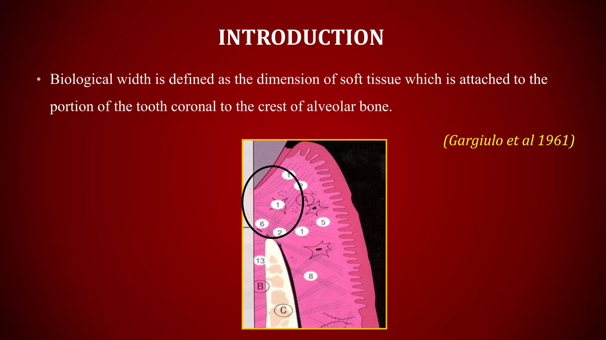
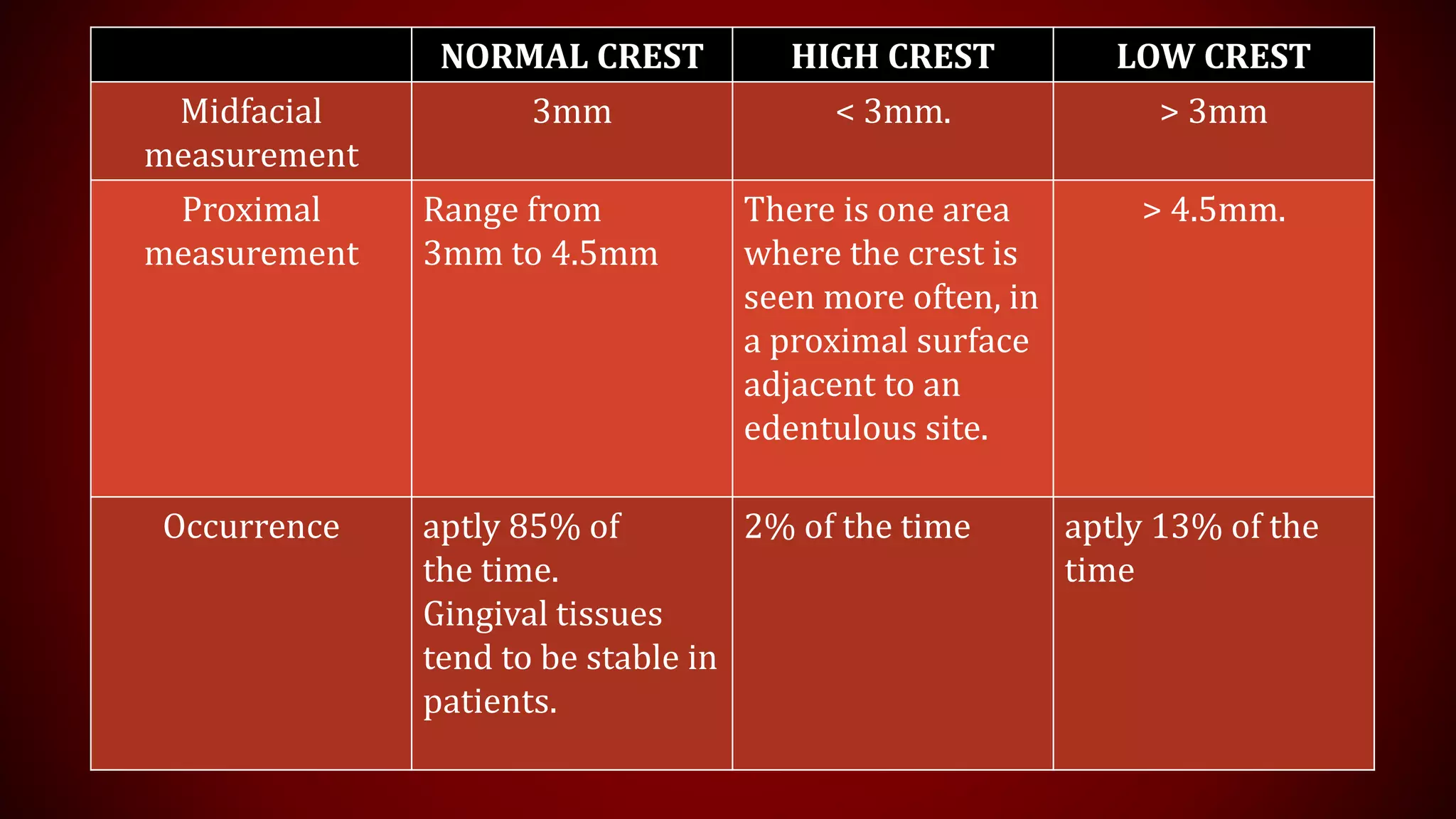
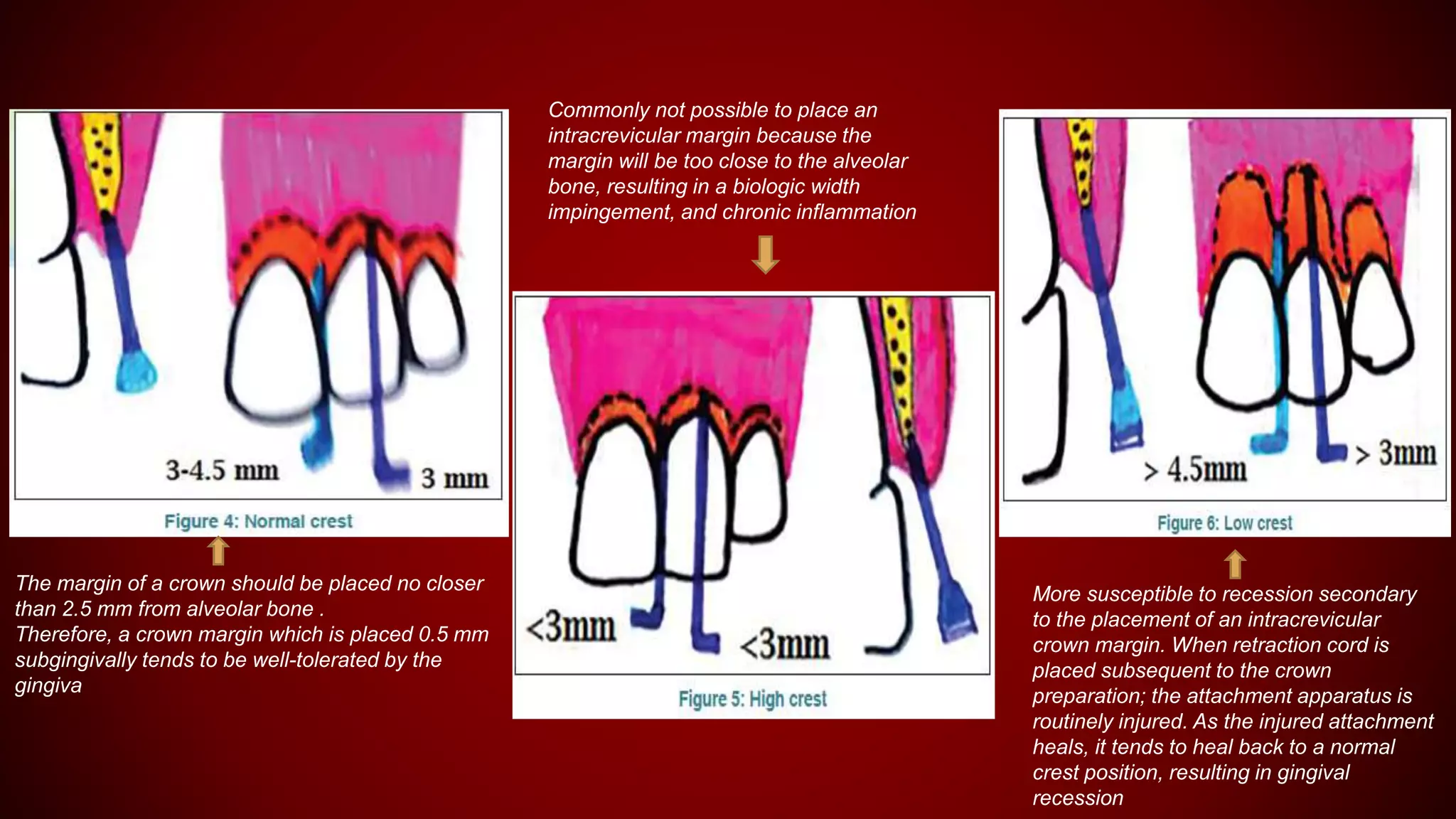
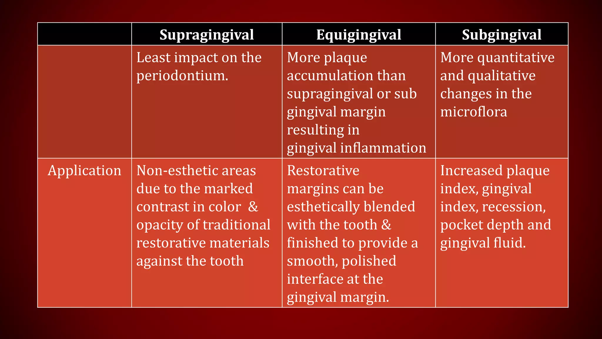
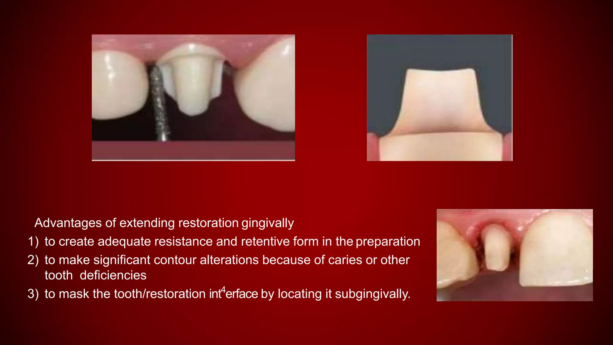
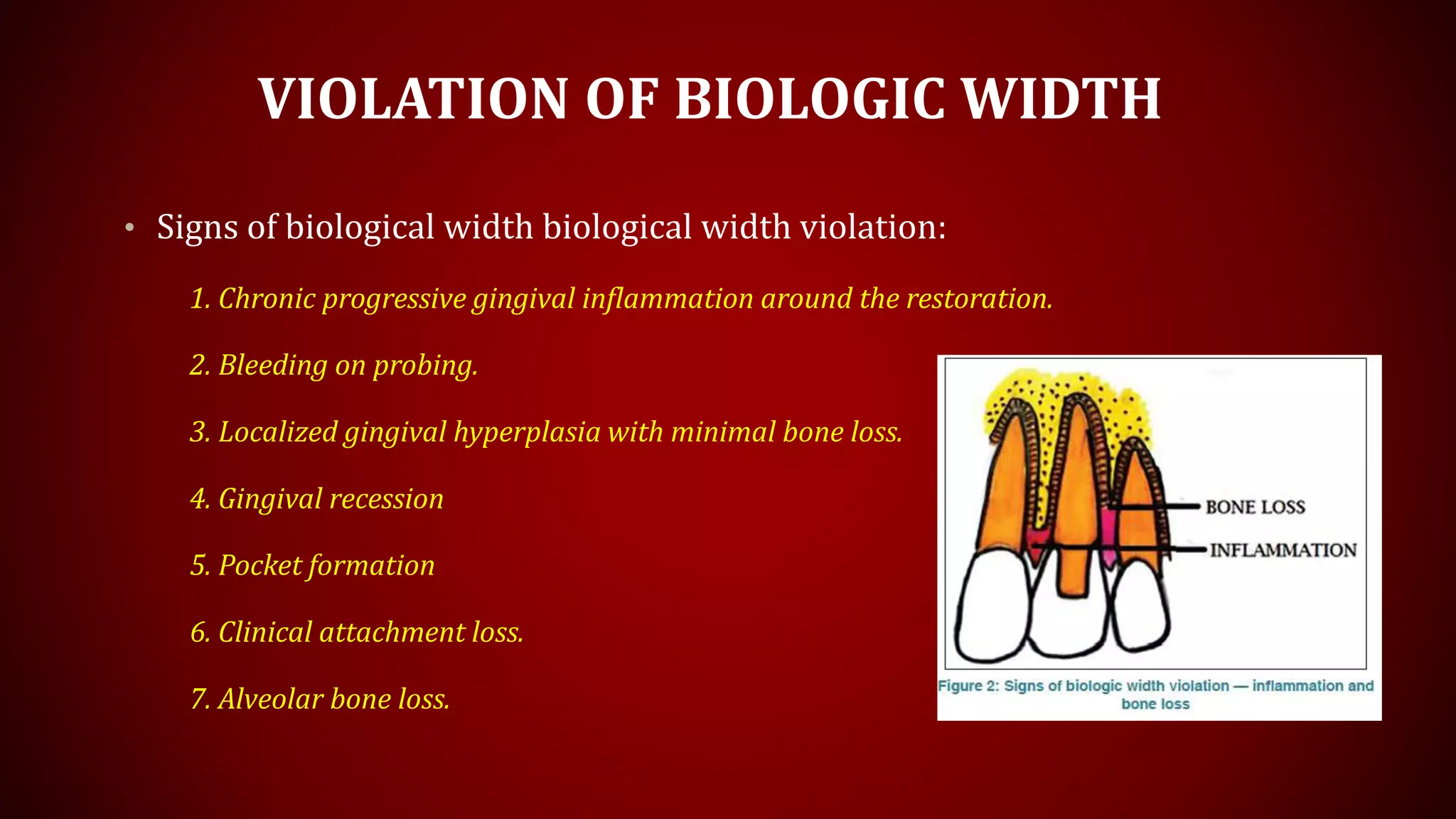
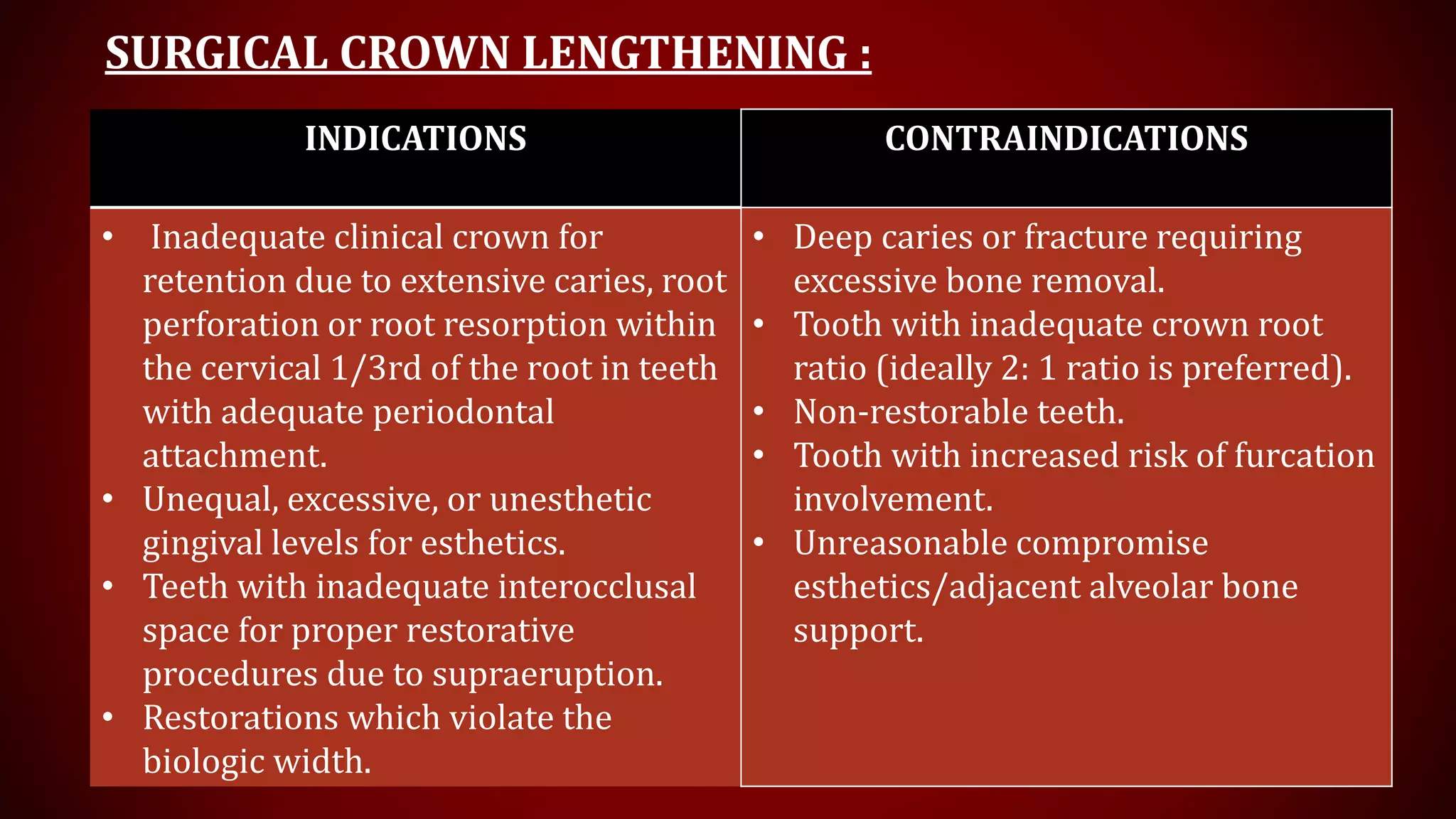
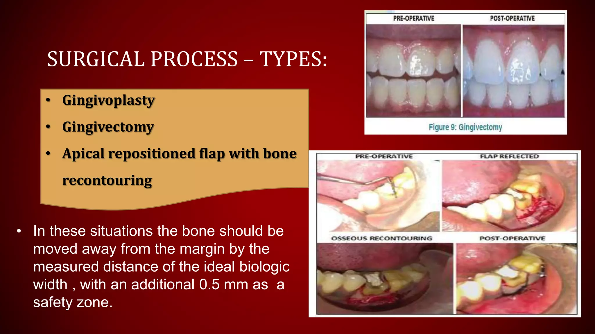
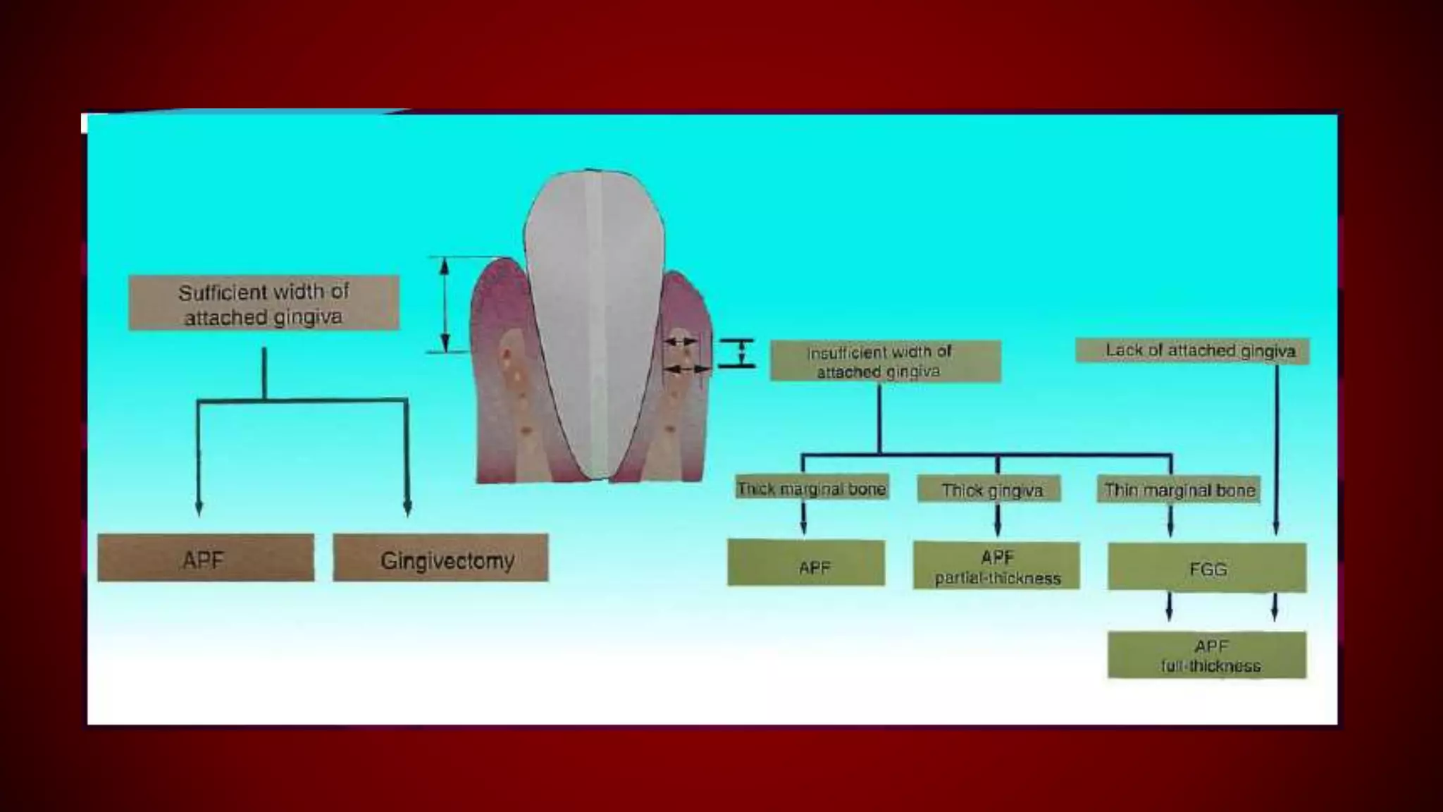
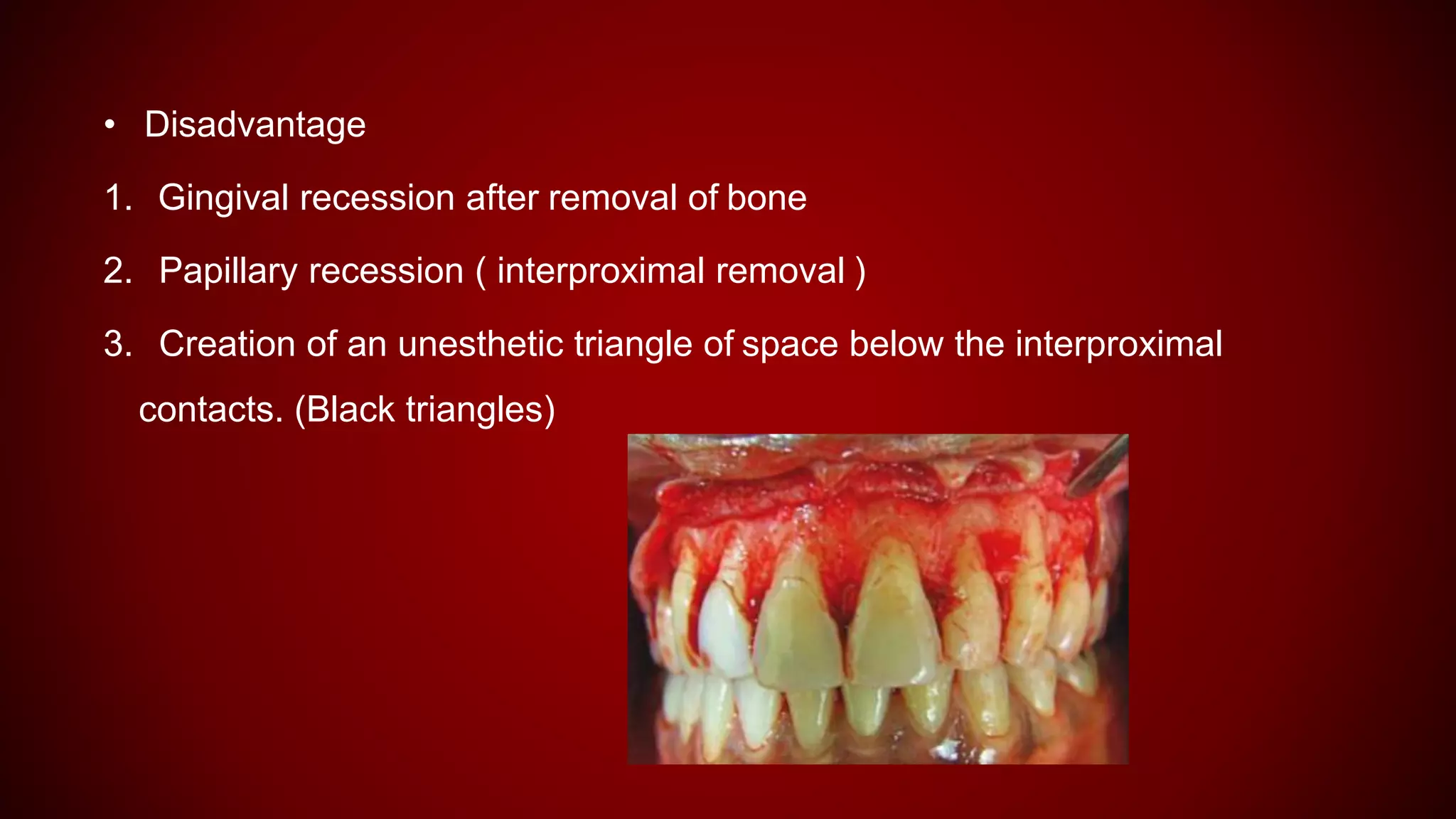
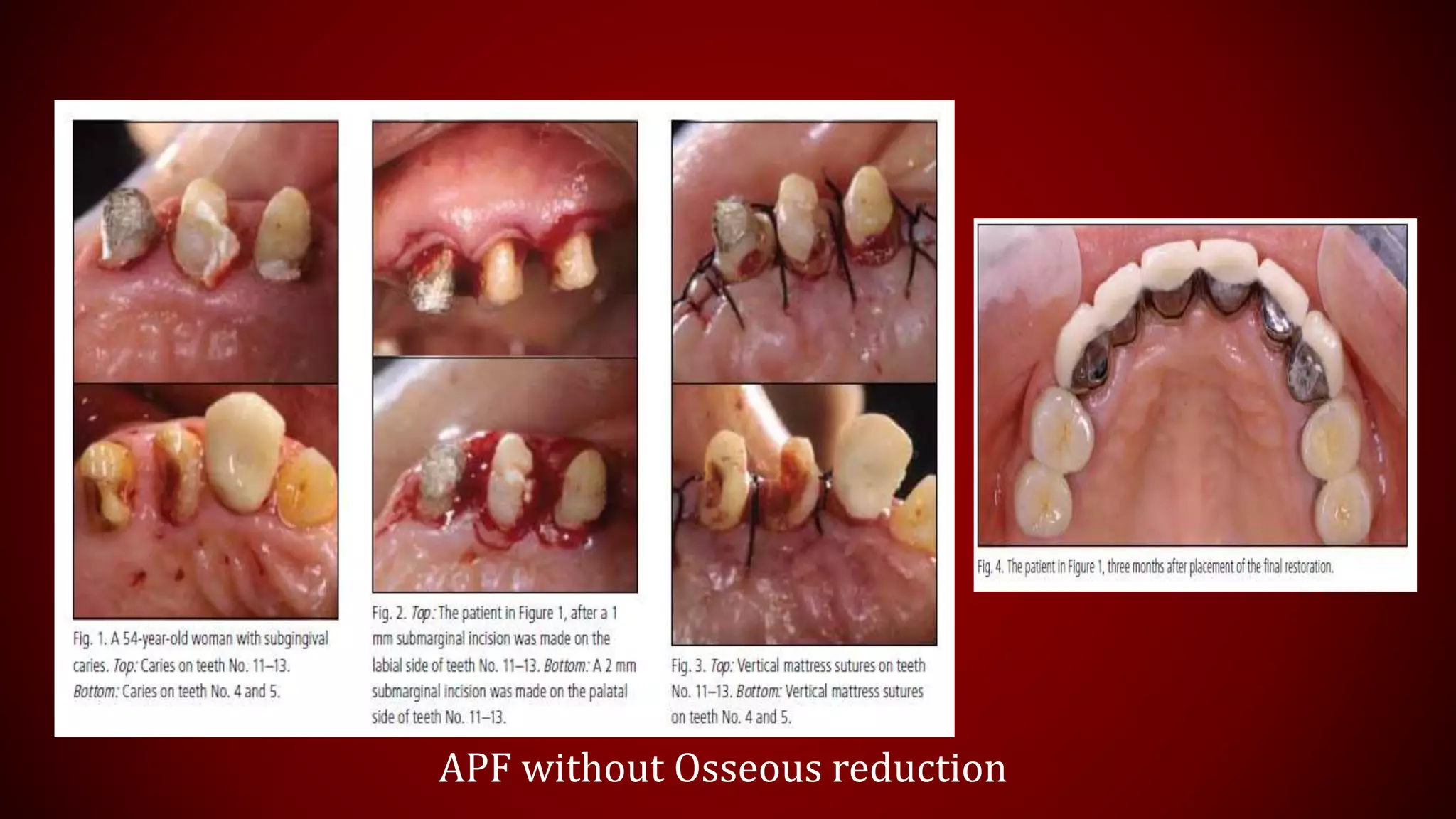
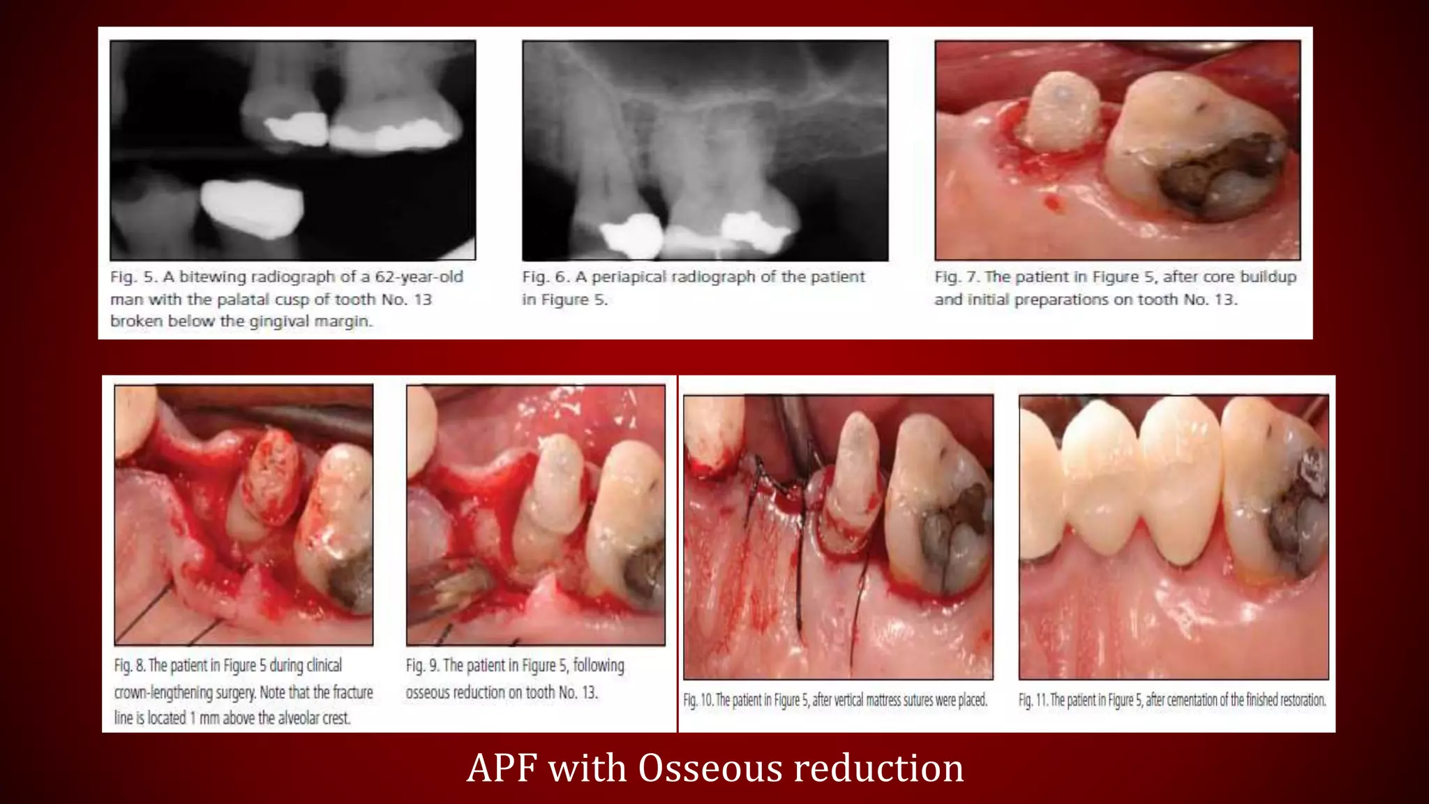
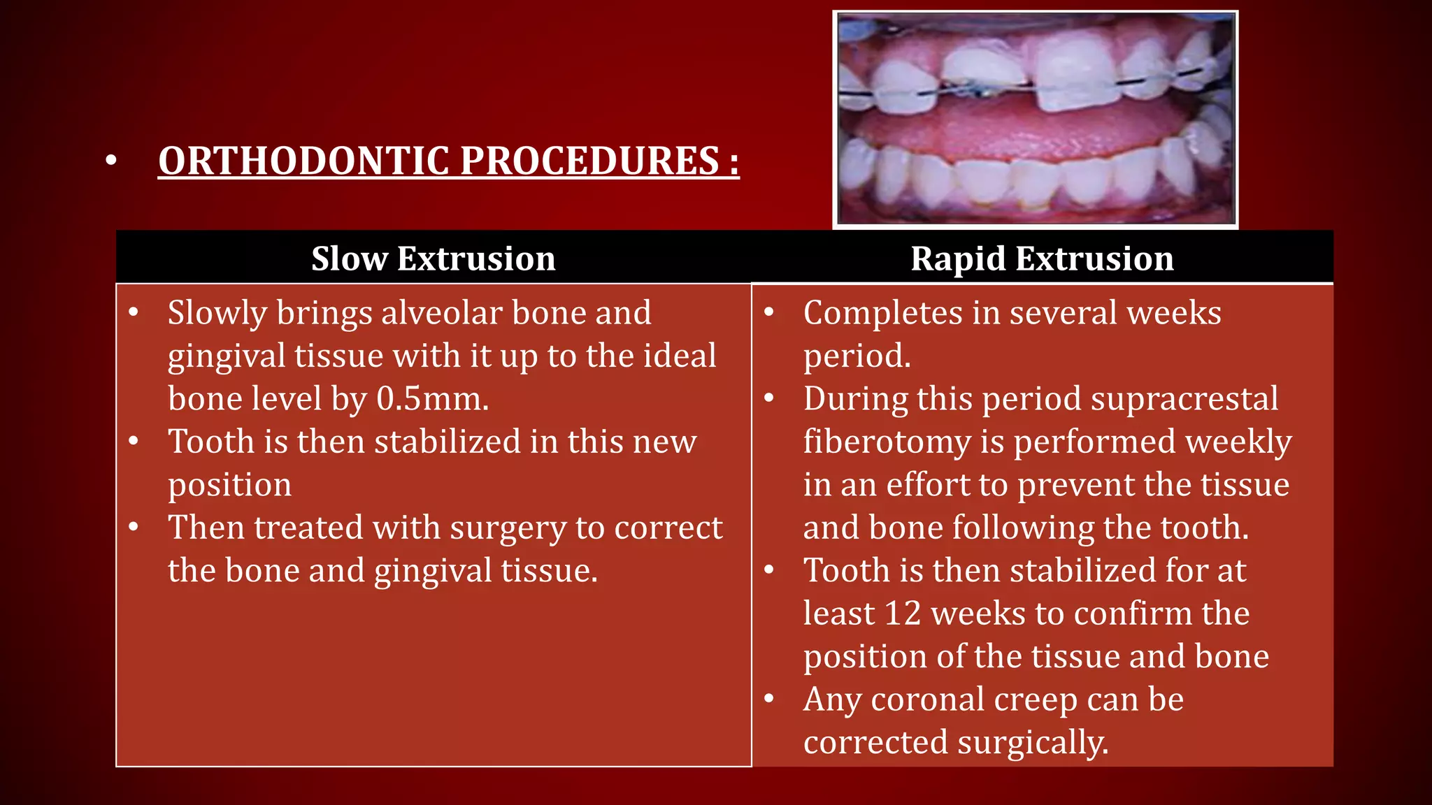
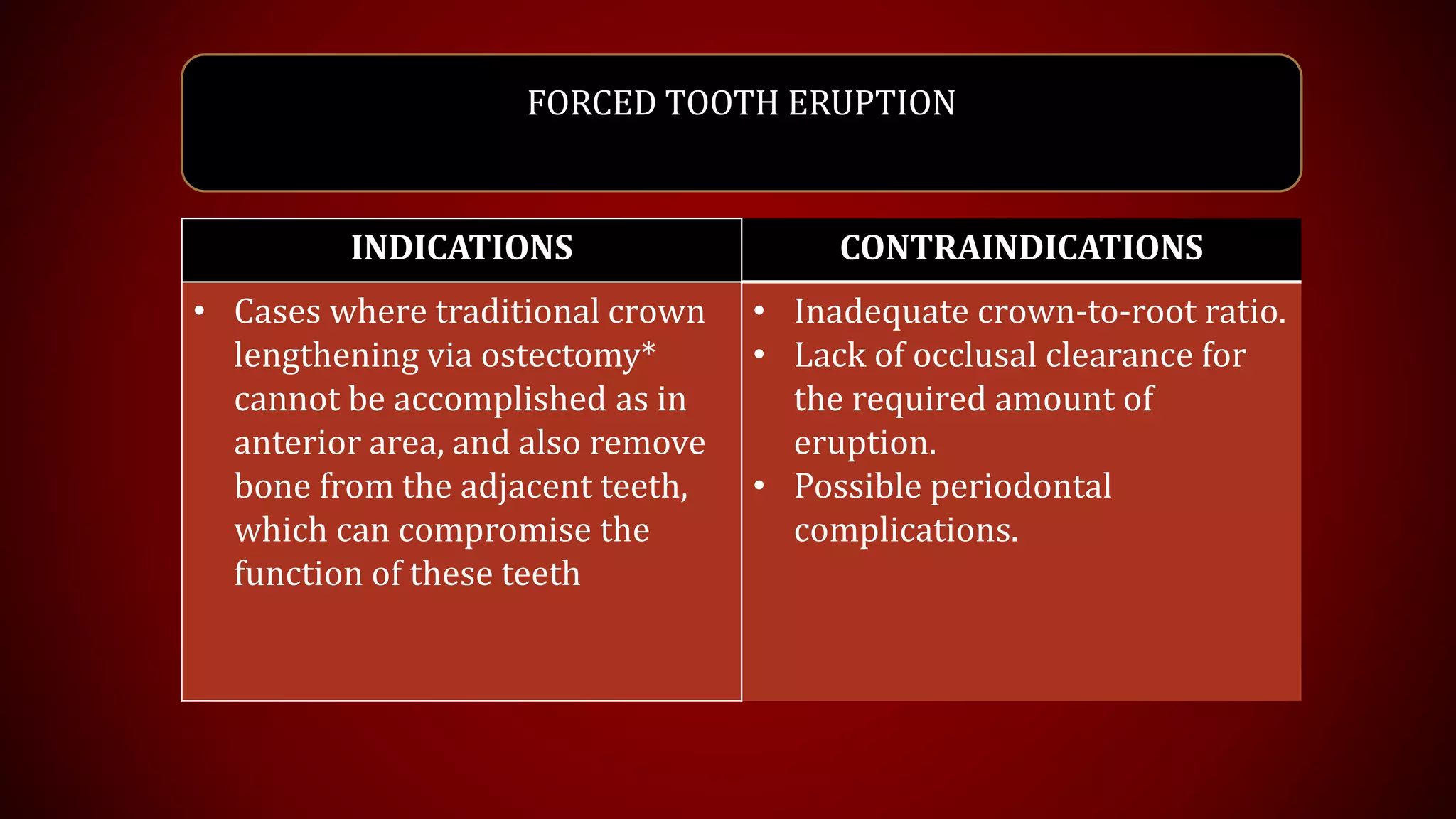
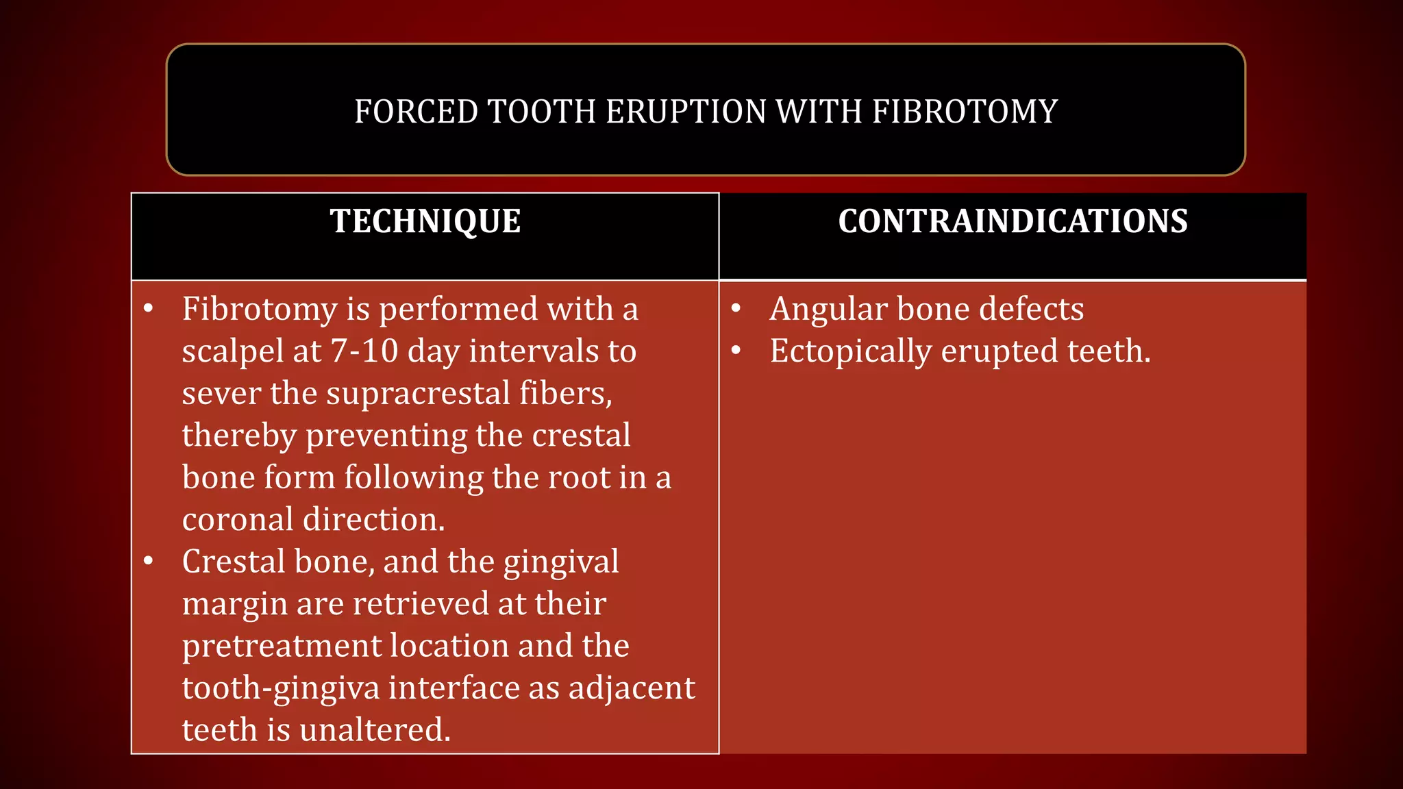
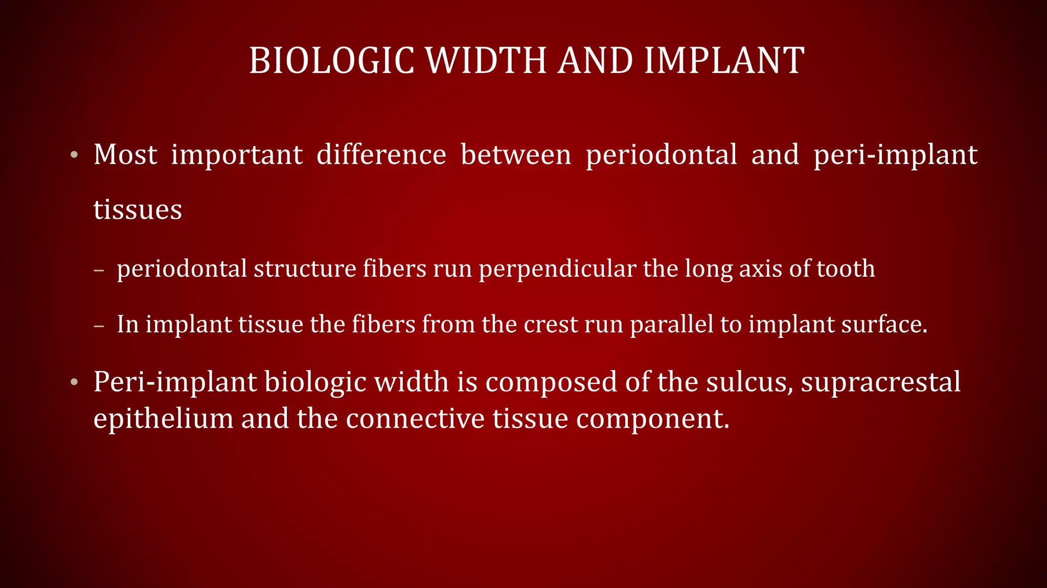
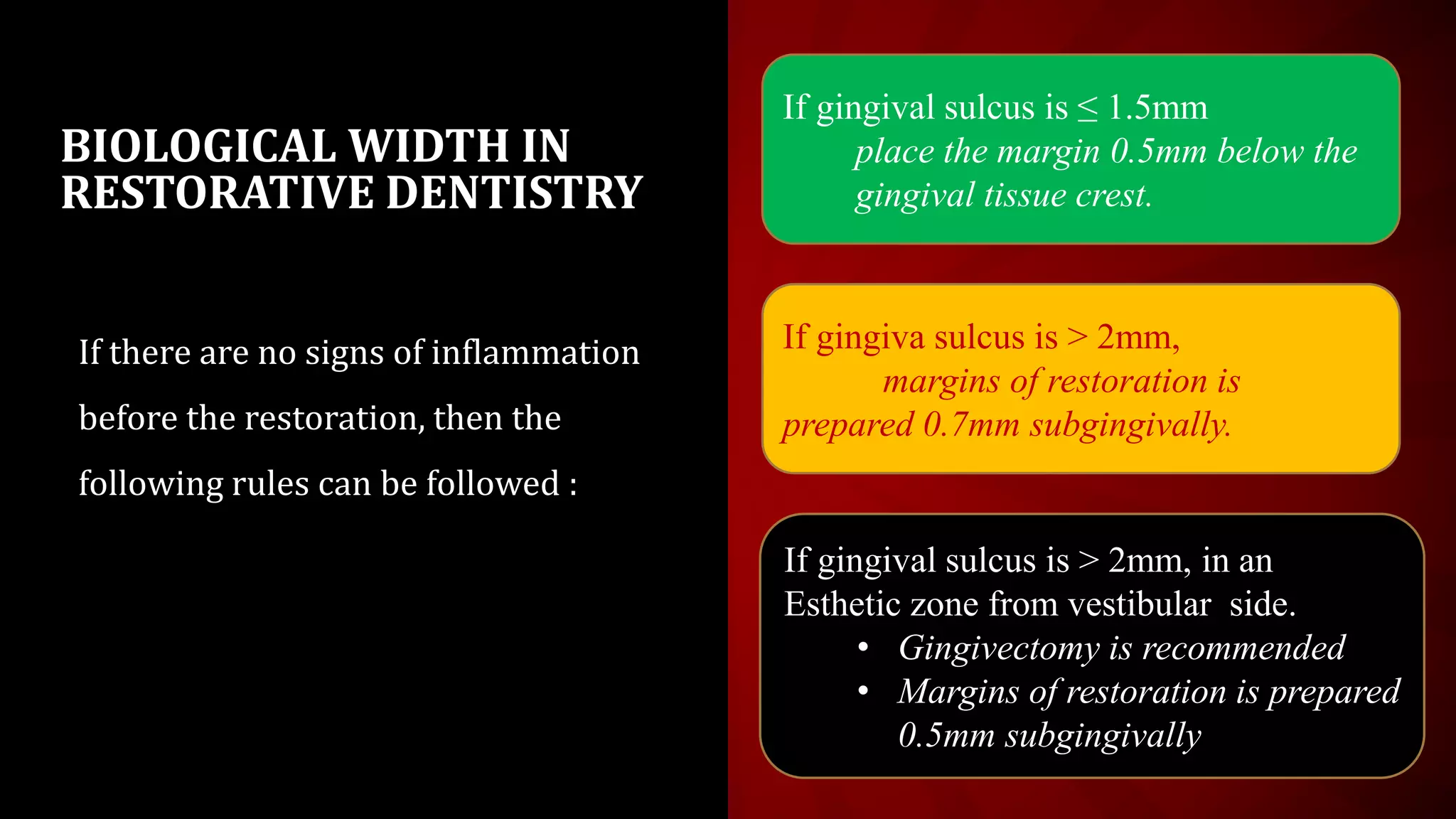
This document discusses biological width, which refers to the dimensions of soft tissue attached to the tooth coronal to the alveolar bone crest. It defines biological width as the connective tissue attachment (1.07mm on average) plus the epithelial attachment (0.97mm on average), totaling 2.04mm. It discusses factors that can lead to biological width violation like subgingival restoration margins and its signs. Methods to evaluate and correct biological width violations like bone sounding, surgical crown lengthening, and forced tooth eruption are also described. The importance of respecting biological width is emphasized in restorative and implant dentistry.
2. CONTENTS
• Introduction
• Interdental BW
• Concept of BW
• Categories/ Profiles of BW
• Margin Placement
• Evaluation of BW Violation
• Signs of BW violation
• Correction of BW Violation
• BW in Implants
• BW in Restorative Dentistry
• Conclusion
• References
3. INTRODUCTION
• Biological width is defined as the dimension of soft tissue which is attached to the
portion of the tooth coronal to the crest of alveolar bone.
(Gargiulo et al 1961)
4. • Average histological width of connective tissue attachment - 1.07mm.
• Mean average length of epithelial attachment - 0.97mm
Range : 0.71mm-1.35mm.
• Sulcus depth : 0.6 mm
• Average combined histological width of connective tissue attachment + Junctional
epithelium = 2.04mm (Biologic Width)
(Gargiulo et al 1961)
5. • On evaluation of cadaver tooth surfaces ,
Connective tissue attachment was the most consistent measurement based
on the mean measurements of :
Sulcus depth - 1.34 mm ,
Epithelial attachment - 1.14 mm and
Connective tissue attachment - 0.77 mm .
Vacek et al 1994
• Further studies by Newcomb (1974),Gunay et al (2000), Maynard and Wilson
(1979),Tal et al (1986)and Nevins and Skurow (1984) suggest that violation of
biological width must be prevented.
6. INTERDENTAL BIOLOGIC WIDTH
Kois and Spear pointed out that the dentogingival complex is 3.0mm facially
and 4.5mm to 5.5mm interproximally.
They noted that the height of interdental papilla can only be explained by
increased scalloping of the bone.
Becker and colleagues (1970) defined variation of gingival scalloping as
flat scalloped and pronounced scalloped.
7. Spear suggested that additional 1.5 to 2.5mm of interproximal gingival tissue
height require the presence of adjacent teeth for maintains of interproximal
gingival volume.
Without the adjacent tooth the interproximal gingival tissue would flatten
out, assuming a normal 3.0mm biologic width.
Tarnow and colleagues found that for the gingival tissue to assume complete
filling of the interdental space, the distance from the contact point to alveolar
crest should not exceeded 5 mm to 5.5mm.
Greater distance result in significant loss of alveolar height.
8. CONCEPT OF BIOLOGIC WIDTH
• Maynard and Wilson (1979) divided the periodontium into 3 dimensions:
– Superficial physiologic
– Crevicular physiologic and
– Subcrevicular physiologic
• The Subcrevicular physiologic space is analogous to the biologic width
described
( Gargiulo et al 1961)
9. CATEGORIES / PROFILES OF B.W
• 3 categories of biological width based on total dimension of
attachment and the sulcus depth following bone sounding
measurements :
• Normal Crest
• High Crest
• Low crest
Kois,2000
10. NORMAL CREST HIGH CREST LOW CREST
Midfacial
measurement
3mm < 3mm. > 3mm
Proximal
measurement
Range from
3mm to 4.5mm
There is one area
where the crest is
seen more often, in
a proximal surface
adjacent to an
edentulous site.
> 4.5mm.
Occurrence aptly 85% of
the time.
Gingival tissues
tend to be stable in
patients.
2% of the time aptly 13% of the
time
11. The margin of a crown should be placed no closer
than 2.5 mm from alveolar bone .
Therefore, a crown margin which is placed 0.5 mm
subgingivally tends to be well-tolerated by the
gingiva
Commonly not possible to place an
intracrevicular margin because the
margin will be too close to the alveolar
bone, resulting in a biologic width
impingement, and chronic inflammation
More susceptible to recession secondary
to the placement of an intracrevicular
crown margin. When retraction cord is
placed subsequent to the crown
preparation; the attachment apparatus is
routinely injured. As the injured attachment
heals, it tends to heal back to a normal
crest position, resulting in gingival
recession
13. Supragingival Equigingival Subgingival
Least impact on the
periodontium.
More plaque
accumulation than
supragingival or sub
gingival margin
resulting in
gingival inflammation
More quantitative
and qualitative
changes in the
microflora
Application Non-esthetic areas
due to the marked
contrast in color &
opacity of traditional
restorative materials
against the tooth
Restorative
margins can be
esthetically blended
with the tooth &
finished to provide a
smooth, polished
interface at the
gingival margin.
Increased plaque
index, gingival
index, recession,
pocket depth and
gingival fluid.
14. Advantages of extending restoration gingivally
1) to create adequate resistance and retentive form in the preparation
2) to make significant contour alterations because of caries or other
tooth deficiencies
3) to mask the tooth/restoration int4
erface by locating it subgingivally.
15. EVALUATION OF BIOLOGICAL WIDTH VIOLATION
• CLINICAL METHOD :
– If a patient experiences tissue discomfort when the restoration margin
levels are being assessed with a periodontal probe
– Margin has extended into the attachment and biologic width violation has
occurred
16. • BONE SOUNDING/TRANSGINGIVAL PROBING
– Biological width can be identified by probing under local anesthesia
(referred to as 'sounding to bone') subtracting the sulcus depth from the
surrounding measurements.
– If this distance is < 2mm at one or more locations, a diagnosis of biological
width violation can be confirmed.
Vavacek et al,1994
17. • RADIOGRAPHIC METHOD :
– Can identify interproximal violations of biologic width.
– However, on the mesiofacial and distofacial line angles of teeth, radiographs
are not diagnostic because of tooth superimposition.
– Parallel profile radiographic technique has been devised which could
be used to measure both length and thickness of the dentogingival unit with
accuracy.[
18. VIOLATION OF BIOLOGIC WIDTH
• Signs of biological width biological width violation:
1. Chronic progressive gingival inflammation around the restoration.
2. Bleeding on probing.
3. Localized gingival hyperplasia with minimal bone loss.
4. Gingival recession
5. Pocket formation
6. Clinical attachment loss.
7. Alveolar bone loss.
19. CORRECTION OF BIOLOGIC WIDTH VIOLATION
• Surgically removing bone away from proximity to the restoration margin.
• Orthodontic extrusion of the tooth and then moving the margin away from
the bone.
• Advantage of surgical process:
• Rapid method.
• Gives more pleasant result if the crown lengthening is done.
20. SURGICAL CROWN LENGTHENING :
INDICATIONS CONTRAINDICATIONS
• Inadequate clinical crown for
retention due to extensive caries, root
perforation or root resorption within
the cervical 1/3rd of the root in teeth
with adequate periodontal
attachment.
• Unequal, excessive, or unesthetic
gingival levels for esthetics.
• Teeth with inadequate interocclusal
space for proper restorative
procedures due to supraeruption.
• Restorations which violate the
biologic width.
• Deep caries or fracture requiring
excessive bone removal.
• Tooth with inadequate crown root
ratio (ideally 2: 1 ratio is preferred).
• Non-restorable teeth.
• Tooth with increased risk of furcation
involvement.
• Unreasonable compromise
esthetics/adjacent alveolar bone
support.
21. SURGICAL PROCESS – TYPES:
• Gingivoplasty
• Gingivectomy
• Apical repositioned flap with bone
recontouring
• In these situations the bone should be
moved away from the margin by the
measured distance of the ideal biologic
width , with an additional 0.5 mm as a
safety zone.
22.
23. • Disadvantage
1. Gingival recession after removal of bone
2. Papillary recession ( interproximal removal )
3. Creation of an unesthetic triangle of space below the interproximal
contacts. (Black triangles)
26. • ORTHODONTIC PROCEDURES :
Slow Extrusion Rapid Extrusion
• Slowly brings alveolar bone and
gingival tissue with it up to the ideal
bone level by 0.5mm.
• Tooth is then stabilized in this new
position
• Then treated with surgery to correct
the bone and gingival tissue.
• Completes in several weeks
period.
• During this period supracrestal
fiberotomy is performed weekly
in an effort to prevent the tissue
and bone following the tooth.
• Tooth is then stabilized for at
least 12 weeks to confirm the
position of the tissue and bone
• Any coronal creep can be
corrected surgically.
28. FORCED TOOTH ERUPTION
INDICATIONS CONTRAINDICATIONS
• Cases where traditional crown
lengthening via ostectomy*
cannot be accomplished as in
anterior area, and also remove
bone from the adjacent teeth,
which can compromise the
function of these teeth
• Inadequate crown-to-root ratio.
• Lack of occlusal clearance for
the required amount of
eruption.
• Possible periodontal
complications.
29. FORCED TOOTH ERUPTION WITH FIBROTOMY
TECHNIQUE CONTRAINDICATIONS
• Fibrotomy is performed with a
scalpel at 7-10 day intervals to
sever the supracrestal fibers,
thereby preventing the crestal
bone form following the root in a
coronal direction.
• Crestal bone, and the gingival
margin are retrieved at their
pretreatment location and the
tooth-gingiva interface as adjacent
teeth is unaltered.
• Angular bone defects
• Ectopically erupted teeth.
30. BIOLOGIC WIDTH AND IMPLANT
• Most important difference between periodontal and peri-implant
tissues
– periodontal structure fibers run perpendicular the long axis of tooth
– In implant tissue the fibers from the crest run parallel to implant surface.
• Peri-implant biologic width is composed of the sulcus, supracrestal
epithelium and the connective tissue component.
31.
32. • The influence of five different factors on implant biologic width has
been evaluated:
– Surgical technique,
– Loading time,
– Abutment material,
– Implant structure and position,
– Immediate post extraction insertion.
• On implant:
• Junctional epithelium + connective tissue = biologic width
1.88mm+1.05mm= 3.08 mm
33. BIOLOGICAL WIDTH IN IMPLANTS
• Implant-abutment connection placement –
– At the gingival level supracrestal to the alveolar bone, the biologic width
measurement was similar to natural dentition.
– At deeper level, the biologic width increased accordingly.
– Far below the gingival tissue crest , it will impinge on the gingiva and constant
inflammation is created.
– Highly scalloped, thin gingiva is more prone to recession than a flat
periodontium with thick fibrous tissue.
• Implant level should always be placed subgingivally to allow development of
desired profile and aesthetics.
34. BIOLOGICAL WIDTH IN
RESTORATIVE DENTISTRY
If there are no signs of inflammation
before the restoration, then the
following rules can be followed :
If gingival sulcus is ≤ 1.5mm
place the margin 0.5mm below the
gingival tissue crest.
If gingiva sulcus is > 2mm,
margins of restoration is
prepared 0.7mm subgingivally.
If gingival sulcus is > 2mm, in an
Esthetic zone from vestibular side.
• Gingivectomy is recommended
• Margins of restoration is prepared
0.5mm subgingivally
35. CONCLUSION :
• The health of periodontal tissue is dependent on properly designed restoration.
Incorrectly placed restorative margin and unadapted restoration violates the
biologic width. If the margin must be placed subgingivally, other factors to be
taken into account are:
1. Correct crown contour in gingival third.
2. Correct polishing and finishing of the margin.
3. Sufficient zone of attached gingival and no biologic width violation by subgingival margin.
• Repeated maintenance visits, patient cooperation and motivation are important
factor for improved success of restoration procedure with positive periodontal
health.
36. REFERENCES:
• Carranza,10th and 11th edition
• Malathi K, Arjun singh. Biologic width: Understanding and its preservation. Int J Med and Dent Sci
2014; 3(1):363-368.
• Linkevicius T et al. Stomatologija, Baltic Dental and Maxillofacial Journal, 2008;10(1):27-35.
• Sharma A, Rahul GR, Gupta B, Hafeez M. Biological width: No violation zone. Eur J Gen Dent
2012;1:137-41.
• Srdjak KJ et al. Periodontal and Prosthetic Aspect of Biological Width Part I: Violation of Biologic
Width.Acta Stomatol Croat,2000;34(2):195-7.
• Nugala B, Santosh Kumar BB, Sahitya S, Krishna PM. Biologic width and its importance in
periodontal and restorative dentistry. J Conserv Dent 2012;15:12-7.
• Dhir S. Significance and clinical relevance of biologic width to implant dentistry. J Interdiscip
Dentistry 2012;2:84-91.
• Aishwarya M, Sivaram G. Biologic width: Concept and violation. SRM J Res Dent Sci 2015;6:250-6.
• Oh SL. Biologic width and crown lengthening: Case reports and review. General Dentistry Sept-Oct
2010;e201-5.
Editor's Notes
Biologic width is the term applied to the dimensional width of dentogingival junction (epithelial attachment and underlying connective tissue). It was first described by Sicher.
It is important from the restorative point of view because its violation leads to complications like gingival enlargement alveolar bone loss and improper fit of the restoration.
essential for preservation of periodontal health and removal of irritation that might damage the periodontium (prosthetic restorations, for example). The dimension of biologic width is not constant, it depends on the location of the tooth in the alveola, varies from tooth to tooth, and also from the aspect of the tooth. Its constancy (is only one - it) can only be found in healthy dentition.
Encroachment of the biological width becomes of particular concern when considering the restoration of a tooth that has fractured or been caries near the alveolar crest.
The superficial physiologic dimension represent the free and attached gingival surrounding the tooth, while the crevicular physiologic dimension represents the gingival dimension from the gingival margin to the junctional epithelium.
The subcrevicular physiologic space is analogous to the biologic width described, consisting of the junctional epithelium and connective tissue attachment.
Authors claimed that to prevent the placement of 'permanent calculus', margin placement into the subcrevicular physiological space should be avoided.
Radiographic interpretation can also be used for identification of inter proximal violations of biological width but they are not diagnostic because of tooth superimposition.
All low crest patients do not react in the same way to an injury to the attachment. Some low crest patients are susceptible to gingival recession while others have a quite
stable attachment apparatus, the difference is based on the depth of the sulcus.
The primary treatment goal according to many clinicians now a days, are to mask the junction of tooth with restoration margin.
Generally clinicians have 3 options for margin placement.
Adv of supragingival margins :
1. Preparation of the tooth and finishing of the margin is easiest.
2. Duplication of the margins with impressions that can be removed past the finish line without tearing
3. Fit and finish of the restoration and removal of excess material is easiest.
4. Verification of the marginal integrity of restoration is easiest.
5. The Supragingival margins are least irritating to the gingival tissues .
This measurement must be performed on teeth with healthy gingiva and should be repeated on more than one tooth to ensure accurate assessment and reduce individual
and site variations.
Biologic width violation occurred during restoration margin placement can be corrected by two methods:
Ind:
Short clinical crowns.
In conjunction with tooth requiring hemisection or root resection.
Teeth with excessive occlusal wear or incisal wear.
Gingivectomy can be done in the case of [Figure 9]:• Hyperplasia or pseudopocketing (>3 mm of biologic width) & Presence of adequate amount of keratinized tissue.
APF is done when there is no adequate width of attached gingiva, and there is a biologic width of >3 mm on multiple teeth
Haling: Nonesthetic areas- Re-eval. Aft 6 weeks post surg. Esthetic Areas- req longer healing period
These six teeth had probing depths of 2–3 mm and 4–7 mm of attached gingiva on the labial side (Fig. 1). No mobility was present on these teeth. Since the teeth had thick
connective tissue attachment and junctional epithelium on the palatal side, an APF with submarginal incision was attempted.
PD:2-3MM palatally.
Ind :
When the biologic width violation is on the interproximal surface.
In condition when biologic width violation is across the facial surface, the gingival level is correct.
Because an APF with osseous reduction would result in bone loss on teeth No. 12 and 14 and a poor crown-to-root ratio on tooth No. 13, forced eruption was planned to
expose the sound tooth structure of tooth No. 13. The tooth erupted approximately 4 mm over a period of five weeks.
*as ostectomy would lead to anegative architecture
Technique : Orthodontic brackets are bonded to the problem tooth and adjacent teeth and are combined with archwire. Power elastic is tied from the bracket to the archwire which pulls the tooth coronally.
If the sulcus probes are more than 1.5 mm, the restorative margin can be placed in half the depth of the sulcus
Reason for maintaining rules 1 & 2 : The margin is far enough below the tissue so that it is still covered if the patient is at higher risk of recession