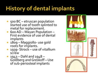
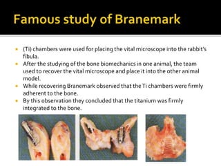
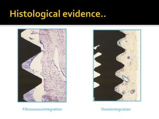
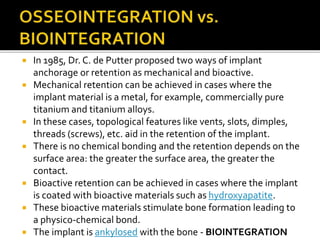
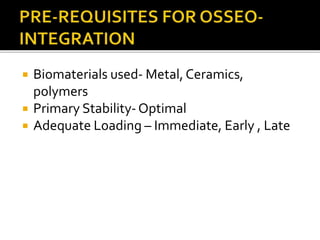
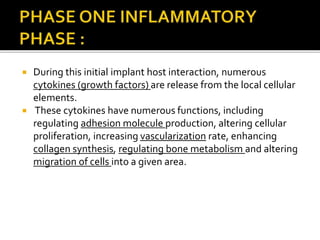
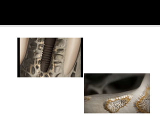
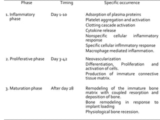
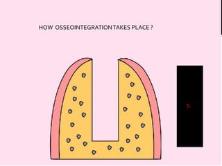
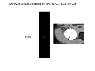
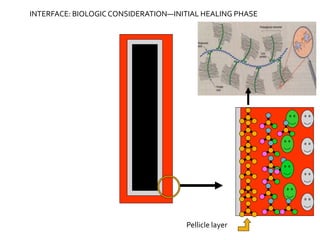
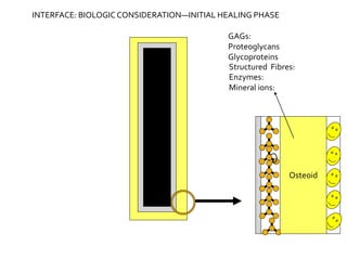
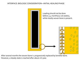
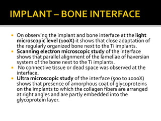
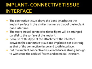
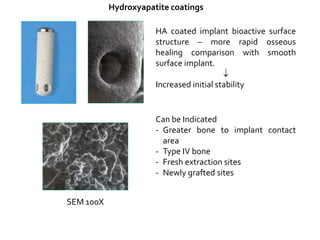
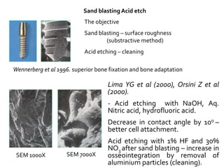
 Since tooth loss from disease and trauma has always been a
feature of mankind’s existence, it is not surprising that the history
of tooth replacement is a long one.
 Evidence from ancient civilizations shows that attempts were
made to replace missing teeth by banding artificial tooth
replacements to remaining teeth with metal many centuries ago.
 For the mechanism of attachment, clinicians have long thought an
analog for periodontal ligament.
 Experiments were made to develop a fibrous attachment that
could serve the same purpose as the periodontal ligament but all
in vain.
 The periodontal ligament in a specialized structure which serves
not only as an efficient attachment mechanism but also as a shock
absorber and sensory organ, so it was impossible to reproduce.
 500 BC – etruscan population
started use of tooth splinted to
metal for replacement.
 600 AD – Mayan Population –
First evidence of use of dental
implants
 1809 – Maggiollo- use gold
roots for implants.
 1939- Strock – use of vitallium
screw
 1943 – Dahl and 1948 –
Goldberg and Greskoff – Use
of sub-periosteal implants
 Consistent failures
• Inflammatory reaction
• Gradual bone loss
• Fibrous encapsulation
 Dr. Per Ingvar Branemark, an anatomist is credited as the person who has
coined the term “osseointegration”.
 Branemark along with his team was working in the laboratory of the vital
microscopy (1952), laboratory of experimental Biology, University of
Goteberg Sweden, (1960), Institute of Applied biotechnology, Goteberg
(1978).
 The main study of his group was to understand the mechanism of bone
healing and bone response to the thermal, mechanical, chemical injuries
by using vital microscopy.
Prof. P.I. Branemark :Father of
modern Dental Implantology
 (Ti) chambers were used for placing the vital microscope into the rabbit’s
fibula.
 After the studying of the bone biomechanics in one animal, the team
used to recover the vital microscope and place it into the other animal
model.
 While recovering Branemark observed that theTi chambers were firmly
adherent to the bone.
 By this observation they concluded that the titanium was firmly
integrated to the bone.
 After ensuring the favourable bone response
to theTi, the team tried to replace the teeth
for the dogs.TheTi implants also showed
good response for the mucosa and skin
penetrating implants.
 The implants, which used for replacement of
the teeth in the dogs showed good
integration upto 10 years and the implants
could bear the load of upto 100 Kgs without
failure at the bone-implant interface.
 By observing this property the integration
between the bone andTi screws was termed
as “osseointegration”.
 In 1965, first human edentulous patient was treated by using
theTi screws (implants) by reconstruction of resorbed
edentulous arches using autologus tibial bone grafts.
 In the mean time Schroeder et al. (1970), the members of the
international team for development of oral implants (I.T.I)
studied theTi plasma sprayed CpTi cylindrical implants in
Monkey models and achieved the firm integration between
the implant and the tissues.
 In their study the bone was joined to implant by fine bridges
of fibrous tissue.
 They termed this union as “Functional Ankylosis.”
 Osseointegration derives from the Greek osteon, bone, and the
Latin integrare, to make whole.
 Definitions :
“The apparent direct attachment or connection of osseous tissue to an inert,
alloplastic material without intervening connective tissue”.
- Glossary of ProstheticTerms
 Structurally oriented definition :
“Direct structural and functional connection between the ordered, living bone and
the surface of a load carrying implants”.
- Branemarks and associates (1977)
 Histologically :
Direct anchorage of an implant by the formation of bone directly on the surface of
an implant without any intervening layer of fibrous tissue.
- Albrektson and Johnson (2001)
 Clinically :
Ankylosis of the implant bone interface. (Schroeder and colleagues 1976)
 “Functional ankylosis”
• “It is a process where by clinically asymptomatic rigid fixation of alloplastic
material is achieved and maintained in bone during functional loading”
- Zarb andT Albrektson 1991
 Biomechanically oriented definition :
“Attachment resistant to shear as well as tensile forces”
- Steinmann et al (1986).
 Contact established without interposition of
non-bone tissue between normal remodeled
bone and an implant entailing a sustained
transfer and distribution of load from implant
to and within the bone tissue.
 Subdivided into based on microscopic level:
 Adaptive Osseointegration : Osseous tissue
approximating the surface of the implant
without apparent soft tissue interface at light
microscopic level.
 Biointegration : Is a direct biochemical bone
surface attachment confirmed at electron
microscopic level.
 It is made of tissue integration around
healed functioning endosteal dental implant
in which the prime load bearing tissue at the
interface is a peri-implant ligament
composed of osteostimulatory collagen. It
limits the further bone resorption.
 Used in case of plate/blade form
endosseous implants and endodontic
stabilizers.
 It is a made of tissue integration around a
healed, functioning, subperiosteal implant in
which the load bearing tissue is the sheath of
dense collagenous tissue constituting the
outer layer of periosteum.
 AAID (1986) defined fibrous integration as “Tissue to implant contact with
interposition of healthy dense collagenous tissue between the implant and bone”.
 “Direct bone to implant interface without any intervening layer of fibrous tissue”.
FIBROINTEGRATION
Vs
Concept of Bony Anchorage
Branemark (1969)
Concept of soft tissue anchorage
Linkow (1970), James (1975),
Weiss (1986).
OSSEOINTEGRATION
 “Pseudoligament”, “Periimplant ligament”, “Periimplant
membrane”.
 Hypothesis – Collagen fibers function similar to the sharpeys fibers
in the natural dentition.
 Fact : The histological difference between the sharpeys fibers and
collagen fibers around the implant.
Natural teeth Implant
Oblique and
horizontal group of
fibers
Parallel, irregular,
complete
encapsulation
Uniform distribution
of load (Shock
absorber)
Difficult to transmit
the load
Fibrosseousintegration Osseointegration
 In 1985, Dr. C. de Putter proposed two ways of implant
anchorage or retention as mechanical and bioactive.
 Mechanical retention can be achieved in cases where the
implant material is a metal, for example, commercially pure
titanium and titanium alloys.
 In these cases, topological features like vents, slots, dimples,
threads (screws), etc. aid in the retention of the implant.
 There is no chemical bonding and the retention depends on the
surface area: the greater the surface area, the greater the
contact.
 Bioactive retention can be achieved in cases where the implant
is coated with bioactive materials such as hydroxyapatite.
 These bioactive materials stimulate bone formation leading to
a physico-chemical bond.
 The implant is ankylosed with the bone - BIOINTEGRATION
 Biomaterials used- Metal, Ceramics,
polymers
 Primary Stability-Optimal
 Adequate Loading – Immediate, Early , Late
 Can be discussed under:
 Endo-osseous healing
 Muco-periosteal healing
 Both can be separated into :
▪ The inflammatory phase,
▪ The proliferative phase, and
▪ The maturation phase.
 The placement of implants into bone involves the creation of
an osseous defects with the subsequent filling of this defect
with an implant device.
 Even with the most careful surgical manipulation of osseous
tissues, the generation of a thin layer of necrotic bone in the
peri-implant region is inevitable.
 In addition, exact microscopic fit between the implant and the
surgical defect is not possible, leaving local areas of dead space
where the implant does not directly contact osseous tissue.
 When the implant is exposed to the surgical site, it comes to
contact with extracellular fluid and cells.
 This initial exposure of the implant to the local tissue
environment results in rapid adsorption of local plasma
proteins to the implant surface.
 These proteins are enzymatically degraded and undergo
conformational changes, degradation, and replacement by
other proteins.
 Platelet contact with synthetic surfaces causes their activation
and liberation of their intracellular granules resulting in release
of serotonin and histamine, leading to further platelet
aggregation and local thrombosis.
 Blood contact with proteins and foreign materials leads to the
initiation of the clotting cascade via the intrinsic and extrinsic
pathways, causing blood coagulation in the peri-implant dead
spaces and within the damaged local microvascular circulation.
 Activation of the clotting cascade also leads to the formation of
bradykinin, which is a strong mediator of vasodilation and
endothelial permeability.
 During this initial implant host interaction, numerous
cytokines (growth factors) are release from the local cellular
elements.
 These cytokines have numerous functions, including
regulating adhesion molecule production, altering cellular
proliferation, increasing vascularization rate, enhancing
collagen synthesis, regulating bone metabolism and altering
migration of cells into a given area.
 Initially, it is nonspecific in nature and consists mainly of
neutrophil emigration into the area of damaged tissue.
 Its duration is variable but generally peaks during the first 3 to 4
days following surgery.
 The role of this cell is primarily phagocytosis and digestion of
debris and damaged tissue.
 Neutrophils are accompanied by smaller numbers of
eosinophils.
 Eosinophils have a similar phagocytic function and they can
also digest antigen antibody complexes.
 These cells are attracted to the local area by chemotactic
stimuli and then migrate from the intravascular space to the
interstitial space by diapedesis.
 End products of this phagocytic process are carried away from
the local area by the lymphatic circulation.
 Toward the end of the first week, the generalized
inflammatory response becomes more specific in
nature.
 Increasing numbers of thymus dependent
lymphocytes (T cells) bursa equivalent lymphocytes (B
cells), killer (K) cells, natural killer (NK) cells and
macrophages are found in the wound at this time.
 These cells respond to foreign antigens such as
bacteria and plaque debris that have been introduced
into the area during the surgical procedure.
 These antigens are processed and presented to the B
andT cell populations by macrophages.
 Macrophages are the predominant phagocytic cell found in the
wound by the fifth to sixth postoperative day.
 These cells are derived from circulating monocytes, which
originate from the bone marrow via monoblast differentiation.
 Macrophages have the ability to ingest immunologic and non-
immunologic particles by phagocytosis and attempt to digest
these particles with lysosomal enzymes.
 The reaction of macrophages on exposure to foreign materials
depends on the physical and chemical nature of the material.
 During this phase, vascular ingrowth occurs from the surrounding vital
tissues, a process called neovascularization.
 In addition, cellular differentiation, proliferation and activation occur
during this phase, resulting in the production of an immature connective
tissue matrix that is eventually remodeled.
 Please Note:This phase of bone repair begins while the inflammatory
phase is still active.
 During the placement of implants into their endosseous locations,
interruption of the local microcirculation occurs in the surgical areas.
 Regeneration of this circulation must eventually occur.
 Metabolism of the local inflammatory cells, fibroblasts, progenitor cells
and other local cells creates an area of relative hypoxia in the wound
area.
 This results in the development of an oxygen gradient with the lowest
oxygen tension near the wound edges.
 This hypoxic state combined with certain cytokines, such as
basic fibroblast growth factor (bFGF) and platelet derived
growth factor (PDGF) is responsible for simulating this
angiogenesis.
 bFGF seems to activate hydrolytic enzymes, such as
collagenase and plasminogen, which help to dissolve the
basement membranes of local blood vessels.
 This initiates the process of endothelial budding, which
progresses along the established chemotactic gradient.
 Once the anastomoses of the capillary buds are developed and
microcirculation is reestablished, the improved tissue oxygen
tension results in a curtailment of the secretion of these
angiogenic growth factors.
 The new circulation provides the delivery of nutrients and
oxygen necessary for connective tissue regeneration.
 Local mesenchymal cells begin to differentiate into
fibroblasts, osteoblasts and chondroblasts in response to
local hypoxia and cytokines released from platelets,
macrophages, and other cellular elements.
 These cells begin to lay down an extracellular matrix
composed of collagen, glycosaminoglycans, glycoproteins
and glycolipids.
 The initial fibrous tissue and ground substance that are laid
down eventually form into a fibrocartilaginous callus and this
callus is eventually transformed into a bone callus with a
process similar to endochondral ossification.
 Ossification centers begin within secretory vesicles that are
liberated from the local osteoblasts.
 These vesicles called matrix vesicles, are rich in phosphate
and calcium ions and also contain the enzymes alkaline
phosphatase and phospholipase A2.
 This callus transformation is aided by improved oxygen
tension and enhanced nutrient delivery that occurs with
improvement of local circulation.
 The initial bone laid down is randomly arranged (Woven
type) bone that is eventually remodeled.
 In vivo studies using an optical chamber (vital chamber)
implanted in along bones of animal models have been
instrumental to the understanding of the healing process
that occurs in the peri-implant space.
 They have revealed that vascular ingrowth precedes
ossification.
 Capillary ingrowth appears initially and it matures to be a
more developed vascular network during the first three
weeks after implant insertion.
 Ossification is initially visualized during the first week, peaks
during the third to fourth week and arrives at a relatively
steady state by the sixth to eight week.
 The necrotic bone in the peri-implant space that resulted
from operative trauma must eventually be replaced with
intact living bone for complete healing to occur.
 Appositional woven bone is laid down on the scaffold of
dead bone trabeculae by differentiated mesenchymal cells in
the advancing granulation tissue mass.
 This process occurs concurrently with the ossification of the
fibrocartilaginous callus.
 Simultaneous resorption of these “composite” trabeculae
and the newly formed bone, coupled with the deposition of
mature concentric lamellae eventually results in complete
bone remodeling, leaving a zone of living lamellar bone that
is continuous with the surrounding basal bone.
 First bone tissue to form within first 4-6
weeks of surgery
 It is a premature type of bone which starts
from the surrounding bone towards the
implant.
 Is of low mineral density with irregularly
shaped osteocytes with the capacity to form
scaffolds of rods and plates.
 Has a random like orientation of collagen
fibres.
 From the 2nd month, microscopic bone forms
towards lamellar bone.
 It is packed with collagen fibres which are
parallelly oriented between woven and lamellar
bone.
 Lamellar bone forms 1 to 1.5 µm/day by
apposition and parallel bone is 3-5 times larger.
 Involves three surfaces namely, woven bone
formation, pre-existing or pristine bone surface
and implant surface.
 Last stage of osseointegration
 Starts around the third month reaches a peak
activity and slows down to continue the rest
of life.
 It is a multicellular unit where remodelling
occurs in discrete units.
 Resorption and formation coupled in space
and time.
 In a histopathologic comparison of loaded and unloaded
implants, Donath et al. showed that unloaded implants
contacted small bone lamellae that were interrupted by
many areas of bone marrow and parts of the haversian canal
system.
 Loaded implants were surrounded by a more compact type
of bone with only small bone free areas near the haversian
canals.
 The lamellae around the implant area remodeled according
to the exposed load, which with passage of time, shows a
characteristic pattern of well organized concentric lamellae
with formation of osteons in the traditional manner.
 Under normal circumstances, healing of implants is usually
associated with a reduction in the height of alveolar marginal
bone.
 Approximately 0.5 to 1.5 mm of vertical bone loss occurs during
the first year after implant insertion.
 After this point, a steady state is reached and normal bone loss
occurs at a rate of approximately 0.1 mm per year.
 The rapid initial bone loss can be attributed to the generalized
healing response resulting from the inevitable surgical trauma,
such as periosteal elevation, removal of marginal bone and bone
damage caused by drilling.
 The later steady state bone loss probably reflects normal
physiologic bone resorption.
 Factors such as excessive surgical trauma, excessive loading or the
presence of peri-implant inflammation may accelerate this normal
resorptive process.
Phase Timing Specific occurrence
1. Inflammatory
phase
Day 1-10 Adsorption of plasma proteins
Platelet aggregation and activation
Clotting cascade activation
Cytokine release
Nonspecific cellular inflammatory
response
Specific cellular inflmmatory response
Macrophage mediated inflammation.
2. Proliferative phase Day 3-42 Neovascularization
Differentiation, Proliferation and
activation of cells.
Production of immature connective
tissue matrix.
3. Maturation phase After day 28 Remodeling of the immature bone
matrix with coupled resorption and
deposition of bone.
Bone remodeling in response to
implant loading
Physiological bone recession.
ti Ti
HOW OSSEOINTEGRATIONTAKES PLACE ?
ti Ti
The implant body is snuggly fitted into
surgically prepared bed
BONE Ti
INTERFACE: BIOLOGICCONSIDERATION—INITIAL HEALING PHASE
BONE Ti
TiO
TiO Ti
INTERFACE: BIOLOGICCONSIDERATION—INITIAL HEALING PHASE
Ti
Potential space
Potential space
in drill-osteotomised
bone-hole
INTERFACE: BIOLOGICCONSIDERATION—INITIAL HEALING PHASE
TiO
Ti
Bone Bio-
liquid
INTERFACE: BIOLOGICCONSIDERATION—INITIAL HEALING PHASE
Small biomolecules,
Water, Ions
Large molecules,
Tissue fragments
UDMCells of
Bone marrow/
Blood cells
Surface Free Energy
(wettability)
Highest amongst all
tested biomaterials
30 kcal/mol
INTERFACE: BIOLOGICCONSIDERATION—INITIAL HEALING PHASE
Macrophage
Osteoclast
Pellicle Layer of 10 nm thickness
The pellicle layer dictates
subsequent adherence of
other formative cells on it
INTERFACE: BIOLOGICCONSIDERATION—INITIAL HEALING PHASE
Pellicle layer
INTERFACE: BIOLOGICCONSIDERATION—INITIAL HEALING PHASE
Too much roughness
on implant surface
invites unnecessary
accumulation of
macrophages
INTERFACE: BIOLOGICCONSIDERATION—INITIAL HEALING PHASE
Macrophage clears up
macromolecules and other
cell debris
INTERFACE: BIOLOGICCONSIDERATION—INITIAL HEALING PHASE
Newly formed
osteoblasts
Oxidation-reduction potential : Vascular Supply & OxygenTension
Undfnd. M Pre-osteoblast
Otherwise, Fibroblasts will be generated leading to fibrous sheath( failure)
INTERFACE: BIOLOGICCONSIDERATION—INITIAL HEALING PHASE
Osteoid
INTERFACE: BIOLOGICCONSIDERATION—INITIAL HEALING PHASE
GAGs:
Proteoglycans
Glycoproteins
Structured Fibres:
Enzymes:
Mineral ions:
INTERFACE: BIOLOGICCONSIDERATION—INITIAL HEALING PHASE
Woven bone is formed
at a pace upto 100µm/day
Osteoblasts will not enter
into less than 100µ porosities;
ONLY GROUND SUBSTANCE IS
FOUND
INTERFACE: BIOLOGICCONSIDERATION—INITIAL HEALING PHASE
Loading should not be done
before 3-4 months(12-16 weeks),
while mostly woven bone is present.
After several months the woven bone is progressively replaced by lamellar bone.
However, a steady state is reached after about 1½ year.
INTERFACE: BIOLOGICCONSIDERATION—INITIAL HEALING PHASE
 The inflammatory phase of wound healing for the
mucoperiosteal complex is essentially the same as that
mentioned in the previous section on bone healing.
 It involves an initial vascular response followed by platelet
aggregation and activation, the clotting cascade and then an
initial non-specific cellular inflammatory response consisting of
infiltrates of predominantly neutrophils.
 This is followed shortly thereafter by a more specific cellular
inflammatory response consisting of infiltrates of
predominantly neutrophils.
 This is followed shortly thereafter by a more specific cellular
inflammatory response marked by increased number of
lymphocytes and macrophages.
 Cytokines also play an important role in the healing of soft
tissue wounds.
 The proliferative phase of wound healing begins within hours
of the injury and is characterized by the establishment of an
active population of epithelial and connective tissue cells and
the beginning of he reestablishment of wound integrity.
 Migration and proliferation of epithelial cells is seen within
the first 24 to 48 hours of wound healing.
 The stimulus for growth and migration of thee cell results
from loss of contact inhibition and from a temporary
decrease in the local level of tissue specific growth inhibitors
called chalones.
 A watertight seal is usually established within the first 24
hours after primary wound closure, but little structural
strength is provided by the seal.
 The main connective tissue cell involved in the proliferative phase
of soft tissue wound healing is the fibroblast.
 Differentiation of mesenchymal cells and proliferation and
migration of the preexisting population of local fibroblasts occur
as a result of hypoxia and the release of cytokines from local
cellular elements, including platelets and macrophages.
 Neovascularization provides the foundation for fibroblastic
proliferation by supplying the local area with the nutritional
support required to maintain this enhanced metabolic state.
 Fibroblasts produce ground substance, collagen and elastic fibers.
 Collagen formation is microscopically detected between the
fourth and sixth days, but biochemical evidence of collagen
formation is noted between the second and fourth days.
 Collagen deposition increases the tensile strength significantly
during this phase and the magnitude is proportional to the
collagen content of the tissues.
 During the final phase of wound healing, maturation of the
deposited collagen occurs.There is no sharp demarcation
between the end of the proliferative phase and the beginning
of the maturation phase because collagen maturation occurs
continuously shortly after initial deposition.
 As time proceeds however, the unorganized fibrils are replaced
larger, thicker and better organized fibers, with the final result
being one of ‘lacing” the wound edges together with a three
dimensional weave.
 The bursting strength of the wound is noted to improve
dramatically from 3 to 9 weeks, reaching a level of 70% of
normal skin by the end of this period. By 6 months, the bursting
strength of the wound is approximately 90% of the level of
normal skin.
 consists of
 Implant and bone interface.
 Implant and connective tissue interface.
 Implant and epithelium interface.
 On observing the implant and bone interface at the light
microscopic level (100X) it shows that close adaptation of
the regularly organized bone next to theTi implants.
 Scanning electron microscopic study of the interface
shows that parallel alignment of the lamellae of haversian
system of the bone next to theTi implants.
 No connective tissue or dead space was observed at the
interface.
 Ultra microscopic study of the interface (500 to 1000X)
shows that presence of amorphous coat of glycoproteins
on the implants to which the collagen fibers are arranged
at right angles and are partly embedded into the
glycoprotein layer.
 As a general rule cells do not bind directly to the
foreign materials.The cells binds to each other or any
other foreign materials by a layer of extracellular
macro molecules (glycoproteins).
 The glycoprotein layer in between the cells or in
between the tissues will be at a thickness of 10 to 20
nm (100 to 200 A0).
 At the interface the glycoprotein layer of normal
thickness (10-20 nm) is adsorbed on the implant
surface within the help of adhesive macromolecules
like Fibronectin, Laminin, Epibiolin, Epinectin,
Vitronectin (serum spreading factor), Osteopontin,
thrombospodin and others.
 At the molecular level the macromolecules contains
Tri-peptides made up of Arginin-glycin-Aspertic acid
(RGD).
 The cells like fibroblasts and other connective tissue
cells contain binding elements called as “integrins”.
The integrins recognizes the RGDs and bind to them.
 The macromolecules are adherent more firmly to the
metallic oxide layer on theTi implants.
 The mode of attachment between the oxide layer and
the macromolecules may be of covalent bonds, ionic
bonds or van-der-walls bonding.
 The connective tissue above the bone attaches to the
implant surface in the similar manner as that of the implant
bone interface.
 The supra crestal connective tissue fibers will be arranged
parallel to the surface of the implant.
 Because of this type of the attachment the interface
between the connective tissue and implant is not as strong
as that of the connective tissue and tooth interface.
 But the implant connective tissue interface is strong enough
to withstand the occlusal forces and microbial invasions
 The implant epithelial interface is considered as Biologic seal
by many authors.
 At this interface the glycoprotein layer is adherent to the
implant surface to which hemidesomosomes are attached.
 The hemidesmosomes connect the interface to the plasma
membrane of the epithelial cells.
 Because of this attachment the implant epithelial interface is
almost similar to the junctional epithelium.
 For the endosseous implants the sulcus depth varies from 3
to 4mm.
 Material- Biocompatibility
 Implant design
 Surface conditions
 Status of host bed
 Surgical conditions
 Loading conditions
 Biotolerant- refers to any material to which
body reacts by encapsulating it. A fibrous
capsule forms around biotolerant implants.
 Bioinert- refers to the materials that once
placed in an organism have minimal
interaction with its surrounding tissue.
 Bioactive- defines a material, which when
placed with in the human body interacts with
the surrounding bone or soft tissue.
 Bioinert
 Most biocompatible material
 Long term clinical function
 Steinman (1988) referred this as Biologically inert
 On Histological investigation  intimate contact between
the titanium surface and the periimplant bone.
 Branemark 1977, Albrektsson et al 1984
 Chemical purity, surface cleanliness  Better
Osseointegration
 2.TITANIUM ALLOYS : Ti6Al4V(90%Ti, 6% Al, 4%V)
Johnson (1992) – Bio-inert, Clinically stable
 3.TANTALUM AND NIOBIUM : High degree of
osseointegration
 There was evidence of exaggerated macrophage reaction
compared to Cp titanium.
 The reason for the good acceptance of these metals
does probably relate to the fact that they are
covered with a very adherent, self-repairing oxide
layer which has an excellent resistance to corrosion.
 B. CERAMICS
 (Calciumphosphate hydroxyapatite,Al2O3,Tricalcium
phosphate)
• Makeup the entire implant
• Applied in the form of coating
• Bio-active..
• Provides better implant-bone interface
• Ceramics such as the calcium phosphate
hydroxyapatite (HA) and various types of aluminium
oxides are proved to be biocompatible and due to
insufficient documentation and very less clinical
trials, they are less commonly used
• Gottlander 1994 – short term and long term reaction
 Short term reaction – Positive, enhanced interfacial bone
formation
 Long term reaction – Cp titanium 50-70% more
interfacial bone compared to HA coated.
• Hahn J (1997) HA coated implant – 97.8%(6 yrs) clinical
success.
 HA coating loosening – macrophage activation and bone
resorption
• Beisbrock,Edgertson et al– Microbial adhesion, Osseous
breakdown, coating failure.
 Not used
• Inferior mechanical properties
• Lack of adhesion to living tissues
• Adverse immunological reaction
 Limited to
• Shock absorbing components – supra structure
component
 Implant design refers to the three dimensional
structure of the implant.
 Form, shape, configuration, geometry, surface
macro structure, macro irregularities.
 Severe bone resorption
 Lack of bone steady state – micro movements
 Albrektsson 1993 – continuing bone
saucerization of 1mm -first year, 0.5mm annually
and thereafter increasing rate of resorption upto
5 year follow-up.
 Alteration in the design, size and pitch of the
threads can influence the long term
osseointegration.
 Advantages of threaded implants
 1. More functional area for stress load
distribution than the cylindrical implants.
 2. Avoids micromovement of the implants till
osseointegration is achieved.
 The threaded implants provide more functional area for stress
distribution than the cylindrical implants.The design of the threads
may also influence the long term osseointegration.
Non threaded
•Tendency for slippage
•Bonding is required
•No slippage tendency
•No bonding is required
Threaded
 Orientation of irregularities on the surface
 Degree of roughness of the surface
 Orientation of irregularities may give :
- Isotopic surface and anisotropic surface
 Wennerberg (1996) Ivanoff (2001) : Better bone
fixation (osseointegration) will be achieved with
implants with an enlarged isotropic surface as
compared to implant with turned anisotropic surface
structure.
1)Turned surface/ machined surface
2) Acid etch surface - HCl and H2SO4
3) Blasted surface –TiO2 / Al2O3 particles
4) Blasted + Acidetch surface
- Al2O3 particles & HCl and H2SO4
5) Hydroxyapatite coated surface (HA)
6)Titanium plasma sprayed surface (TPS)
 Additive surface treatment :
 Titanium plasma spraying (TPS)
 hydroxyapatite (HA) coating
 Substractive surface treatment :
 Blasting with titanium oxide / aluminum oxide and acid etching
 Modified surface treatment :
 Oxidized surface treatment
 Laser treatment
 Ion implantation
 Moderately rough implant surfaces
• Roughness parameter (Sa)
 0.04 –0.4 m - smooth
 0.5 – 1.0 m – minimally rough
 1.0 –2.0 m – moderately rough
 > 2.0 m – rough
• Wennerberg (1996) – moderately rough implants developed the
best bone fixation as described by peak removal torque and bone to
implant contact.
• In vivo studies
 Smooth surface < 0.2 m will – soft tissue no bone cell adhesion 
clinical failure.
 Moderately rough surface more bone in contact with implant  better
osseointegration.
 Carlsson et al 1988, Gotfredsen (2000) – positive correlation between
increasing surface roughness and degree of implant incorporation
(osseointegration).
 Advantages of moderately rough surface :
 Faster osseointegration, retention of the fibrin clot, osteoconductive
scaffold, osteoprogenator cell migration.
 Increase rate and extent of bone accumulation  contact osteogenesis
 Increased surface area renders greater osteoblastic proliferation,
differentiation of surface adherent cells.
 Increased cell attachment growth and differentiation.
 Increased rough surfaces :
Increased risk of periimplantitis
Increased risk of ionic leakage / corrosion
Machined / turned surface
SEM x 1000 SEM x 4700
CpTitanium
Surface roughness profile 5 m
Titanium plasma sprayed coating (TPS)
The first rough titanium surface
introduced
Coated with titanium powder
particles in the form of titanium
hydride
Plasma flame spraying
technique
 6-10 times increase surface
area. Steinemann 1988, Tetsch
1991
Roughness Depth profile of about 15m
Hydroxyapatite coatings
HA coated implant bioactive surface
structure – more rapid osseous
healing comparison with smooth
surface implant.

Increased initial stability
Can be Indicated
- Greater bone to implant contact
area
- Type IV bone
- Fresh extraction sites
- Newly grafted sites
SEM 100X
Sand blasting Acid etch
The objective
Sand blasting – surface roughness
(substractive method)
Acid etching – cleaning
SEM 1000X SEM 7000X
Lima YG et al (2000), Orsini Z et al
(2000).
- Acid etching with NaOH, Aq.
Nitric acid, hydrofluoric acid.
Decrease in contact angle by 100 –
better cell attachment.
Acid etching with 1% HF and 30%
NO3 after sand blasting – increase in
osseointegration by removal of
aluminium particles (cleaning).
Wennerberg et al 1996. superior bone fixation and bone adaptation
Laser induced surface roughening
Eximer laser – “Used to create roughness”
Regularly oriented surface roughness configuration compared to TPS
coating and sandblasting
SEM x 300
SEM x 300
SEM x 70
 If available, the ideal host bed is healthy and
with an adequate bone stock. However, in the
clinical reality, the host bed may suffer from
previous irradiation, ridge height resorption
and osteoporosis, to mention some
undesirable states for implantation.
 Previous irradiation need not be an absolute
contraindication for the insertion of oral implants.
 However, it is preferable that some delay is allowed before
an implant is inserted into a previously irradiated bed.
 Furthermore, some 10-15% poorer clinical results must be
anticipated after a therapeutical dose of irradiation.
 The explanation for less satisfactory clinical outcome
found in irradiated beds could be vascular damage, at
least in part.
 One attempt to increase the healing conditions in a
previously irradiated bed is by using hyperbaric oxygen, as
a low oxygen tension definitely has negative effects on
tissue repair.
 Branemark system (5 year documentation)
 Mandible – 95% success
 Maxilla – 85-90% success
 Aden et al (1981) – 10% greater success rate in anterior
mandible compared to anterior maxilla.
 Schnitman et al (1988) – lower success rate in posterior
mandible compared to anterior mandible
 posterior maxilla higher failure rates.
LIKHOM AND ZARB CLASSIFICATION 1985
Class I : Jaw consist
almost exclusively
of homogeneous
compact bone
Class II :Thick
compact bone
surrounds highly
trabecular core
Class III :Thin
cortical bone
surrounds highly
trabecular core
Class IV :Thin
cortical bone
surrounds loose,
spongy core
D1 D2 D3 D4
MISCH CLASSIFICATION 1988
 According to Branemark and Misch
 D1 and D2 bone  initial stability / better osseointegration
 D3 and D4  poor prognosis
 D1 bone – least risk
 D4 bone - most at risk
 Jaffin and Berman (1991) – 44% failure in type IV bone
 Selection of implant
 D1 and D2 – conventional threaded implants
 D3 and D4 – HA coated orTitanium plasma coated implants
 The main aim of the careful surgical preparation of the implant
bed is to promote regenerative type of the bone healing rather
than reparative type of the bone healing.
 If too violent a surgical technique is used, frictional heat will
cause a temperature rise in the bone and the cells that should
be responsible for bone repair will be destroyed.
 Bone tissue is more sensitive to heat than previously believed.
 In the past the critical temperature was regarded to be in the
560C range, as this temperature will cause denaturation of one
of the bone enzymes, alkaline phosphatase.
 However, the critical time / temperature relationship for bone
tissue necrosis is around 470C applied for one minute.
 Erickson R.A. recommended the importance of using
well sharpened drills, slow drill speeds, a graded series
of drills (avoid making, for instance, a 4mm hole in
one step) and adequate cooling by profuse irrigation.
By using such a controlled technique it has been
demonstrated in clinical studies that overheating may
be totally avoided.
 The mechanical injury will of course remain and is
quite sufficient to trigger a proper healing response.
 Erickson also recommended bone cutting speed of
less than 2000 rpm and tapping at a speed of 15 rpm
with irrigation.
 Another surgical parameter of relevance is
the power used at implant insertion.
 Too strong a hand will use in bone tension
and a resorption response will be stimulated.
 This means that the holding power of the
implant will fall to dangerous levels after a
strong insertion torque.
 A moderate power at the screwing home of
an implant is therefore recommended.
 LoadedVs. Unloaded Implants
 Immediate loading
 Early loading
 Delayed loading (Preferred)
 The safe procedure remains two stage
surgery with unloaded period of 3-6 months.
 With a controlled two stage technique, very
good clinical results with steady state bone
have been reported. (Frieberg et al 2005)
 Branemark, Albrektson – two stage implant
insertion.
 First stage – Installation of fixture into bone
 Second stage – Connection of abutment to the
fixtures
 Maxilla 6 months
 Mandible 3 months
 Gradual/Progressive loading – Misch
 Suggested in
 Softer bone
 less number of implants to be used
 Immediate functional loading protocol
 Clinical trials successful osseointegration
 (95-100% success rate- Completely
edentulous patients)
 Bone quality is good
 Functional forces are controlled
 More favourable in mandible compared to
maxilla
 Age
 Smoking
 Diabetes
 Microbiologically comprimized host
 Hematological disorders
 Radiotherapy
 InadequateOral Hygiene
Old age – no poorer result
Extreme young age - Relative
contraindication to insertion of implants.
Infrapositioning of implant because of alveolar
growth
 Wait till the completion of growth
 Heavy smoking, causing a local oral
vasoconstriction, is one factor that will lower
the expected outcome of an implantation
procedure.
 Clin Oral implant Res.2010 Dec;21 (12): 1353-9
 Yamano S et al. Effect of nicotine on gene expression and
osseointegration in rats.
 Nicotine inhibit bone matrix related gene expressions
required for wound healing and thereby diminish implant
osseointegration.
 J Oral MaxillofacialSurg 2011 Aug ; 69 (8)
 Rodriguez-Argueta OF et al. Restrospective study on
post-operative complications in smoking patients treated with
implants
 Concluded that smokers had an increased risk of
complications, implant loss, mucositis and peri-implantitis
 IJDR 2007 Oct-Dec 18 (4) 190-5
 Baig MR et al. Effect of smoking on outcome of
implant treatment
 Findings:
 Increased complications
 Statistical significance in failure rates in smokers than
non-smokers.
 Increased incidence of peri-implantitis
 Increased marginal bone loss around implant
 Failure rate of implant placed in grafted maxillary sinus
of smoker is more than twice than seen in non-smokers
 Med Oral Pathol Oral Cir Buccal. 2011 Jul
16(4)
 PalmaCarrio et al. Risk factors associated with early
failure of dental implants
 In majority of studies, singnificant factor associated
with early implant failure were smoking followed by
bone quality and quantity.
 J Oral Implantol. 2010 ; 36 (2) 85-90
 D’Aliva S et al. Sand blasted acid etched surface
presented better results than the machined
surface after the healing period in smokers
 Clin. Oral Implant Res. 2009 ; jun 20 (6) 588-93
 Correa MG et al.
 Al2O3 blasted surface implants gained better BIC
in smokers.
 Clin Oral Implant Res. 2012 Apr. 18
 De Molon et al. Impact of DM and metabolic control on
bone healing around osseo-integrated implants: removal
torque and histomorphometric analysis in rats
 DM impaired bone healing around dental implants
 DM gained lowest torque value for implant removal
 Diabetes metab. 2012 Feb; 38(1): 14-9
 Marchand F et al. Dental implants and diabetes
condition for success
 Success rate for only 85 % in a well controlled diabetic
patient with HbA1C at around 7 %
 Implant Dent. 2010 Aug ; 19(4) : 323-9
 Turkyilmaz et al.
 Supports the use of dental implant in patients with well or
moderately well controlled type 2 DM
 J Mass Dent Soc. 2010 spring; 59(1) :12-4
 Burku et al. Dental Implant placement in type2 diabetes: a
review of literature
 Success of dental implant seen in cases with HbA1C <8%
along with prophylactic antibiotic administration.
 J Periodontol 2009 ; 80(11) : 1719-30
 Javed F et al. Impact of DM and glycemic control on the
osseointegration of dental implants- a systematic literature
review
 33 articles included (1982- july 2009)
 Reported poorly controlled diabetes negatively
affects osseointegration
 However, optimal serum glycemic control can
predict the success in osseointegration
 Clin. Implant Dent Relat. Res. 2009, Mar
 Tabanella et al. Clinical and microbiological
determinants of ailing dental implants
 Microbiological identification- presence of putative
periodontopathogens
 Tf, campylobacter sps., peptostreptococcus prevailing around
ailing implants.
 J Periodontol 2007 ; 78 (12) : 2229-37
 Mengel R et al. Osseointegrated implants in subjects
treated for GAP: 10 year results of a prospective, long term
cohort study
 GAP subjects showed 83.33% success rate and successful
osseointegration.
 Clin. Oral Implant Res. 2006
 De BoeverAL et al. Early colonization of non-submerged dental
implants in patients with a history of advanced aggressive periodontitis.
 22 patients
 Presence of Aa, Pg, Pi,Tf,Td confirmed by DNA probe
 The microbiota remain unchanged over 6 month period and
did not hamper ossseointegration clinically or
radigraphically.
 J Peridontol 2005 Apr ; 76 (4) : 534-43
 Mengel et al. Implants in patient treated for GAP and
chronic periodontitis: 3 year prospective longitudinal study.
 Slight attachment loss and bone loss were registered at the
implant in patients with aggressive periodontitis
 Acta Odontol Latinoam 2000, 13(2)
 Giglio et al. Bone healing under experimental anemia
in rats.
 Osteogenesis is sensitive to anemia and/or the associated
conditions, causing a delay in bone healing.
 J Clin Periodontol 2007 Jul; 34(7)
 Alsaadi G et al.
 Retrospective study
 6946 Branemark implants (1982-2003)
 Patient with coagulation problems presented with
failure rate of 3.6%.
 Radiation induces cellular changes in bone
where osteocytes in direct pathway of
irradiation are killed
 regenerative potential of the periosteum is
compromised because of reduced cellularity,
vascularity and osteoid formation potential.
 Blood vessels patency is reduced leading to
diminished hematopoietic turnover .
 Irradiation therapy in which more that 55
gray(Gy) has been associated with increased risk
of implant failure .
 Aust Dent J 2011 Jun; 56 (2) : 160-5
 Barrowman et al. Oral rehabilitation with dental
implants after cancer treatment.
 Retrospective study with 31 patients with total of 115 implants
 Concluded that there is a increased risk of implant failure in free
flap bone that has been irradiated.
 J Oral Maxillofacial Surg. 2012 may ; 70 (5)
 Mancha de La Plata et al. Osseointegrated implant
rehabilitation of irradiated oral cancer patients.
 Irradiated patients had a marginally significantly
increased implant loss than non-irradiated patients
(p=0.063)
 Oral Oncol. 2010 Dec 46 (12) : 854-9
 Javed F et al. Implant survival rate after oral
cancer therapy.
 1986- sep 2010
 21 clinical studies included.
 16 studies – DI can osseointegrate and remain
functionally stable in patient having undergone
radiotherapy
 3 studies- Showed irradiation to have negative
effect on survival rate.
 Hyperbaric oxygen therapy (HBO) :
• HBO  Elevates the partial pressure of oxygen in the tissues.
• Granstrom G (1998)  HBO can counteract some of the
negative effect from irradiation and act as a stimulator for
osseointegration.
• Role of HBO in osseointegration
• – Bone cell metabolism
• - Bone turnover
• - Implant interface and the capillary network in the implant
bed (angiogenesis) .
 Invasive methods:
 Histological section
 Histomorphometric analysis
 Torque gauges
 Transmission electron microscopy
 Pull-out tests
 Non-Invasive methods:
 Tapping with a metallic instruments :The fixture produces ringing
sound, it osseointegrated, produces dull sound if fibrous
integration.
 The radiographs
 Perio test : Checks mobility and damping system. Normal values : -
5 to + 5 PTV
 Dental fine tester : evaluates the mobility, should be less than 5.
 Reverse torque test with 20 N cm.
 Resonance frequency analysis : this method gives the idea of
amount, rate of osseointegration.This method can be utilized for
healing or failing implants.
 Dynamic model testing
 Impulse tesing
•Not Practical clinically
•Used for study purpose,
particularly in animal
studies
•HME staining
•Can be viewed at various
microscopic levels for
viewing BIC
 The quantitative study of the microscopic
organization and structure of a tissue (as
bone) especially by computer-assisted
analysis of images formed by a microscope
 Can be used to measure implant insertion torque in
Ncm .
 Insertion torque values have been used to measure
bone quality in various parts of jaw during implant
placement. ( O Sullaivan et al. 2004)
 Transmission electron microscopy (TEM) is
a microscopy technique whereby a beam of electrons is
transmitted through an ultra-thin specimen, interacting with the
specimen as it passes through.
 An image is formed from the interaction of the electrons
transmitted through the specimen; the image is magnified
and focused onto an imaging device, such as a fluorescent screen,
on a layer of photographic film, or to be detected by a sensor such
as a CCD camera.
 Non-invasive method
 Can be performed at ant stage of healing.
 Bitewing view used to measure crestal bone
level, check implant success (Attard et al. 2004)
 1.5mm- mean value of crestal bone loss.
 Changes in radiographic bone level alone cannot
precisely predict stability.
 Impossible to detect changes in radigraph at 0.1
mm resolution
 Distortion is frequent, requires standardization.
 No information on facial bone level
 Bone quality , density – limitation.
 Originally developed by Johansson and Strid,
later improved by Friberg et al.
 The energy required (J/mm3) for a current
fed electric motor in cutting off a unit volume
of bone during implant surgery is measured.
 Corelates with bone density.
 Can be used to identify area of low density
bone and to quantify bone hardness during
low- speed threading of implant osteotomy
sites.
 Proposed by Roberts et al. and
developed by Johansson and
Albrektsson.
 Measures the critical torque threshold
where bone-implant contact was
destroyed.
 Range- 45 to 48 Ncm
 RTV greater than 20 Ncm may be
acceptable as a criterion for successful
osseointegration. (Sullivan et al.)
 RTT – relaible diagnostic method for
verification ofosseointegration.
 Measures the natural frequency or
displacement signal of a system in resonance,
which is initiated by external steady state
waves or transient impulse force.
 Vibration analysis
 Effective test method for structural analysis.
 Quatification of osseointegration.
 Theoritical Modal Analysis
 Finite element method
 Experimental/ Dynamic Modal Analysis
 Percussion test
 Impact Hammer method
 RFA
 Others (Pulsed oscillation waveform by kaneko)
 2D or 3D FEM- example of computer stimulated
theoritical modal analysis, which is
mathematically constructed using known
biomechanical properties (Eg.Young
modulus,Poisson ratio)
 Useful in investigation of vibrational
characteristics of objects that are difficult to
excite because of a damping effect from
boundary conditions .
 Calculate anticipated stress and strain in various
simulated peri-implant bone levels.
 Measures structural changes and dynamic
characteristics of a system that is excited via
vibration testing (eg, impactor or hammer)
 Reliable assessment
 Quantify the degree of osseointegration and
implant stability.
 Simplest method
 Based on vibrational- acoustic science and
impact-response theory.
 Clinical judgement on osseointegration can be
made based on the sound heard up on
percussion with a metallic instrument.
 A clear ringing crystal sound indicates successful
osseointegration, whereas a dull sound may
indicate no osseointegration.
 Relies on clinician’s experience and subjective
belief.
 Not standardized.
 Improved version of the percussion test
except that the sound generated from a
contact between a hammer and an object is
processed through fast Fourier transform
(FFT) .
 Response detection using various other
devices.Viz. microphone, accelerometer,
strain gauge
 Originally developed by Aoki and Hirakawa et
al.
 Measures mobility with an impact hammer
method using transient impact force.
 Microphone as reciever.
 Processed by FFT
 Duration of first wave generated by the
impact detected.
 Difficulty in attaining constant excitation.
 Not standardized.
 Reliable method
 Uses an electromagnetically driven and electronically
controlled tapping metallic rod in a handpiece.
 Response to striking or barking is measured by a small
accelerometer incorporated into the head.
 Contact time between the test object and tapping rod is
measured on the time axis as a signal for analysis.
 The signals are then converted to a unique value called the
Periotest value (PTV).
 PTV range -8 to +50
 PTV of an osseointegrated implant falls in a relatively
narrow zone ( -5 to +5) with in a wide scale.
 Less sensitivity in implant mobility measurement
 Mostly for natural tooth mobility
 Aparicio et al.
 Used periotest to measure
implant stability and
found a direct correlation
between PTV and the
degree of initial
osseointegration.
 PTV should be included in
the current success
criteria .
 Kaneko et al.
 Analyse mechanical vibrational
characteristics of the implant-bone
interface using forced excitation of a
steady state wave.
 POWF based on estimation of
frequency and amplitude of vibration
of the implant induced by a small
pulsed force.
 Consists of acousto-electric driver
(AED), acoustoelectric reciever (AER),
pulse generator, and a oscilloscope.
 Low sensitivity in assesing implant
rigidity.
 Non-invasive diagnostic method.
 Measures implant stabity and bone density at various
time points using vibration and a pinciple of structural
analysis.
 Utilizes a small L-shaped transducer that is tightened
to the implant or abutment by a screw.
 The transducer comprises 2 piezoceramic elements,
one of which is vibrated by a sinusoidal signal (5 to 15
kHz).
 The other serves as a receptor for the signal.
 Resonance peaks from received signal indicate the
flectural (bending) resonance frequency of the object.
 Currently, 2 RFA machines are in use’
 Osstell (Integration Diagnostics)
 Implomates (Biotech One)
 Measured in ISQ (Implant Stability Quotient)
 Range – o to 100 ISQ ?(same as 3500-8500 Hz)
 A higher value indicates greater stability.
 Successful implant should have ISQ greater than
65.
 ISQ < 50 indicate potential failure or incresed
risk of failure.
Schuitman and Schulman criteria (1979)
1) The mobility of the implant must be less than 1mm when
tested clinically.
2) There must be no evidence of radiolucency
3) Bone loss should be less than 1/3rd of the height of the
implant
4) There should be an absence of infection, damage to
structure or violation of body cavity, inflammation present
must be amneable to treatment.
5) The success rate must be 75% or more after 5 years of
functional service.
 THE SUCCESS CRITERIA (ALBERKTSSON ET AL) :1980
 The individual unattached implant should be immobile
when tested clinically.
 The radiographic evaluation should not show any evidence
of radiolucency.
 The vertical bone loss around the fixtures should be less
than 0.2 mm per year after first year of implant loading.
 The implant should not show any signs of pain, infection,
neuropathies, parasthesia, violation of mandible canals
and sinus drainage.
 The success rate of 85% at the end of 5 year and 80% at
the end of 10 service.
1. Early- failure to establish a close bone to
implant
2. Late- disruption of established contact
Biologic – Bacterial
Mechanical(Aseptic) – due to overload and
fracture
 Sign and symptoms
1. Horizontal mobility beyond 0.5 mm or any clinically
observed vertical movement under less than 500 gm
force
2. Rapid progressive bone loss regardless of the stress
reduction and periimplant therapy .
3. Pain during percussion or function .
4. Continued uncontrolled exudate in spite of surgical
attempts at correction.
5. Generallised radiolucency around an implant
6. More than one half of the surrounding bone is lost
around an implant
7. Implant insertion in poor position, making them
useless for prosthetic support
 We know from extensive research and clinical trails
that osseointegration is possible ; and succeeds when
prudent patient selection, use of biocompatible
materials, and meticulous adherence to the
recommended surgical protocol, skilful prosthetic
managements and long term maintenance is carried
out .
 The surgical and prosthetic stages are highly
technique-are highly sensitive.
 Hence, proper understanding and careful patient
selection could help the patient as well as the doctor
to go ahead with a successful osseointegrated
functional dental implant.
 Osseointegration in clinical dentistry – Branemark, Zarb,
Albrektsson
 Osseointegration and occlusal rehabilitation – Sumiya Hobo
 Contemporary Implant Dentistry – Carl. Misch
 Carranza 10th edition
 Endosseous implants for Maxillofacial reconstruction – Block
and Kent
 Implants in Dentistry –Block and Kent
 Dental and Maxillofacial Implantology – John. A. Hobkrik,
RogerWatson
 Osseointegration in dentistry : an introduction : Philip
Worthington, Brein. R. Lang,W.E. Lavelle.
 D.C.N.A., 1986 ; 10-34, 151-160
 D.C.N.A., 1992 ; 36, 1-17
osseointegration seminar.pptx

osseointegration seminar.pptx

  • 2.
     Since toothloss from disease and trauma has always been a feature of mankind’s existence, it is not surprising that the history of tooth replacement is a long one.  Evidence from ancient civilizations shows that attempts were made to replace missing teeth by banding artificial tooth replacements to remaining teeth with metal many centuries ago.  For the mechanism of attachment, clinicians have long thought an analog for periodontal ligament.  Experiments were made to develop a fibrous attachment that could serve the same purpose as the periodontal ligament but all in vain.  The periodontal ligament in a specialized structure which serves not only as an efficient attachment mechanism but also as a shock absorber and sensory organ, so it was impossible to reproduce.
  • 3.
     500 BC– etruscan population started use of tooth splinted to metal for replacement.  600 AD – Mayan Population – First evidence of use of dental implants  1809 – Maggiollo- use gold roots for implants.  1939- Strock – use of vitallium screw  1943 – Dahl and 1948 – Goldberg and Greskoff – Use of sub-periosteal implants
  • 4.
     Consistent failures •Inflammatory reaction • Gradual bone loss • Fibrous encapsulation
  • 5.
     Dr. PerIngvar Branemark, an anatomist is credited as the person who has coined the term “osseointegration”.  Branemark along with his team was working in the laboratory of the vital microscopy (1952), laboratory of experimental Biology, University of Goteberg Sweden, (1960), Institute of Applied biotechnology, Goteberg (1978).  The main study of his group was to understand the mechanism of bone healing and bone response to the thermal, mechanical, chemical injuries by using vital microscopy. Prof. P.I. Branemark :Father of modern Dental Implantology
  • 6.
     (Ti) chamberswere used for placing the vital microscope into the rabbit’s fibula.  After the studying of the bone biomechanics in one animal, the team used to recover the vital microscope and place it into the other animal model.  While recovering Branemark observed that theTi chambers were firmly adherent to the bone.  By this observation they concluded that the titanium was firmly integrated to the bone.
  • 7.
     After ensuringthe favourable bone response to theTi, the team tried to replace the teeth for the dogs.TheTi implants also showed good response for the mucosa and skin penetrating implants.  The implants, which used for replacement of the teeth in the dogs showed good integration upto 10 years and the implants could bear the load of upto 100 Kgs without failure at the bone-implant interface.  By observing this property the integration between the bone andTi screws was termed as “osseointegration”.
  • 8.
     In 1965,first human edentulous patient was treated by using theTi screws (implants) by reconstruction of resorbed edentulous arches using autologus tibial bone grafts.  In the mean time Schroeder et al. (1970), the members of the international team for development of oral implants (I.T.I) studied theTi plasma sprayed CpTi cylindrical implants in Monkey models and achieved the firm integration between the implant and the tissues.  In their study the bone was joined to implant by fine bridges of fibrous tissue.  They termed this union as “Functional Ankylosis.”
  • 9.
     Osseointegration derivesfrom the Greek osteon, bone, and the Latin integrare, to make whole.  Definitions : “The apparent direct attachment or connection of osseous tissue to an inert, alloplastic material without intervening connective tissue”. - Glossary of ProstheticTerms  Structurally oriented definition : “Direct structural and functional connection between the ordered, living bone and the surface of a load carrying implants”. - Branemarks and associates (1977)  Histologically : Direct anchorage of an implant by the formation of bone directly on the surface of an implant without any intervening layer of fibrous tissue. - Albrektson and Johnson (2001)
  • 10.
     Clinically : Ankylosisof the implant bone interface. (Schroeder and colleagues 1976)  “Functional ankylosis” • “It is a process where by clinically asymptomatic rigid fixation of alloplastic material is achieved and maintained in bone during functional loading” - Zarb andT Albrektson 1991  Biomechanically oriented definition : “Attachment resistant to shear as well as tensile forces” - Steinmann et al (1986).
  • 11.
     Contact establishedwithout interposition of non-bone tissue between normal remodeled bone and an implant entailing a sustained transfer and distribution of load from implant to and within the bone tissue.
  • 12.
     Subdivided intobased on microscopic level:  Adaptive Osseointegration : Osseous tissue approximating the surface of the implant without apparent soft tissue interface at light microscopic level.  Biointegration : Is a direct biochemical bone surface attachment confirmed at electron microscopic level.
  • 13.
     It ismade of tissue integration around healed functioning endosteal dental implant in which the prime load bearing tissue at the interface is a peri-implant ligament composed of osteostimulatory collagen. It limits the further bone resorption.  Used in case of plate/blade form endosseous implants and endodontic stabilizers.
  • 14.
     It isa made of tissue integration around a healed, functioning, subperiosteal implant in which the load bearing tissue is the sheath of dense collagenous tissue constituting the outer layer of periosteum.
  • 15.
     AAID (1986)defined fibrous integration as “Tissue to implant contact with interposition of healthy dense collagenous tissue between the implant and bone”.  “Direct bone to implant interface without any intervening layer of fibrous tissue”. FIBROINTEGRATION Vs Concept of Bony Anchorage Branemark (1969) Concept of soft tissue anchorage Linkow (1970), James (1975), Weiss (1986). OSSEOINTEGRATION
  • 16.
     “Pseudoligament”, “Periimplantligament”, “Periimplant membrane”.  Hypothesis – Collagen fibers function similar to the sharpeys fibers in the natural dentition.  Fact : The histological difference between the sharpeys fibers and collagen fibers around the implant. Natural teeth Implant Oblique and horizontal group of fibers Parallel, irregular, complete encapsulation Uniform distribution of load (Shock absorber) Difficult to transmit the load
  • 17.
  • 18.
     In 1985,Dr. C. de Putter proposed two ways of implant anchorage or retention as mechanical and bioactive.  Mechanical retention can be achieved in cases where the implant material is a metal, for example, commercially pure titanium and titanium alloys.  In these cases, topological features like vents, slots, dimples, threads (screws), etc. aid in the retention of the implant.  There is no chemical bonding and the retention depends on the surface area: the greater the surface area, the greater the contact.  Bioactive retention can be achieved in cases where the implant is coated with bioactive materials such as hydroxyapatite.  These bioactive materials stimulate bone formation leading to a physico-chemical bond.  The implant is ankylosed with the bone - BIOINTEGRATION
  • 19.
     Biomaterials used-Metal, Ceramics, polymers  Primary Stability-Optimal  Adequate Loading – Immediate, Early , Late
  • 20.
     Can bediscussed under:  Endo-osseous healing  Muco-periosteal healing  Both can be separated into : ▪ The inflammatory phase, ▪ The proliferative phase, and ▪ The maturation phase.
  • 22.
     The placementof implants into bone involves the creation of an osseous defects with the subsequent filling of this defect with an implant device.  Even with the most careful surgical manipulation of osseous tissues, the generation of a thin layer of necrotic bone in the peri-implant region is inevitable.  In addition, exact microscopic fit between the implant and the surgical defect is not possible, leaving local areas of dead space where the implant does not directly contact osseous tissue.  When the implant is exposed to the surgical site, it comes to contact with extracellular fluid and cells.  This initial exposure of the implant to the local tissue environment results in rapid adsorption of local plasma proteins to the implant surface.
  • 23.
     These proteinsare enzymatically degraded and undergo conformational changes, degradation, and replacement by other proteins.  Platelet contact with synthetic surfaces causes their activation and liberation of their intracellular granules resulting in release of serotonin and histamine, leading to further platelet aggregation and local thrombosis.  Blood contact with proteins and foreign materials leads to the initiation of the clotting cascade via the intrinsic and extrinsic pathways, causing blood coagulation in the peri-implant dead spaces and within the damaged local microvascular circulation.  Activation of the clotting cascade also leads to the formation of bradykinin, which is a strong mediator of vasodilation and endothelial permeability.
  • 24.
     During thisinitial implant host interaction, numerous cytokines (growth factors) are release from the local cellular elements.  These cytokines have numerous functions, including regulating adhesion molecule production, altering cellular proliferation, increasing vascularization rate, enhancing collagen synthesis, regulating bone metabolism and altering migration of cells into a given area.
  • 25.
     Initially, itis nonspecific in nature and consists mainly of neutrophil emigration into the area of damaged tissue.  Its duration is variable but generally peaks during the first 3 to 4 days following surgery.  The role of this cell is primarily phagocytosis and digestion of debris and damaged tissue.  Neutrophils are accompanied by smaller numbers of eosinophils.  Eosinophils have a similar phagocytic function and they can also digest antigen antibody complexes.  These cells are attracted to the local area by chemotactic stimuli and then migrate from the intravascular space to the interstitial space by diapedesis.  End products of this phagocytic process are carried away from the local area by the lymphatic circulation.
  • 26.
     Toward theend of the first week, the generalized inflammatory response becomes more specific in nature.  Increasing numbers of thymus dependent lymphocytes (T cells) bursa equivalent lymphocytes (B cells), killer (K) cells, natural killer (NK) cells and macrophages are found in the wound at this time.  These cells respond to foreign antigens such as bacteria and plaque debris that have been introduced into the area during the surgical procedure.  These antigens are processed and presented to the B andT cell populations by macrophages.
  • 27.
     Macrophages arethe predominant phagocytic cell found in the wound by the fifth to sixth postoperative day.  These cells are derived from circulating monocytes, which originate from the bone marrow via monoblast differentiation.  Macrophages have the ability to ingest immunologic and non- immunologic particles by phagocytosis and attempt to digest these particles with lysosomal enzymes.  The reaction of macrophages on exposure to foreign materials depends on the physical and chemical nature of the material.
  • 28.
     During thisphase, vascular ingrowth occurs from the surrounding vital tissues, a process called neovascularization.  In addition, cellular differentiation, proliferation and activation occur during this phase, resulting in the production of an immature connective tissue matrix that is eventually remodeled.  Please Note:This phase of bone repair begins while the inflammatory phase is still active.  During the placement of implants into their endosseous locations, interruption of the local microcirculation occurs in the surgical areas.  Regeneration of this circulation must eventually occur.  Metabolism of the local inflammatory cells, fibroblasts, progenitor cells and other local cells creates an area of relative hypoxia in the wound area.  This results in the development of an oxygen gradient with the lowest oxygen tension near the wound edges.
  • 29.
     This hypoxicstate combined with certain cytokines, such as basic fibroblast growth factor (bFGF) and platelet derived growth factor (PDGF) is responsible for simulating this angiogenesis.  bFGF seems to activate hydrolytic enzymes, such as collagenase and plasminogen, which help to dissolve the basement membranes of local blood vessels.  This initiates the process of endothelial budding, which progresses along the established chemotactic gradient.  Once the anastomoses of the capillary buds are developed and microcirculation is reestablished, the improved tissue oxygen tension results in a curtailment of the secretion of these angiogenic growth factors.  The new circulation provides the delivery of nutrients and oxygen necessary for connective tissue regeneration.
  • 30.
     Local mesenchymalcells begin to differentiate into fibroblasts, osteoblasts and chondroblasts in response to local hypoxia and cytokines released from platelets, macrophages, and other cellular elements.  These cells begin to lay down an extracellular matrix composed of collagen, glycosaminoglycans, glycoproteins and glycolipids.  The initial fibrous tissue and ground substance that are laid down eventually form into a fibrocartilaginous callus and this callus is eventually transformed into a bone callus with a process similar to endochondral ossification.
  • 33.
     Ossification centersbegin within secretory vesicles that are liberated from the local osteoblasts.  These vesicles called matrix vesicles, are rich in phosphate and calcium ions and also contain the enzymes alkaline phosphatase and phospholipase A2.  This callus transformation is aided by improved oxygen tension and enhanced nutrient delivery that occurs with improvement of local circulation.  The initial bone laid down is randomly arranged (Woven type) bone that is eventually remodeled.
  • 35.
     In vivostudies using an optical chamber (vital chamber) implanted in along bones of animal models have been instrumental to the understanding of the healing process that occurs in the peri-implant space.  They have revealed that vascular ingrowth precedes ossification.  Capillary ingrowth appears initially and it matures to be a more developed vascular network during the first three weeks after implant insertion.  Ossification is initially visualized during the first week, peaks during the third to fourth week and arrives at a relatively steady state by the sixth to eight week.
  • 36.
     The necroticbone in the peri-implant space that resulted from operative trauma must eventually be replaced with intact living bone for complete healing to occur.  Appositional woven bone is laid down on the scaffold of dead bone trabeculae by differentiated mesenchymal cells in the advancing granulation tissue mass.  This process occurs concurrently with the ossification of the fibrocartilaginous callus.  Simultaneous resorption of these “composite” trabeculae and the newly formed bone, coupled with the deposition of mature concentric lamellae eventually results in complete bone remodeling, leaving a zone of living lamellar bone that is continuous with the surrounding basal bone.
  • 37.
     First bonetissue to form within first 4-6 weeks of surgery  It is a premature type of bone which starts from the surrounding bone towards the implant.  Is of low mineral density with irregularly shaped osteocytes with the capacity to form scaffolds of rods and plates.  Has a random like orientation of collagen fibres.
  • 39.
     From the2nd month, microscopic bone forms towards lamellar bone.  It is packed with collagen fibres which are parallelly oriented between woven and lamellar bone.  Lamellar bone forms 1 to 1.5 µm/day by apposition and parallel bone is 3-5 times larger.  Involves three surfaces namely, woven bone formation, pre-existing or pristine bone surface and implant surface.
  • 40.
     Last stageof osseointegration  Starts around the third month reaches a peak activity and slows down to continue the rest of life.  It is a multicellular unit where remodelling occurs in discrete units.  Resorption and formation coupled in space and time.
  • 42.
     In ahistopathologic comparison of loaded and unloaded implants, Donath et al. showed that unloaded implants contacted small bone lamellae that were interrupted by many areas of bone marrow and parts of the haversian canal system.  Loaded implants were surrounded by a more compact type of bone with only small bone free areas near the haversian canals.  The lamellae around the implant area remodeled according to the exposed load, which with passage of time, shows a characteristic pattern of well organized concentric lamellae with formation of osteons in the traditional manner.
  • 43.
     Under normalcircumstances, healing of implants is usually associated with a reduction in the height of alveolar marginal bone.  Approximately 0.5 to 1.5 mm of vertical bone loss occurs during the first year after implant insertion.  After this point, a steady state is reached and normal bone loss occurs at a rate of approximately 0.1 mm per year.  The rapid initial bone loss can be attributed to the generalized healing response resulting from the inevitable surgical trauma, such as periosteal elevation, removal of marginal bone and bone damage caused by drilling.  The later steady state bone loss probably reflects normal physiologic bone resorption.  Factors such as excessive surgical trauma, excessive loading or the presence of peri-implant inflammation may accelerate this normal resorptive process.
  • 44.
    Phase Timing Specificoccurrence 1. Inflammatory phase Day 1-10 Adsorption of plasma proteins Platelet aggregation and activation Clotting cascade activation Cytokine release Nonspecific cellular inflammatory response Specific cellular inflmmatory response Macrophage mediated inflammation. 2. Proliferative phase Day 3-42 Neovascularization Differentiation, Proliferation and activation of cells. Production of immature connective tissue matrix. 3. Maturation phase After day 28 Remodeling of the immature bone matrix with coupled resorption and deposition of bone. Bone remodeling in response to implant loading Physiological bone recession.
  • 45.
  • 46.
    ti Ti The implantbody is snuggly fitted into surgically prepared bed
  • 47.
  • 48.
    BONE Ti TiO TiO Ti INTERFACE:BIOLOGICCONSIDERATION—INITIAL HEALING PHASE
  • 49.
    Ti Potential space Potential space indrill-osteotomised bone-hole INTERFACE: BIOLOGICCONSIDERATION—INITIAL HEALING PHASE
  • 50.
  • 51.
    Small biomolecules, Water, Ions Largemolecules, Tissue fragments UDMCells of Bone marrow/ Blood cells Surface Free Energy (wettability) Highest amongst all tested biomaterials 30 kcal/mol INTERFACE: BIOLOGICCONSIDERATION—INITIAL HEALING PHASE
  • 52.
    Macrophage Osteoclast Pellicle Layer of10 nm thickness The pellicle layer dictates subsequent adherence of other formative cells on it INTERFACE: BIOLOGICCONSIDERATION—INITIAL HEALING PHASE
  • 53.
  • 54.
    Too much roughness onimplant surface invites unnecessary accumulation of macrophages INTERFACE: BIOLOGICCONSIDERATION—INITIAL HEALING PHASE
  • 55.
    Macrophage clears up macromoleculesand other cell debris INTERFACE: BIOLOGICCONSIDERATION—INITIAL HEALING PHASE
  • 56.
    Newly formed osteoblasts Oxidation-reduction potential: Vascular Supply & OxygenTension Undfnd. M Pre-osteoblast Otherwise, Fibroblasts will be generated leading to fibrous sheath( failure) INTERFACE: BIOLOGICCONSIDERATION—INITIAL HEALING PHASE
  • 57.
    Osteoid INTERFACE: BIOLOGICCONSIDERATION—INITIAL HEALINGPHASE GAGs: Proteoglycans Glycoproteins Structured Fibres: Enzymes: Mineral ions:
  • 58.
  • 59.
    Woven bone isformed at a pace upto 100µm/day Osteoblasts will not enter into less than 100µ porosities; ONLY GROUND SUBSTANCE IS FOUND INTERFACE: BIOLOGICCONSIDERATION—INITIAL HEALING PHASE
  • 60.
    Loading should notbe done before 3-4 months(12-16 weeks), while mostly woven bone is present. After several months the woven bone is progressively replaced by lamellar bone. However, a steady state is reached after about 1½ year. INTERFACE: BIOLOGICCONSIDERATION—INITIAL HEALING PHASE
  • 63.
     The inflammatoryphase of wound healing for the mucoperiosteal complex is essentially the same as that mentioned in the previous section on bone healing.  It involves an initial vascular response followed by platelet aggregation and activation, the clotting cascade and then an initial non-specific cellular inflammatory response consisting of infiltrates of predominantly neutrophils.  This is followed shortly thereafter by a more specific cellular inflammatory response consisting of infiltrates of predominantly neutrophils.  This is followed shortly thereafter by a more specific cellular inflammatory response marked by increased number of lymphocytes and macrophages.  Cytokines also play an important role in the healing of soft tissue wounds.
  • 64.
     The proliferativephase of wound healing begins within hours of the injury and is characterized by the establishment of an active population of epithelial and connective tissue cells and the beginning of he reestablishment of wound integrity.  Migration and proliferation of epithelial cells is seen within the first 24 to 48 hours of wound healing.  The stimulus for growth and migration of thee cell results from loss of contact inhibition and from a temporary decrease in the local level of tissue specific growth inhibitors called chalones.  A watertight seal is usually established within the first 24 hours after primary wound closure, but little structural strength is provided by the seal.
  • 65.
     The mainconnective tissue cell involved in the proliferative phase of soft tissue wound healing is the fibroblast.  Differentiation of mesenchymal cells and proliferation and migration of the preexisting population of local fibroblasts occur as a result of hypoxia and the release of cytokines from local cellular elements, including platelets and macrophages.  Neovascularization provides the foundation for fibroblastic proliferation by supplying the local area with the nutritional support required to maintain this enhanced metabolic state.  Fibroblasts produce ground substance, collagen and elastic fibers.  Collagen formation is microscopically detected between the fourth and sixth days, but biochemical evidence of collagen formation is noted between the second and fourth days.  Collagen deposition increases the tensile strength significantly during this phase and the magnitude is proportional to the collagen content of the tissues.
  • 66.
     During thefinal phase of wound healing, maturation of the deposited collagen occurs.There is no sharp demarcation between the end of the proliferative phase and the beginning of the maturation phase because collagen maturation occurs continuously shortly after initial deposition.  As time proceeds however, the unorganized fibrils are replaced larger, thicker and better organized fibers, with the final result being one of ‘lacing” the wound edges together with a three dimensional weave.  The bursting strength of the wound is noted to improve dramatically from 3 to 9 weeks, reaching a level of 70% of normal skin by the end of this period. By 6 months, the bursting strength of the wound is approximately 90% of the level of normal skin.
  • 67.
     consists of Implant and bone interface.  Implant and connective tissue interface.  Implant and epithelium interface.
  • 68.
     On observingthe implant and bone interface at the light microscopic level (100X) it shows that close adaptation of the regularly organized bone next to theTi implants.  Scanning electron microscopic study of the interface shows that parallel alignment of the lamellae of haversian system of the bone next to theTi implants.  No connective tissue or dead space was observed at the interface.  Ultra microscopic study of the interface (500 to 1000X) shows that presence of amorphous coat of glycoproteins on the implants to which the collagen fibers are arranged at right angles and are partly embedded into the glycoprotein layer.
  • 69.
     As ageneral rule cells do not bind directly to the foreign materials.The cells binds to each other or any other foreign materials by a layer of extracellular macro molecules (glycoproteins).  The glycoprotein layer in between the cells or in between the tissues will be at a thickness of 10 to 20 nm (100 to 200 A0).  At the interface the glycoprotein layer of normal thickness (10-20 nm) is adsorbed on the implant surface within the help of adhesive macromolecules like Fibronectin, Laminin, Epibiolin, Epinectin, Vitronectin (serum spreading factor), Osteopontin, thrombospodin and others.
  • 70.
     At themolecular level the macromolecules contains Tri-peptides made up of Arginin-glycin-Aspertic acid (RGD).  The cells like fibroblasts and other connective tissue cells contain binding elements called as “integrins”. The integrins recognizes the RGDs and bind to them.  The macromolecules are adherent more firmly to the metallic oxide layer on theTi implants.  The mode of attachment between the oxide layer and the macromolecules may be of covalent bonds, ionic bonds or van-der-walls bonding.
  • 71.
     The connectivetissue above the bone attaches to the implant surface in the similar manner as that of the implant bone interface.  The supra crestal connective tissue fibers will be arranged parallel to the surface of the implant.  Because of this type of the attachment the interface between the connective tissue and implant is not as strong as that of the connective tissue and tooth interface.  But the implant connective tissue interface is strong enough to withstand the occlusal forces and microbial invasions
  • 72.
     The implantepithelial interface is considered as Biologic seal by many authors.  At this interface the glycoprotein layer is adherent to the implant surface to which hemidesomosomes are attached.  The hemidesmosomes connect the interface to the plasma membrane of the epithelial cells.  Because of this attachment the implant epithelial interface is almost similar to the junctional epithelium.  For the endosseous implants the sulcus depth varies from 3 to 4mm.
  • 73.
     Material- Biocompatibility Implant design  Surface conditions  Status of host bed  Surgical conditions  Loading conditions
  • 75.
     Biotolerant- refersto any material to which body reacts by encapsulating it. A fibrous capsule forms around biotolerant implants.  Bioinert- refers to the materials that once placed in an organism have minimal interaction with its surrounding tissue.  Bioactive- defines a material, which when placed with in the human body interacts with the surrounding bone or soft tissue.
  • 76.
     Bioinert  Mostbiocompatible material  Long term clinical function  Steinman (1988) referred this as Biologically inert  On Histological investigation  intimate contact between the titanium surface and the periimplant bone.  Branemark 1977, Albrektsson et al 1984  Chemical purity, surface cleanliness  Better Osseointegration
  • 77.
     2.TITANIUM ALLOYS: Ti6Al4V(90%Ti, 6% Al, 4%V) Johnson (1992) – Bio-inert, Clinically stable  3.TANTALUM AND NIOBIUM : High degree of osseointegration  There was evidence of exaggerated macrophage reaction compared to Cp titanium.  The reason for the good acceptance of these metals does probably relate to the fact that they are covered with a very adherent, self-repairing oxide layer which has an excellent resistance to corrosion.
  • 78.
     B. CERAMICS (Calciumphosphate hydroxyapatite,Al2O3,Tricalcium phosphate) • Makeup the entire implant • Applied in the form of coating • Bio-active.. • Provides better implant-bone interface • Ceramics such as the calcium phosphate hydroxyapatite (HA) and various types of aluminium oxides are proved to be biocompatible and due to insufficient documentation and very less clinical trials, they are less commonly used
  • 79.
    • Gottlander 1994– short term and long term reaction  Short term reaction – Positive, enhanced interfacial bone formation  Long term reaction – Cp titanium 50-70% more interfacial bone compared to HA coated. • Hahn J (1997) HA coated implant – 97.8%(6 yrs) clinical success.  HA coating loosening – macrophage activation and bone resorption • Beisbrock,Edgertson et al– Microbial adhesion, Osseous breakdown, coating failure.
  • 80.
     Not used •Inferior mechanical properties • Lack of adhesion to living tissues • Adverse immunological reaction  Limited to • Shock absorbing components – supra structure component
  • 82.
     Implant designrefers to the three dimensional structure of the implant.  Form, shape, configuration, geometry, surface macro structure, macro irregularities.
  • 83.
     Severe boneresorption  Lack of bone steady state – micro movements  Albrektsson 1993 – continuing bone saucerization of 1mm -first year, 0.5mm annually and thereafter increasing rate of resorption upto 5 year follow-up.
  • 84.
     Alteration inthe design, size and pitch of the threads can influence the long term osseointegration.  Advantages of threaded implants  1. More functional area for stress load distribution than the cylindrical implants.  2. Avoids micromovement of the implants till osseointegration is achieved.
  • 85.
     The threadedimplants provide more functional area for stress distribution than the cylindrical implants.The design of the threads may also influence the long term osseointegration. Non threaded •Tendency for slippage •Bonding is required •No slippage tendency •No bonding is required Threaded
  • 86.
     Orientation ofirregularities on the surface  Degree of roughness of the surface  Orientation of irregularities may give : - Isotopic surface and anisotropic surface  Wennerberg (1996) Ivanoff (2001) : Better bone fixation (osseointegration) will be achieved with implants with an enlarged isotropic surface as compared to implant with turned anisotropic surface structure.
  • 87.
    1)Turned surface/ machinedsurface 2) Acid etch surface - HCl and H2SO4 3) Blasted surface –TiO2 / Al2O3 particles 4) Blasted + Acidetch surface - Al2O3 particles & HCl and H2SO4 5) Hydroxyapatite coated surface (HA) 6)Titanium plasma sprayed surface (TPS)
  • 88.
     Additive surfacetreatment :  Titanium plasma spraying (TPS)  hydroxyapatite (HA) coating  Substractive surface treatment :  Blasting with titanium oxide / aluminum oxide and acid etching  Modified surface treatment :  Oxidized surface treatment  Laser treatment  Ion implantation
  • 89.
     Moderately roughimplant surfaces • Roughness parameter (Sa)  0.04 –0.4 m - smooth  0.5 – 1.0 m – minimally rough  1.0 –2.0 m – moderately rough  > 2.0 m – rough • Wennerberg (1996) – moderately rough implants developed the best bone fixation as described by peak removal torque and bone to implant contact. • In vivo studies  Smooth surface < 0.2 m will – soft tissue no bone cell adhesion  clinical failure.  Moderately rough surface more bone in contact with implant  better osseointegration.
  • 90.
     Carlsson etal 1988, Gotfredsen (2000) – positive correlation between increasing surface roughness and degree of implant incorporation (osseointegration).  Advantages of moderately rough surface :  Faster osseointegration, retention of the fibrin clot, osteoconductive scaffold, osteoprogenator cell migration.  Increase rate and extent of bone accumulation  contact osteogenesis  Increased surface area renders greater osteoblastic proliferation, differentiation of surface adherent cells.  Increased cell attachment growth and differentiation.  Increased rough surfaces : Increased risk of periimplantitis Increased risk of ionic leakage / corrosion
  • 91.
    Machined / turnedsurface SEM x 1000 SEM x 4700 CpTitanium Surface roughness profile 5 m
  • 92.
    Titanium plasma sprayedcoating (TPS) The first rough titanium surface introduced Coated with titanium powder particles in the form of titanium hydride Plasma flame spraying technique  6-10 times increase surface area. Steinemann 1988, Tetsch 1991 Roughness Depth profile of about 15m
  • 93.
    Hydroxyapatite coatings HA coatedimplant bioactive surface structure – more rapid osseous healing comparison with smooth surface implant.  Increased initial stability Can be Indicated - Greater bone to implant contact area - Type IV bone - Fresh extraction sites - Newly grafted sites SEM 100X
  • 94.
    Sand blasting Acidetch The objective Sand blasting – surface roughness (substractive method) Acid etching – cleaning SEM 1000X SEM 7000X Lima YG et al (2000), Orsini Z et al (2000). - Acid etching with NaOH, Aq. Nitric acid, hydrofluoric acid. Decrease in contact angle by 100 – better cell attachment. Acid etching with 1% HF and 30% NO3 after sand blasting – increase in osseointegration by removal of aluminium particles (cleaning). Wennerberg et al 1996. superior bone fixation and bone adaptation
  • 95.
    Laser induced surfaceroughening Eximer laser – “Used to create roughness” Regularly oriented surface roughness configuration compared to TPS coating and sandblasting SEM x 300 SEM x 300 SEM x 70
  • 99.
     If available,the ideal host bed is healthy and with an adequate bone stock. However, in the clinical reality, the host bed may suffer from previous irradiation, ridge height resorption and osteoporosis, to mention some undesirable states for implantation.
  • 100.
     Previous irradiationneed not be an absolute contraindication for the insertion of oral implants.  However, it is preferable that some delay is allowed before an implant is inserted into a previously irradiated bed.  Furthermore, some 10-15% poorer clinical results must be anticipated after a therapeutical dose of irradiation.  The explanation for less satisfactory clinical outcome found in irradiated beds could be vascular damage, at least in part.  One attempt to increase the healing conditions in a previously irradiated bed is by using hyperbaric oxygen, as a low oxygen tension definitely has negative effects on tissue repair.
  • 102.
     Branemark system(5 year documentation)  Mandible – 95% success  Maxilla – 85-90% success  Aden et al (1981) – 10% greater success rate in anterior mandible compared to anterior maxilla.  Schnitman et al (1988) – lower success rate in posterior mandible compared to anterior mandible  posterior maxilla higher failure rates.
  • 103.
    LIKHOM AND ZARBCLASSIFICATION 1985 Class I : Jaw consist almost exclusively of homogeneous compact bone Class II :Thick compact bone surrounds highly trabecular core Class III :Thin cortical bone surrounds highly trabecular core Class IV :Thin cortical bone surrounds loose, spongy core
  • 104.
    D1 D2 D3D4 MISCH CLASSIFICATION 1988
  • 106.
     According toBranemark and Misch  D1 and D2 bone  initial stability / better osseointegration  D3 and D4  poor prognosis  D1 bone – least risk  D4 bone - most at risk  Jaffin and Berman (1991) – 44% failure in type IV bone  Selection of implant  D1 and D2 – conventional threaded implants  D3 and D4 – HA coated orTitanium plasma coated implants
  • 107.
     The mainaim of the careful surgical preparation of the implant bed is to promote regenerative type of the bone healing rather than reparative type of the bone healing.  If too violent a surgical technique is used, frictional heat will cause a temperature rise in the bone and the cells that should be responsible for bone repair will be destroyed.  Bone tissue is more sensitive to heat than previously believed.  In the past the critical temperature was regarded to be in the 560C range, as this temperature will cause denaturation of one of the bone enzymes, alkaline phosphatase.  However, the critical time / temperature relationship for bone tissue necrosis is around 470C applied for one minute.
  • 108.
     Erickson R.A.recommended the importance of using well sharpened drills, slow drill speeds, a graded series of drills (avoid making, for instance, a 4mm hole in one step) and adequate cooling by profuse irrigation. By using such a controlled technique it has been demonstrated in clinical studies that overheating may be totally avoided.  The mechanical injury will of course remain and is quite sufficient to trigger a proper healing response.  Erickson also recommended bone cutting speed of less than 2000 rpm and tapping at a speed of 15 rpm with irrigation.
  • 109.
     Another surgicalparameter of relevance is the power used at implant insertion.  Too strong a hand will use in bone tension and a resorption response will be stimulated.  This means that the holding power of the implant will fall to dangerous levels after a strong insertion torque.  A moderate power at the screwing home of an implant is therefore recommended.
  • 111.
     LoadedVs. UnloadedImplants  Immediate loading  Early loading  Delayed loading (Preferred)  The safe procedure remains two stage surgery with unloaded period of 3-6 months.  With a controlled two stage technique, very good clinical results with steady state bone have been reported. (Frieberg et al 2005)
  • 112.
     Branemark, Albrektson– two stage implant insertion.  First stage – Installation of fixture into bone  Second stage – Connection of abutment to the fixtures  Maxilla 6 months  Mandible 3 months
  • 113.
     Gradual/Progressive loading– Misch  Suggested in  Softer bone  less number of implants to be used
  • 114.
     Immediate functionalloading protocol  Clinical trials successful osseointegration  (95-100% success rate- Completely edentulous patients)  Bone quality is good  Functional forces are controlled  More favourable in mandible compared to maxilla
  • 116.
     Age  Smoking Diabetes  Microbiologically comprimized host  Hematological disorders  Radiotherapy  InadequateOral Hygiene
  • 117.
    Old age –no poorer result Extreme young age - Relative contraindication to insertion of implants. Infrapositioning of implant because of alveolar growth  Wait till the completion of growth
  • 118.
     Heavy smoking,causing a local oral vasoconstriction, is one factor that will lower the expected outcome of an implantation procedure.  Clin Oral implant Res.2010 Dec;21 (12): 1353-9  Yamano S et al. Effect of nicotine on gene expression and osseointegration in rats.  Nicotine inhibit bone matrix related gene expressions required for wound healing and thereby diminish implant osseointegration.
  • 119.
     J OralMaxillofacialSurg 2011 Aug ; 69 (8)  Rodriguez-Argueta OF et al. Restrospective study on post-operative complications in smoking patients treated with implants  Concluded that smokers had an increased risk of complications, implant loss, mucositis and peri-implantitis
  • 120.
     IJDR 2007Oct-Dec 18 (4) 190-5  Baig MR et al. Effect of smoking on outcome of implant treatment  Findings:  Increased complications  Statistical significance in failure rates in smokers than non-smokers.  Increased incidence of peri-implantitis  Increased marginal bone loss around implant  Failure rate of implant placed in grafted maxillary sinus of smoker is more than twice than seen in non-smokers
  • 121.
     Med OralPathol Oral Cir Buccal. 2011 Jul 16(4)  PalmaCarrio et al. Risk factors associated with early failure of dental implants  In majority of studies, singnificant factor associated with early implant failure were smoking followed by bone quality and quantity.
  • 122.
     J OralImplantol. 2010 ; 36 (2) 85-90  D’Aliva S et al. Sand blasted acid etched surface presented better results than the machined surface after the healing period in smokers  Clin. Oral Implant Res. 2009 ; jun 20 (6) 588-93  Correa MG et al.  Al2O3 blasted surface implants gained better BIC in smokers.
  • 123.
     Clin OralImplant Res. 2012 Apr. 18  De Molon et al. Impact of DM and metabolic control on bone healing around osseo-integrated implants: removal torque and histomorphometric analysis in rats  DM impaired bone healing around dental implants  DM gained lowest torque value for implant removal  Diabetes metab. 2012 Feb; 38(1): 14-9  Marchand F et al. Dental implants and diabetes condition for success  Success rate for only 85 % in a well controlled diabetic patient with HbA1C at around 7 %
  • 124.
     Implant Dent.2010 Aug ; 19(4) : 323-9  Turkyilmaz et al.  Supports the use of dental implant in patients with well or moderately well controlled type 2 DM  J Mass Dent Soc. 2010 spring; 59(1) :12-4  Burku et al. Dental Implant placement in type2 diabetes: a review of literature  Success of dental implant seen in cases with HbA1C <8% along with prophylactic antibiotic administration.
  • 125.
     J Periodontol2009 ; 80(11) : 1719-30  Javed F et al. Impact of DM and glycemic control on the osseointegration of dental implants- a systematic literature review  33 articles included (1982- july 2009)  Reported poorly controlled diabetes negatively affects osseointegration  However, optimal serum glycemic control can predict the success in osseointegration
  • 126.
     Clin. ImplantDent Relat. Res. 2009, Mar  Tabanella et al. Clinical and microbiological determinants of ailing dental implants  Microbiological identification- presence of putative periodontopathogens  Tf, campylobacter sps., peptostreptococcus prevailing around ailing implants.  J Periodontol 2007 ; 78 (12) : 2229-37  Mengel R et al. Osseointegrated implants in subjects treated for GAP: 10 year results of a prospective, long term cohort study  GAP subjects showed 83.33% success rate and successful osseointegration.
  • 127.
     Clin. OralImplant Res. 2006  De BoeverAL et al. Early colonization of non-submerged dental implants in patients with a history of advanced aggressive periodontitis.  22 patients  Presence of Aa, Pg, Pi,Tf,Td confirmed by DNA probe  The microbiota remain unchanged over 6 month period and did not hamper ossseointegration clinically or radigraphically.  J Peridontol 2005 Apr ; 76 (4) : 534-43  Mengel et al. Implants in patient treated for GAP and chronic periodontitis: 3 year prospective longitudinal study.  Slight attachment loss and bone loss were registered at the implant in patients with aggressive periodontitis
  • 128.
     Acta OdontolLatinoam 2000, 13(2)  Giglio et al. Bone healing under experimental anemia in rats.  Osteogenesis is sensitive to anemia and/or the associated conditions, causing a delay in bone healing.  J Clin Periodontol 2007 Jul; 34(7)  Alsaadi G et al.  Retrospective study  6946 Branemark implants (1982-2003)  Patient with coagulation problems presented with failure rate of 3.6%.
  • 129.
     Radiation inducescellular changes in bone where osteocytes in direct pathway of irradiation are killed  regenerative potential of the periosteum is compromised because of reduced cellularity, vascularity and osteoid formation potential.  Blood vessels patency is reduced leading to diminished hematopoietic turnover .  Irradiation therapy in which more that 55 gray(Gy) has been associated with increased risk of implant failure .
  • 130.
     Aust DentJ 2011 Jun; 56 (2) : 160-5  Barrowman et al. Oral rehabilitation with dental implants after cancer treatment.  Retrospective study with 31 patients with total of 115 implants  Concluded that there is a increased risk of implant failure in free flap bone that has been irradiated.  J Oral Maxillofacial Surg. 2012 may ; 70 (5)  Mancha de La Plata et al. Osseointegrated implant rehabilitation of irradiated oral cancer patients.  Irradiated patients had a marginally significantly increased implant loss than non-irradiated patients (p=0.063)
  • 131.
     Oral Oncol.2010 Dec 46 (12) : 854-9  Javed F et al. Implant survival rate after oral cancer therapy.  1986- sep 2010  21 clinical studies included.  16 studies – DI can osseointegrate and remain functionally stable in patient having undergone radiotherapy  3 studies- Showed irradiation to have negative effect on survival rate.
  • 132.
     Hyperbaric oxygentherapy (HBO) : • HBO  Elevates the partial pressure of oxygen in the tissues. • Granstrom G (1998)  HBO can counteract some of the negative effect from irradiation and act as a stimulator for osseointegration. • Role of HBO in osseointegration • – Bone cell metabolism • - Bone turnover • - Implant interface and the capillary network in the implant bed (angiogenesis) .
  • 133.
     Invasive methods: Histological section  Histomorphometric analysis  Torque gauges  Transmission electron microscopy  Pull-out tests
  • 134.
     Non-Invasive methods: Tapping with a metallic instruments :The fixture produces ringing sound, it osseointegrated, produces dull sound if fibrous integration.  The radiographs  Perio test : Checks mobility and damping system. Normal values : - 5 to + 5 PTV  Dental fine tester : evaluates the mobility, should be less than 5.  Reverse torque test with 20 N cm.  Resonance frequency analysis : this method gives the idea of amount, rate of osseointegration.This method can be utilized for healing or failing implants.  Dynamic model testing  Impulse tesing
  • 135.
    •Not Practical clinically •Usedfor study purpose, particularly in animal studies •HME staining •Can be viewed at various microscopic levels for viewing BIC
  • 136.
     The quantitativestudy of the microscopic organization and structure of a tissue (as bone) especially by computer-assisted analysis of images formed by a microscope
  • 137.
     Can beused to measure implant insertion torque in Ncm .  Insertion torque values have been used to measure bone quality in various parts of jaw during implant placement. ( O Sullaivan et al. 2004)
  • 138.
     Transmission electronmicroscopy (TEM) is a microscopy technique whereby a beam of electrons is transmitted through an ultra-thin specimen, interacting with the specimen as it passes through.  An image is formed from the interaction of the electrons transmitted through the specimen; the image is magnified and focused onto an imaging device, such as a fluorescent screen, on a layer of photographic film, or to be detected by a sensor such as a CCD camera.
  • 139.
     Non-invasive method Can be performed at ant stage of healing.  Bitewing view used to measure crestal bone level, check implant success (Attard et al. 2004)  1.5mm- mean value of crestal bone loss.  Changes in radiographic bone level alone cannot precisely predict stability.  Impossible to detect changes in radigraph at 0.1 mm resolution  Distortion is frequent, requires standardization.  No information on facial bone level  Bone quality , density – limitation.
  • 140.
     Originally developedby Johansson and Strid, later improved by Friberg et al.  The energy required (J/mm3) for a current fed electric motor in cutting off a unit volume of bone during implant surgery is measured.  Corelates with bone density.  Can be used to identify area of low density bone and to quantify bone hardness during low- speed threading of implant osteotomy sites.
  • 141.
     Proposed byRoberts et al. and developed by Johansson and Albrektsson.  Measures the critical torque threshold where bone-implant contact was destroyed.  Range- 45 to 48 Ncm  RTV greater than 20 Ncm may be acceptable as a criterion for successful osseointegration. (Sullivan et al.)  RTT – relaible diagnostic method for verification ofosseointegration.
  • 142.
     Measures thenatural frequency or displacement signal of a system in resonance, which is initiated by external steady state waves or transient impulse force.  Vibration analysis  Effective test method for structural analysis.  Quatification of osseointegration.
  • 143.
     Theoritical ModalAnalysis  Finite element method  Experimental/ Dynamic Modal Analysis  Percussion test  Impact Hammer method  RFA  Others (Pulsed oscillation waveform by kaneko)
  • 144.
     2D or3D FEM- example of computer stimulated theoritical modal analysis, which is mathematically constructed using known biomechanical properties (Eg.Young modulus,Poisson ratio)  Useful in investigation of vibrational characteristics of objects that are difficult to excite because of a damping effect from boundary conditions .  Calculate anticipated stress and strain in various simulated peri-implant bone levels.
  • 145.
     Measures structuralchanges and dynamic characteristics of a system that is excited via vibration testing (eg, impactor or hammer)  Reliable assessment  Quantify the degree of osseointegration and implant stability.
  • 146.
     Simplest method Based on vibrational- acoustic science and impact-response theory.  Clinical judgement on osseointegration can be made based on the sound heard up on percussion with a metallic instrument.  A clear ringing crystal sound indicates successful osseointegration, whereas a dull sound may indicate no osseointegration.  Relies on clinician’s experience and subjective belief.  Not standardized.
  • 147.
     Improved versionof the percussion test except that the sound generated from a contact between a hammer and an object is processed through fast Fourier transform (FFT) .  Response detection using various other devices.Viz. microphone, accelerometer, strain gauge
  • 148.
     Originally developedby Aoki and Hirakawa et al.  Measures mobility with an impact hammer method using transient impact force.  Microphone as reciever.  Processed by FFT  Duration of first wave generated by the impact detected.  Difficulty in attaining constant excitation.  Not standardized.
  • 149.
     Reliable method Uses an electromagnetically driven and electronically controlled tapping metallic rod in a handpiece.  Response to striking or barking is measured by a small accelerometer incorporated into the head.  Contact time between the test object and tapping rod is measured on the time axis as a signal for analysis.  The signals are then converted to a unique value called the Periotest value (PTV).  PTV range -8 to +50  PTV of an osseointegrated implant falls in a relatively narrow zone ( -5 to +5) with in a wide scale.  Less sensitivity in implant mobility measurement  Mostly for natural tooth mobility
  • 150.
     Aparicio etal.  Used periotest to measure implant stability and found a direct correlation between PTV and the degree of initial osseointegration.  PTV should be included in the current success criteria .
  • 151.
     Kaneko etal.  Analyse mechanical vibrational characteristics of the implant-bone interface using forced excitation of a steady state wave.  POWF based on estimation of frequency and amplitude of vibration of the implant induced by a small pulsed force.  Consists of acousto-electric driver (AED), acoustoelectric reciever (AER), pulse generator, and a oscilloscope.  Low sensitivity in assesing implant rigidity.
  • 152.
     Non-invasive diagnosticmethod.  Measures implant stabity and bone density at various time points using vibration and a pinciple of structural analysis.  Utilizes a small L-shaped transducer that is tightened to the implant or abutment by a screw.  The transducer comprises 2 piezoceramic elements, one of which is vibrated by a sinusoidal signal (5 to 15 kHz).  The other serves as a receptor for the signal.  Resonance peaks from received signal indicate the flectural (bending) resonance frequency of the object.
  • 153.
     Currently, 2RFA machines are in use’  Osstell (Integration Diagnostics)  Implomates (Biotech One)  Measured in ISQ (Implant Stability Quotient)  Range – o to 100 ISQ ?(same as 3500-8500 Hz)  A higher value indicates greater stability.  Successful implant should have ISQ greater than 65.  ISQ < 50 indicate potential failure or incresed risk of failure.
  • 155.
    Schuitman and Schulmancriteria (1979) 1) The mobility of the implant must be less than 1mm when tested clinically. 2) There must be no evidence of radiolucency 3) Bone loss should be less than 1/3rd of the height of the implant 4) There should be an absence of infection, damage to structure or violation of body cavity, inflammation present must be amneable to treatment. 5) The success rate must be 75% or more after 5 years of functional service.
  • 156.
     THE SUCCESSCRITERIA (ALBERKTSSON ET AL) :1980  The individual unattached implant should be immobile when tested clinically.  The radiographic evaluation should not show any evidence of radiolucency.  The vertical bone loss around the fixtures should be less than 0.2 mm per year after first year of implant loading.  The implant should not show any signs of pain, infection, neuropathies, parasthesia, violation of mandible canals and sinus drainage.  The success rate of 85% at the end of 5 year and 80% at the end of 10 service.
  • 157.
    1. Early- failureto establish a close bone to implant 2. Late- disruption of established contact Biologic – Bacterial Mechanical(Aseptic) – due to overload and fracture
  • 158.
     Sign andsymptoms 1. Horizontal mobility beyond 0.5 mm or any clinically observed vertical movement under less than 500 gm force 2. Rapid progressive bone loss regardless of the stress reduction and periimplant therapy . 3. Pain during percussion or function . 4. Continued uncontrolled exudate in spite of surgical attempts at correction. 5. Generallised radiolucency around an implant 6. More than one half of the surrounding bone is lost around an implant 7. Implant insertion in poor position, making them useless for prosthetic support
  • 159.
     We knowfrom extensive research and clinical trails that osseointegration is possible ; and succeeds when prudent patient selection, use of biocompatible materials, and meticulous adherence to the recommended surgical protocol, skilful prosthetic managements and long term maintenance is carried out .  The surgical and prosthetic stages are highly technique-are highly sensitive.  Hence, proper understanding and careful patient selection could help the patient as well as the doctor to go ahead with a successful osseointegrated functional dental implant.
  • 160.
     Osseointegration inclinical dentistry – Branemark, Zarb, Albrektsson  Osseointegration and occlusal rehabilitation – Sumiya Hobo  Contemporary Implant Dentistry – Carl. Misch  Carranza 10th edition  Endosseous implants for Maxillofacial reconstruction – Block and Kent  Implants in Dentistry –Block and Kent  Dental and Maxillofacial Implantology – John. A. Hobkrik, RogerWatson  Osseointegration in dentistry : an introduction : Philip Worthington, Brein. R. Lang,W.E. Lavelle.  D.C.N.A., 1986 ; 10-34, 151-160  D.C.N.A., 1992 ; 36, 1-17