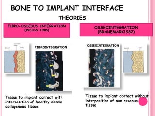
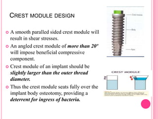
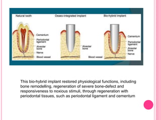
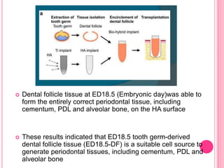
OSSEOINTEGRATION
Guided By
DR.S.ANILKUMAR
HOD
Presented By
DR.JOTHIPRIYA.B
FINAL YR PG
CONTENTS
 Introduction
 Definitions
 History
 Theories of integration
 Mechanism of osseointegration
 Factors affecting osseointegration
 Methods to determine osseointegration
 Conclusion
 References & articles
INTRODUCTION
 A successful replacement of missing natural tooth by dental
implant supported prosthesis is a major clinical advance in dental
science.
 The successful outcome of the treatment depends upon the
degree of Osseointegration.
 The science of Osseointegration has evolved over the last few
decades in both experimentally or clinically due to extensive
multidisciplinary approach.
o The present surge in the use of implants
was initiated by Branemark (1952)
o He described the relationship between
titanium and bone for which they coined
the term OSSEOINTEGRATION.
Osseointegration
 "osteon” -the Greekword for "bone"
 “integrare” -the Latinword for " to make
whole" meaning the state of being
combined into a complete whole
HISTORICAL BACKGROUND
The concept of Osseo integration based on research that began by Branemark
in 1952.
Early stage (1965-1968)
Developmental stage (1968-1971)
Production stage (1971-Present)
 To observe the microcirculation of both soft and hard
tissues under various phases of injuries
 He implanted titanium optic chamber in to rabbits fibula and
carried out the investigation with a vital microscopic
(essentially made of titanium) and when he tried to remove the
titanium chamber he found that bone was normally adhered
to the metal.
 This led to an idea of using titanium as an implant material
in the oral cavity.
 Researchers like Schroeder et al (1976),Alberktsson et al (1986)
proposed fibrous theory of implant fixation, Robert et al (1987),
Cork et al (1987), William (1986), Bebbush (1986), Meffert et
al (1992) and may more have dose extensive studies on retention
of dental implants.
 But it is Dr. Per Ingvar Branemark, who is considered as the
prime proponent for the philosophy that the absence of
connective tissues at bone implant interface is the key to clinical
success in dental implantology
DEFINITIONS &
TERMINOLOGIES
1969- “As a direct contact between the bone and metallic implants
without interposed soft tissues layers”.
1977 – “Direct structural and functional connection between
ordered, living bone and the surface of a load carrying implant.”
According to GPT-8
“ the apparent direct attachment or connection of osseous tissue
to an inert, alloplastic material without intervening
connective tissue”
OSTEOPRESERVATION (STALLARD R.E )
Tissue integration around healed functioning endosteal dental
implant in which the prime load bearing tissue at the interface
is a periimplant ligament composed of osteostimulatory
collagen.
PERIOSTEAL INTEGRATION
Tissue integration around a healed functioning subperiosteal
implant in which the load bearing tissue is the sheath of dense
collagenous tissue constituting the outer layer of periosteum.
THEORIES ON
BONE TO IMPLANT
INTERFACE
There are two basic theories regarding the bone implant interface.
Fibro-osseous
integration
(Linkow 1970, James
1975, and Weiss
1986)
Osseointegration
(supported by Branemark,
Zarb, and Albrektsson
1985)
BONE TO IMPLANT INTERFACE
THEORIES
FIBRO-OSSEOUS INTEGRATION
(WEISS 1986)
OSSEOINTEGRATION
(BRANEMARK1982)
Tissue to implant contact with
interposition of healthy dense
collagenous tissue
Tissue to implant contact without
interposition of non osseous
tissue
FIBRO-OSSEOUS INTEGRATION
“PSEUDOLIGAMENT”, “PERIIMPLANT LIGAMENT”,
“PERIIMPLANT MEMBRANE”.
 Presence of connective tissue between the implant and bone .
 Collagen fibers functions similarly to Sharpey’s fibers found
in natural dentition.
 The fibers are arranged irregularly, parallel to the implant
body, when forces are applied they are not transmitted through
the fibers.
FAILURE OF FIBRO OSSEOUS THEORY
 No evidence to support this theory.
 Since fibres are arranged parallel to the implant surface, do not
help in force transfer.
 Forces applied resulted in widening of fibrous encapsulation,
inflammatory reaction and gradual bone resorption thereby leading
to failure.
Meffert et al (1987) redefined and subdivided
osseointegration into :
ADAPTIVE OSSEOINTEGRATION
Osseous tissue approximating the surface of the implant
without apparent soft tissue interface at light microscopic
level.
BIOINTEGRATION
Is a direct biochemical bone surface attachment confirmed
at electron microscopic level.
THEORY OF OSSEOINTEGRATION
 Unlike fibro-osseous integration, osseointegration was able to
distribute vertical and slightly inclined loads more equally in to
surrounding bone.
 If osseointegration does not occur or osseointegration is lost for
some reason, a fibrous connective tissue forms around the implant.
 In such condition, the organization process continues against the
implant material, possibly resulting from chronic inflammation and
granulation tissue formation & osseointegration will never occur
(Albrektsson et al, 1983).
MECHANISM
OF
OSSEOINTEGRATION
 Healing process may be primary bone healing or secondary
bone healing.
 In primary bone healing, there is well organized bone
formation with minimal granulation tissue formation – ideal
 Secondary bone healing may have granulation tissue
formation and infection at the site, prolonging healing period.
(Fibrocartilage is sometimes formed instead of bone –
undesirable
EARLY BONE FORMATION ADJACENT TO ROUGH AND TURNED ENDOSSEOUS
IMPLANT SURFACES -AN EXPERIMENTAL STUDY IN THE DOG
ABRAHAMSSON ET AL (2004).
 screw-shaped titanium implant,
diameter: 4.1mm, length 10mm
with circumferential trough in
the endosseous part. Cross-
section of the wound chamber;
 (a) pitches engaging the bone
tissue walls;
 (b) inner U-shaped wound
chamber proper.
 The dotted line indicates the
lateral wall of the chamber, that
is, the position of the cut bone
surface.
The pitches engaged the hard
tissue walls of the cylindrical
canal prepared in the jawbone
and provided primary mechanical
stability for the devices.
 HEALING OF WOUND CHAMBER
FOLLOWING 2HR OF IMPLANT
INSTALLATION:
Thus, the chamber was first
filled with a blood clot characterized by
the presence of a high proportion of
erythrocytes entrapped in a fibrin network.
 DAY 4: the clot in its become penetrated
by vascular structures surrounded by
fibroblast-like mesenchymal cells forming
an early granulation tissue remains.
 The presence of PMN cells and
macrophages/monocytes at that time
documented the ongoing wound cleansing
process.
Bone modelling
 1 week of healing
 Vascular structures with few inflammatory cells.
 Cell rich immature (WOVEN) bone(centre of chamber and
direct contact of implant surface.)
 1st phase of osseointegration.
2 weeks: the amount of newly formed bone had formed
 Woven bone extends from parent bone into connective tissue.
 Mature osseointegration
 Pitch – ongoing new bone formation
 Recipient site immediate to implant in direct contact – bone resorption
with new bone formation.
4 weeks:
The large volume of the woven bone had been replaced by
lamellar bone
 Cell-rich woven bone covered most of titanium.
 Central portion – primary spongiosa – vascular structures and
mesenchymal cells.
Remodelling
 After 6-12 weeks – mineralised bone.
 Primary and secondary osteons
 Bone marrow, adipocytes and mesenchymal cells.
Presence of secondary
osteon is a sign of
remodelling
The damage caused during the surgical procedure and
the interlocking of the implant to the hard and soft tissues initiate
the process of healing.
The wound healing at the implant site depends on the –
Presence of adequate cells
Their adequate nutrition
Adequate stimulus for bone repair
The three main phases of bone healing necessary for
osseointegration are –
Phase 1 Inflammation
Phase 2 Proliferation
Phase3 Maturation
 The surgical invasion of the natural bone causes vascular trauma
at the osteotomy site which is instantly filled with blood and
subsequently the implant surfaces.
 A series of cellular and molecular events is initiated as a response
to surgical trauma that includes
a. Injury phase (0 to 2 weeks)
b. Granulation phage (2 to 3 weeks)
c. Callus phase (4 to 16 weeks)
BIOLOGICAL PROCESS OF
OSSEOINTEGRATION(BRANEMARK)
OSTEOPHYLIC
OSTEOINDUCTIVE
OSTEO CONDUCTIVE
STAGES OF OSSEOINTEGRATION
 According to Misch, there are two stages in osseointegration,
Each stage been again divided into two substages.
STAGE 1:
woven callus (0-
6 weeks)
STAGE 2:
lamellar
compaction
(6-18 weeks)
SURFACE
MODELLING
STAGE 3:
interface
remodeling
(6-18 weeks)
STAGE 4:
compact
maturation (18-
54 weeks)
REMODELLING
AND MATURATION
STAGE 1:WOVEN CALLUS
 Woven bone is formed at implant site.
 Primitive type of bone tissue and characterized random, felt-
like orientation of collagen fibrils
 Numerous irregularly shaped osteocytes
 Relatively low mineral density
STAGE 2:LAMELLAR COMPACTION
 Woven callus matures as it is replaced by lamellar bone.
 This stage helps in achieving sufficient strength for loading.
STAGE 3:INTERFACE REMODELING
 This stage begins at the same time when woven callus is
completing lamellar compaction.
 During this stage callus starts to resorb, and remodeling of
devitalized interface begins.
 The interface remodeling helps in establishing a viable
interface between the implant and original bone.
STAGE 4:COMPACT BONE MATURATION
 During this stage compact bone matures by series of modeling
and remodeling processes.
 The callus volume is decreased and interface remodeling
continues.
 Thus mechanism of osseointegration can be subdivided
into three biologic phenomena as described by J.E.Davies
(1998), they are
1. Osteoconduction
2. New bone formation
3. Bone remodeling
MECHANISM OF
OSSEOINTEGRATION(DAVIES 1998)
EARLY PHASES OF
OSTEOGENIC CELL MIGRATION
(OSTEOCONDUCTION)
DE NOVO BONE FORMATION
BONE REMODELING AT
DISCRETE SITES.
BONE TISSUE RESPONSE
DISTANCE OSTEOGENESIS
 A gradual process of bone
healing inward from the edge of
the osteotomy toward the
implant.
 Bone does not grow directly on
the implant surface
CONTACT OSTEOGENESIS
 The direct migration of bone-
building cells through the clot
matrix to the implant surface.
 Bone is quickly formed directly
on the implant surface.
IMPLANT TISSUE INTERFACE
Implant – CT
Interface
Implant – Bone
Interface
Implant – Epithelium
Interface
IMPLANT AND BONE INTERFACE
Light microscopic level (100X)
 Close adaptation of the regularly organized bone next to the Ti
implants.
Scanning electron microscopic level
 Parallel alignment of the lamellae of haversian system of the
bone next to the Ti implants. No connective tissue or dead
space at the interface.
Ultramicroscopic(500 to 1000X) level
Amorphous coat of glycoproteins on the implants to which the
collagen fibers are arranged at right angles and are partly
embedded into the glycoprotein layer.
IMPLANT CONNECTIVE TISSUE
INTERFACE
 Supracrestal connective tissue fibers will be arranged parallel to the
surface of the implant
 Not as strong as that of the connective tissue and tooth interface.
 An implant has no connective tissue fibers in the connective tissue
zone that insert into the implant .
IMPLANT EPITHELIAL INTERFACE
Biologic seal”
 Hemidesmosomes attached to glycoprotein layer
 Connect the interface to the plasma membrane of the
epithelial cells
 Similar to the junctional epithelium, Sulcus depth varies from 3
to 4mm
“
IMPLANT MATERIALS
IMPLANT MATERIALS
 To date, a multinational survey by ISO indicated that titanium and
its alloy are mainly used.
 The most widely used non-metallic implants are oxidic, carbonic or
graphitic oxide like materials.
 The major groups of implantable materials for dentistry are titanium
and alloys, cobalt chromium alloys, austenitic Fe-Cr-Ni-Mo steels,
tantalum, niobium and zirconium alloys, precious metals, ceramics
and polymeric materials.
TITANIUM AND TITANIUM ALLOYS
 Titanium is a metal that presents low weight high strength/weight ratio, low
modulus of elasticity, excellent corrosion resistance, excellent
biocompatibility and easy shaping and finishing.
 The most frequently used alloy (titanium.6 aluminum-4 vanadium)
 Titanium oxidizes (passivates) on contact with room temperature air and
normal tissue fluids.
 This reactivity is favourable for dental implant devices as it minimizes bio-
corrosion phenomena
 Cobalt-Chromium-Molybdenum based alloys
 Iron-Chromium-Nickel Based alloys
 Ceramics
 Aluminum, Titanium and Zirconium oxide
 Zirconia
 Carbon compounds
KEY FACTORS FOR SUCCESSFUL IMPLANT
OSSEOINTEGRATION
The successful outcome of any implant procedure is dependent on the
interrelationship of the following:
1. Biocompatibility of the Implant material
2. Macroscopic and microscopic nature of the implant surface
3. The status of the implant bed in both a health (non-infected) and a
morphologic (bone quality) context
4. The surgical technique
5. The undisturbed healing phase
6. The subsequent prosthetic design and long term loading phase.
FACTORS THAT INFLUENCE OSSEOINTEGRATION
PATIENT
RELATED
FACTORS
SURGICAL
RELATED
FACTORS
IMPLANT
RELATED
FACTORS
Implant material
biocompatibility
Loading conditions
Implant design
characteristic
Implant surface
characteristic
State of the implantation or host bed
Surgical
considerations
IMPLANT RELATED FACTORS
 Implant Biomaterial(Biocompatibility)
 Implant Biomechanics
 Implant Design
 Implant Taper
 Apical Design
 Implant Width
 Crest module design
 Implant Surface Topography(Surface roughness)
 Implant Surface Modifications
 Contamination
 Heat Production
 Implant Loading
IMPLANT BIOCOMPATIBILITY
Commercially pure titanium is widely used as an implant
material as
 highly biocompatible
 good resistance to corrosion, and
 no toxicity on macrophages or fibroblasts,
 lack of inflammatory response in peri-implant tissues and
 it’s composed of an oxide layer and has the ability to repair
itself by reoxidation when damaged.
 Another material used for implants,
Titanium -6 Aluminum-4 Vanadium (TI-6AL-4 V) alloy
exhibits soft tissue reactions very similar to those reported to
cp Ti
IMPLANT DESIGN(MACROSTRUCTURE):THE MACRODESIGN OR
SHAPE OF AN IMPLANT HAS AN IMPORTANT BEARING ON THE BONE
RESPONSE
Threaded or screw design implants
 Demonstrated to function for decades
without clinical problems.
 provide more functional area for stress
distribution than the cylindrical implants.
 minimal - <0.2 mm/year bone loss
Cylindrical implants
 Press fit root form implants depend on
coating or surface condition to provide
microscopic retention and bonding to the
bone
Combination root forms
 Macroscopic features of cylinder and screw
root forms
THE DESIGN OF THE THREADS
The dental implant applications dictate the need for a thread shape
optimized for long term function , load transmission under occlusal ,
intrusive and shear loading
 Functional surface area per unit length of implant may be modified
by the three thread geometry parameters
 Thread shape
 Thread pitch
 Thread depth
SMOOTH SURFACED & THREADED IMPLANTS
A smooth sided tapered implant results in essentially a
shear type of force at the implant – bone interface.
 provide for ease of surgical placement and
allow for greater functional surface area).
 In addition, a threaded implant is easily
rigidly fixated to limit micromovement
during wound healing
Threaded (or plateaued) implants with circular cross-section
 Unlike a cylinder implant, a tapered
threaded implant serves no functional
surface area advantage.
 The thread shape bears the compressive
and tensile loads.
 The tapered thread has less surface area
than a parallel threaded implant body.
 The tapered threaded implant cannot be
unthreaded once seated to place the crest
module in a more ideal prosthetic position.
 Non- threaded implants
 Tendency for slippage
 Bonding is required
 Threaded implants
 No tendency of slippage
 No bonding required.
IMPLANT TAPER
 A smooth sided tapered implant allows for a component of
compressive load to be delivered to the bone to implant
interface, depending on the degree of taper.
 The larger the taper, the greater the component of
compressive load delivered to the interface
 The amount of taper cannot be greater than 30º
 Tapering of threaded implant has no advantage
APICAL DESIGN OF IMPLANT
 Round cross sections at apex of the implant
do not resist torsional or shear forces when
abutments screws tightened.
 As a result , an anti rotational feature are
incorporated ,usually in the apical region of
implant body,with a hole or vent being the
most common design.
 Another anti rotational features are flat sides
or grooves at apical region
IMPLANT WIDTH
 Most implants fall within the range of 3.5 to 6 mm
 Increased implant width adequately increases the area
over which occlusal forces may be dissipated.
CREST MODULE DESIGN
 A smooth paralled sided crest module will
result in shear stresses.
 An angled crest module of more than 20º
will impose beneficial compressive
component.
 Crest module of an implant should be
slightly larger than the outer thread
diameter.
 Thus the crest module seats fully over the
implant body osteotomy, providing a
deterrent for ingress of bacteria.
IMPLANT SURFACE CHARACTERISTICS
 The Surface Quality will determine tissue reaction to an oral
implant.
 Surface quality may be dived into three categories:
(1) Mechanical properties, (2) Topographic properties
(3) physiochemical properties.
MECHANICAL PROPERTIES
 Mechanical properties of implant surfaces relate to potential
stresses in the surface that may result in increased corrosion rate
and wear relating to the hardness of the material.
 Wear is related to the strength of the material, but also to the
surface roughness.
 One technique to minimize the wear is ion implantation
 Ion implantation is clean, versatile,highly controllable and
reproducible,and induces intrinsic modifications within the
more superficial layers, while preserving the structure and
characteristics of the background material.
The benefits derived from these surface changes are
 mechanical (with increased resistance to wear and friction;
increased material hardness)
 chemical (increased resistance to corrosion, less lixiviation),
 electrochemical (enhanced ionic stability) and
 biological (better tissue adsorption)
Clin. Oral Impl. Res, 14, 2003; 57–62
TOPOGRAPHIC PROPERTIES
 The surface topography relates to the degree of roughness of the
surface and the orientation of the surface irregularities.
 The chemical composition of the implant interface on the implant
surface was shown to affect initial cell attachment
 This has stimulated a great interest on implant surface modification
as a way to accelerate the rate of osseointegration
SURFACE ROUGHNESS OF IMPLANT
 Depending on the scale of the features and based on the proposal
of Wennerberg and Albrektsson, surface roughness can be divided
into four categories:
Smooth surfaces:
Sa value <0.5 µm
(e.g. polished abutment
surface)
Rough surfaces:
Sa value >2.0 µm
(e.g. plasma sprayed
surfaces).
Moderately rough
surfaces:
Sa value 1.0 to <2.0 µm
(e.g. most commonly
used types
Minimally rough
surfaces:
Sa value 0.5 to <1.0 µm
(e.g. turned implants).
Moderate roughness
and roughness is
associated with
implant geometry, such
as screw structure,
and macroporous
surface treatments
Study evaluated the effects of phosphated titanium and
EMD on osteoblast function.
CONCLUSION:
 Phosphated titanium has the potential to accelerate
implant osseointegration by increased Tgf-ß1
production.
 EMD did not accelerate osteoblast function
Int J Oral Maxillofac Implants. 2007 ; 22(5): 701–709
IMPLANT SURFACE MODIFICATION
 To increase the surface area.
 To remove surface contaminants
 To bring better bonding
 To increase surface roughness of metal
 To increase corrosion resistance of metal.
 To make the metal more passive .
NEED FOR SURFACE MODIFICATION
TECHNIQUES FOR SURFACE MODIFICATION OF
IMPLANTS
 The implant modifications can be achieved either
by additive or subtractive methods.
 The additive methods employed the treatment in which other
materials are added to the surface, either superficial or
integrated, categorized into coating and impregnation,
respectively
Impregnation implies that the
material/chemical agent is fully integrated
into the titanium core, such as calcium
phosphate crystals within TiO2 layer or
incorporation of fluoride ions to surface,
Coating : is addition of material/agent of
various thicknesses superficially on the
surface of core material
titanium plasma spraying
(TPS), plasma sprayed
hydroxyapatite (HA) coating,
alumina coating, and biomimetic
calcium phosphate (CaP)
coating.
 The subtractive techniques are the procedure to either remove
the layer of core material or plastically deform the superficial
surface and thus roughen the surface of core material.
. The common subtractive techniques are large-grit
sands or ceramic particle blasts, acid etch, and
anodization
PLASMA SPRAYED SURFACE
 are prepared by spraying molten metal on the titanium
base which results in a surface with irregularly sized and
shaped valleys, pores and crevices, increasing the
microscopic surface area by 6 to 10 times.
 This topography may improve the fixation of implants by
the growth of bone into the coating, forming a
mechanical interlock.
TITANIUM PLASMA SPRAY
 The titanium plasma spray (TPS) surface has been reported to
increase the surface area of the bone-implant interface and
acts similarly to a three-dimensional surface, which may
stimulate adhesion osteogenesis.
 The surface area increase has been reported to be as great as
600%.
 One disadvantage of using the plasma-sprayed implants is the
detachment of titanium after implant insertion.
Osseointegration- Key Factors Affecting Its Success-An Overview Dr. Naveen
SAND BLASTING
 Sandblasting the metal core with gritting agents creates these
modified surfaces
 The blasting procedure is performed with the aim of increasing the
irregularity of the surface of the implant, using agents such as
aluminum oxide (Al2O3, also called alumina) and titanium
dioxide (TiO2).
 Blasting a surface with TiO2 particles was proposed to promote a
modification on the implant by using a component of the oxide layer
naturally formed around titanium implants.
ACID-ETCHING
 a titanium base was proposed to modify the implant surface
without leaving the residues found after the sandblasting
procedure, to avoid the non-uniform treatment of the surface,
and to control the loss of metallic substance from the body of
the implant.
 This is performed using baths of hydrochloric acid (HCl),
sulfuric acid (H2SO4), HF and nitric acid (HNO3) in
different combinations.
 A dual acid-etched technique has been proposed to produce
a microtextured (instead of a macrotextured) surface.
Osseotite Implant: is treated in a dual acid etching
procedure using hydrochloric and sulphuric acids
ANODIZED SURFACE
 The oxidation process has been used in dental implants to
change the characteristics of the oxide layer and
consequently to improve the biocompatibility of the
surface.
 The advantage is to modify the surface without depositing
grit particles.
 Anodized surfaces are prepared by applying a voltage on the
titanium specimen immersed in an electrolyte.
 The resultant surface presents micropores of variable
diameters and demonstrates lack of cytotoxicity;
LASERS
 An advantage of lasers in surface modification is that laser
has the property of melting surface layer locally.
 In addition, laser processing is contactless and the thermal,
mechanical deformation of the substrate is generally low.
 CO2 lasers and Nd-YAG laser
TRICALCIUM PHOSPHATE COATINGS
 By coating a metallic implant with Tricalcium phosphate
(TCP), an implant is produced that is biocompatible,
bioreactive and partially biodegradable.
 While TCP does not induce new bone formation, it does
have Osteoconductive properties that act as a scaffold or
nidus for new bone in growth.
 It also forms a chemical bio reactive bone with the
calcium and phosphorus in bone
HYDROXYAPATITE COATINGS
 Hydroxyapatite coatings have a similar roughness
and increase in functional surface area as TPS.
 A direct bone bond shown with HA coating and the
strength of the HA-to-bone interface is greater than
titanium to bone and even greater than TPS to
bone.
 Gap healing may be enhanced by the HA coating
The study investigated the effects of different chemically modified
titanium surfaces on protien adsortion and the osteoblastic
differentiation of human palatal mesenchyml cells(HEPM)
Three different surfaces were evaluated.
 The first ,machined surface(Ti-M) as control
 The second surface was acid etched(Ti-AE)
 The third surface was – Ti-AE sample exposed to sodium hydroxide
(NaOH) solution (Ti-AAE)
Concluded that
 Ti-AAE surfaces exhibited greater efficiency in promoting the
expression of osteoblastic differentiation markers.
 Serve as a suitable substratum for enhancing the process of
osseointegration.
CAPCITIVELY COUPLED ELECTRIC FIELD
(CCEF)
 CCEF treatment effectively stimulated osteogenesis near the
implant by generating undifferentiated mesenchymal cells.
 It has been believed that functional loading on an implant
restoration in the early period after implant placement prevents
osseointegration in the nearby bone.
 However, by the applicaiton of CCEF after implant placement,
shortens the recovery period of normal occlusal function.
BOVINE OSTEOGENIC PROTEIN OSTEOGENIC
PROTEIN
 inserted into unmodified sockets with implants may
significantly shorten the time interval between tooth
extraction and osseointegration of the implant and thereby
reduce the necessary period of total or partial edentulism.
 In addition, this treatment may expand the use of implant
therapy and enhance success rates by eliminating a
surgical procedure, reducing the amount of bone lost
after tooth extraction, permitting the insertion of longer
implants and minimizing prosthetic compromises
associated with alveolar ridge resorption
STATE OF HOST BED
Ideal host bed
 Healthy and with an adequate bone stock
 Bone height
 Bone width
 Bone length
 Bone density
 Undesirable host bed states for implantation
 Previous irradiation
 Ridge height resorption
 Osteoporosis
 According to Branemark and Misch
 D1 and D2 bone initial stability / better osseointegration
 D3 and D4  poor prognosis
 D1 bone – least risk
 D4 bone - most at risk
Jaffin and Berman (1991) – 44% failure in type IV bone
Selection of implant
D1 and D2 – conventional threaded implants
D3 and D4 – HA coated or Titanium plasma coated implants
Initial implant stability
 Bone quality  bone with well formed cortex and densely
trabeculated medullary spaces
 Bone quantity  Refers to the dimension of available bone
in reference to length, width and depth.
Branemark system (5 year documentation)
 Mandible – 95% success
 Maxilla – 85-90% success
 Aden et al (1981) – 10% greater success rate in anterior mandible
compared to anterior maxilla.
 Schnitman et al (1988)
 lower success rate in posterior mandible compared to anterior
mandible
 posterior maxilla higher failure rates
LEKHOLM AND ZARB CLASSIFICATION 1985
Class I :
Jaw consist
almost
exclusively
of
homogeneou
s compact
bone
Class II :
Thick
compact
bone
surrounds
highly
trabecular
core
Class III :
Thin cortical
bone
surrounds
highly
trabecular
core
Class IV :
Thin cortical
bone
surrounds
loose,
spongy core
FACTORS COMPROMISING BONE
QUALITY
SURGICAL FACTORS THAT AFFECT
OSSEOINTEGRATION
 Minimal tissue violence at surgery is essential for
osseointegration.
 This objective depends on continuous and careful cooling while
surgical drilling is performed at low speed.
 The critical time/temperature relationship for bone tissue
necrosis is around 47 ºC applied for 1 min.
 Use of sharp drills with a drill speed of less than 2000rpm is
desired.
 56ºC is the critical temperature to prevent bone overheating .
 Use of torque wrench with moderate torque of 45 N/cm is ideal.
PATIENT RELATED FACTORS
 Age :
 Old age – no poorer result
 Extreme young age - Relative contraindication to
insertion of implants
 Smoking and osseointegration :
 History of smoking affect the healing response in
osseointegration.
 Lower success rates with oral implants
 Mean failure rates in smokers is about twice
than in non smoker.
 Radiation therapy and osseointegration :
 Jacobson (1985) previous irradiation – relative
contraindication for implant placement.
 Expected success rate 10-15% lower than the non irradiated
patients.
 Granstrom G (1998)  HBO(Hyperbaric oxygen therapy)
can counteract some of the negative effect from irradiation
and act as a stimulator for osseointegration.
o Compromised oral hygiene
o Vitamin C deficiency
o Uncontrolled Diabetes.
o Bone density
o Available bone
LOADING CONDITIONS
 The primary factor for success at the time of placement is
achieving primary stability.
 Any micromotion during initial phases of bone healing will cause
a lack of integration.
 Objective : “No loading while healing”  successful osseointegration.
Premature loading
leads to implant
movement
The end result
“Soft tissue
interface”
“Bony
interface”
DIFFERENT PHILOSOPHIES REGARDING
LOADING CONDITIONS
 Branemark, Albrektson
– two stage implant
insertion.
 First stage – Installation
of fixture into bone
 Second stage–
Connection of abutment
to the fixtures
 Maxilla: 6 months
 Mandible: 3 months
Misch – Progressive /
Gradual loading
Suggested in
Softer bone
less number of
implants to be used
 Immediate functional loading protocol
 Clinical trials successful osseointegration
 (95-100% success rate- Completely edentulous
patients)
 Bone quality is good
 Functional forces are controlled
 More favourable in mandible compared to maxilla
Over loading – Stress concentration, undermining bone
resorption without apposition (Branemark 1984)
Prosthetic design considerations
Cantilever length may be shortened or eliminated
Narrow occlusal table
Minimizing the offset load
Increasing the implant number
Use of wider implant with D4 bone compared to D1 & D2
To decrease the bio-mechanical load
ENDOPORE DENTAL IMPLANT
The Endopore dental implant incorporates a unique, truncated
cone-shaped design that uses a multilayered porous surface
geometry over most of its length to achieve integration by
three dimensional bone ingrowth.
Endopore's surgical Advantages
 A secure, three-dimensional interlocking interface with
bone
 Predictable and minimal crestal bone remodeling
 Uncomplicated surgical sequence
 Minimal instrumentation and inventory
 Self seating , tapered, pressfit, prosthetically
friendly design
 Good resistance to torsional forces
 Shorter initial healing time.
METHODS OF EVALUATION OF
OSSEOINTEGRATION
Invasive methods
Which interferes with
osseointegration
process of implant
Non- invasive methods:
which does not interfere
with osseointegration
process
INVASIVE METHODS
HISTOLOGIC
ANALYSIS
HISTOMORPHOMETRIC
ANALYSIS
USE OF TORQUE
GAUGES
NON-INVASIVE METHODS :
 Radiographs
 Cutting Torque resistance analysis
 Reverse torque test
 Insertion torque analysis
 Percussion test
 Impact Hammer method
 Pulsed oscillation waveform
 Resonance frequency analysis
RADIOGRAPHIC ANALYSIS
 Radiographic evaluation is a noninvasive method
that can be performed at any stage of healing.
CUTTING TORQUE RESISTANCE ANALYSIS
 In cutting resistance analysis (CRA), originally developed by
Johansson and Strid and later improved by Friberg et al
 the energy (J/mm3) required for a current fed electric
motor in cutting off a unit volume of bone during implant
surgery is measured.
 This energy was shown to be significantly correlated with bone
density, which is the primary indicator implant stability
Advantages
1. Detect bone density
2. High correlation between cutting
resistance and bone quality
3. Reliable method to assess bone quality
4. Identify bone density during surgery
5. Can be used in daily practice
Disadvantages
1. Can only be used during surgery
THE REVERSE TORQUE
TEST (RTT),
 proposed by Roberts et al and developed by Johansson and
Albrektsson measures the “critical” torque threshold where
bone-implant contact (BIC) was destroyed.
 a 20-Ncm threshold RTV for successful osseointegration
 it cannot quantify degree of osseointegration.
 Hence, RTT is mainly used in experiments.
PERIOTEST
 Periotest has been thoroughly studied and advocated as a
reliable method to determine implant stability.
 Periotest uses an electromagnetically driven and electronically
controlled tapping metallic rod in a handpiece.,that hammers
an object at a rate of 4 times /sec.
 Response to a striking or “barking” is measured by a small
accelerometer incorporated into the head.
DENTAL MOBILITY CHECKER
 The DMC, which was originally developed by Aoki and
Hirakawa measures tooth mobility with an impact hammer
method using transient impact force.
 A microphone used as a receiver and signals transferred is
processes by FFT for analysis.
A PERCUSSION TEST
 is one of the simplest methods that can be used to estimate
the level of osseointegration.
 This test is based upon vibrational acoustic science and
impact-response theory.
 ringing “crystal” sound indicates successful osseointegration
 a “dull” sound- indicate no osseointegration.
 it cannot be used experimentally as a standardized testing
method.
IMPACT HAMMER METHOD
 It is an improved version of the percussion test except that
sound generated from a contact between a hammer and an
object is processed through fast Fourier transform (FFT ) for
analysis of transfer characteristics
 Periotest (Siemens, Bensheim,Germany) and Dental Mobility
Checker (DMC; J. Morita, Suita, Japan) are currently available
mobility testers designed according to the impact hammer method.
PULSED OSCILLATION WAVEFORM
 Kaneko et al described the use of a pulsed oscillation
waveform (POWF) to analyze the mechanical vibrational
characteristics of the implant-bone interface using forced
excitation of a steady-state wave.
 POWF is based on estimation of frequency and amplitude
of the vibration of the implant induced by a small pulsed
force
RFA
 It is a noninvasive diagnostic method that measures implant
stability and bone density at various time points using
vibration and a principle of structural analysis.
 Currently, 2 RFA machines are in clinical use: Osstell
(Integration Diagnostics) and Implomates (Bio Tech One).
Methods Used to Assess Implant Stability: Current Status
Mihoko Atsumi et al INT J ORAL MAXILLOFAC IMPLANTS
2007;22:743–754
 Resonance frequency values ranging from 3,500 to
8,500 Hz are translated into an ISQ of 0 to 100.
 A high value indicates greater stability, whereas a
low value implies instability
.
OTHER METHODS TO ASSESS
OSSEOINTEGRATION
 Cone beam CT Periotest
 Dynamic model testing
 Impulse testing
FAILURES OF OSSEOINTEGRATION
EARLY:failure to establish a
close bond to implant
BIOLOGIC:bacteria
LATE:disruption of established
contact
MECHANICAL:due to overload and
fracture
SUCCESS CRITERIA OF IMPLANTS
 Albrektson and Zarb G (1980)
The individual unattached implant should be
immobile when tested clinically
The radiographic evaluation should not show
any peri - implant radiolucency
Vertical bone loss around the fixtures should be
less than 0.2mm annually after first year of
implant loading
The implant should not show any sign and symptom
of pain, infection, neuropathies, parastehsia, violation
of mandibular canal and sinus drainage.
Success rate of 85% at the end of 5 year
observation period and 80% at the end of 10
year service.
FUTURISTIC CONCEPTS OF
OSSEOINTEGRATION
Nanoscale modification of titanium
endosseous implant surfaces can
alter cellular and tissue responses that
may benefit osseointegration and
dental implant therapy.
Biomaterials 29 (2008) 3822–3835
 Biomimetic dental implants may be the next development in the
field.
 Coating implants with factors
 Induce endothelial cell differentiation and proliferation may
 promote greater vascularity in highly cortical bone, thereby
 improving conditions for early and long-term (in response
 to functional loading) bone remodelling.
 Coating implants with BMPs may also accelerate initial healing
times during integration of the dental implant, thereby reducing
overall treatment times and improving implant success rates.
OSSEOPERCEPTION
 Osseoperception is the term given to the patient reported
with feeling of heightened perception of the environment with
osseointegrated prostheses
 Implantation of Schwann cells, neural stem cells and
mesenchymal cells can contribute to nerve regeneration
surrounding the implant
Osseoperception in Dental Implants Rupinder Singh Dhall International Journal of
Periodontology and Implantology, October-December 2017;2(4):130-135
 Guided tissue regeneration can
be applied to reconstruct
periodontal tissue.
 This technique implants
periodontal ligament stem cells
that express high levels of
bone morphogenetic protein,
and platelet-derived growth factor
and has achieved some success
 have enveloped with embryonic dental follicle tissue
around a HA-coated dental implant, and transplanted
into the lower first molar region of a murine tooth-loss
model.
 Had successfully developed a novel fibrous connected
tooth implant using a HA-coated dental implant and
dental follicle stem cells as a bio-hybrid organ.
This bio-hybrid implant restored physiological functions, including
bone remodelling, regeneration of severe bone-defect and
responsiveness to noxious stimuli, through regeneration with
periodontal tissues, such as periodontal ligament and cementum
 Dental follicle tissue at ED18.5 (Embryonic day)was able to
form the entirely correct periodontal tissue, including
cementum, PDL and alveolar bone, on the HA surface
 These results indicated that ED18.5 tooth germ-derived
dental follicle tissue (ED18.5-DF) is a suitable cell source to
generate periodontal tissues, including cementum, PDL and
alveolar bone
CONCLUSION
AS THE CONCEPT OF OSSEOINTEGRATION HAS DEVELOPED AND
SPREAD GLOBALLY , IT HAS HAD A DRAMATIC IMPACT ON THE
PRACTICES OF DENTISTRY.
 In implant dentistry , an undisturbed healing period is
always required to ensure osseointegration.
 A modified protocol with early or immediate loading has been
tested to satisfy the demand of more rapid treatment and to
reduce discomfort of wearing removable appliances during the
healing period.
 Provided that the implant has primary stability , studies have
shown that the survival of loaded implants can be analogous
to the unloaded protocol.
THANK YOU
REFERENCES
 Kasemo B (1983) Biocompatibity of titanium implants: surface science aspects. J
Prosthet Dent 49:832–837
 Osseointegration- Key Factors Affecting Its Success-An Overview Dr. Naveen
Reddy Vootla1,Dr. K. Varun Reddy2
 Methods Used to Assess Implant Stability: Current Status Mihoko Atsumi et al
INT J ORAL MAXILLOFAC IMPLANTS 2007;22:743–754
 Osseoperception in Dental Implants Rupinder Singh Dhall International Journal of
Periodontology and Implantology, October-December 2017;2(4):130-135
 Hobo, Ichida, Garcia “Osseointegration and occlusal rehabilitation” Quintessence
Publishing.
 Jan Lindhe “Clinical periodontology and implant dentistry” 4th edition, Blackwell
Publishing.
 Elaine McClarence “Branemark and the development of osseointegration”
Quintessence publication
 Carl E. Misch “Implant dentistry” 2nd edition, Mosby. Charles M.Weis “Principles
and practice of implant dentistry” Mosby.
 Per Ingvar Branemark “Osseointegration and its experimental background” JPD
1983 Vol. 50, 399-410.
 Hanson, Alberktson “Structural aspects of the interface between tissue and
titanium implants” JPD 1983 vol. 50, 108-113.

Osseointegration final

  • 1.
  • 2.
    CONTENTS  Introduction  Definitions History  Theories of integration  Mechanism of osseointegration  Factors affecting osseointegration  Methods to determine osseointegration  Conclusion  References & articles
  • 3.
    INTRODUCTION  A successfulreplacement of missing natural tooth by dental implant supported prosthesis is a major clinical advance in dental science.  The successful outcome of the treatment depends upon the degree of Osseointegration.  The science of Osseointegration has evolved over the last few decades in both experimentally or clinically due to extensive multidisciplinary approach.
  • 4.
    o The presentsurge in the use of implants was initiated by Branemark (1952) o He described the relationship between titanium and bone for which they coined the term OSSEOINTEGRATION. Osseointegration  "osteon” -the Greekword for "bone"  “integrare” -the Latinword for " to make whole" meaning the state of being combined into a complete whole
  • 5.
    HISTORICAL BACKGROUND The conceptof Osseo integration based on research that began by Branemark in 1952. Early stage (1965-1968) Developmental stage (1968-1971) Production stage (1971-Present)
  • 6.
     To observethe microcirculation of both soft and hard tissues under various phases of injuries  He implanted titanium optic chamber in to rabbits fibula and carried out the investigation with a vital microscopic (essentially made of titanium) and when he tried to remove the titanium chamber he found that bone was normally adhered to the metal.  This led to an idea of using titanium as an implant material in the oral cavity.
  • 7.
     Researchers likeSchroeder et al (1976),Alberktsson et al (1986) proposed fibrous theory of implant fixation, Robert et al (1987), Cork et al (1987), William (1986), Bebbush (1986), Meffert et al (1992) and may more have dose extensive studies on retention of dental implants.  But it is Dr. Per Ingvar Branemark, who is considered as the prime proponent for the philosophy that the absence of connective tissues at bone implant interface is the key to clinical success in dental implantology
  • 8.
  • 9.
    1969- “As adirect contact between the bone and metallic implants without interposed soft tissues layers”. 1977 – “Direct structural and functional connection between ordered, living bone and the surface of a load carrying implant.” According to GPT-8 “ the apparent direct attachment or connection of osseous tissue to an inert, alloplastic material without intervening connective tissue”
  • 11.
    OSTEOPRESERVATION (STALLARD R.E) Tissue integration around healed functioning endosteal dental implant in which the prime load bearing tissue at the interface is a periimplant ligament composed of osteostimulatory collagen. PERIOSTEAL INTEGRATION Tissue integration around a healed functioning subperiosteal implant in which the load bearing tissue is the sheath of dense collagenous tissue constituting the outer layer of periosteum.
  • 12.
    THEORIES ON BONE TOIMPLANT INTERFACE
  • 13.
    There are twobasic theories regarding the bone implant interface. Fibro-osseous integration (Linkow 1970, James 1975, and Weiss 1986) Osseointegration (supported by Branemark, Zarb, and Albrektsson 1985)
  • 14.
    BONE TO IMPLANTINTERFACE THEORIES FIBRO-OSSEOUS INTEGRATION (WEISS 1986) OSSEOINTEGRATION (BRANEMARK1982) Tissue to implant contact with interposition of healthy dense collagenous tissue Tissue to implant contact without interposition of non osseous tissue
  • 15.
    FIBRO-OSSEOUS INTEGRATION “PSEUDOLIGAMENT”, “PERIIMPLANTLIGAMENT”, “PERIIMPLANT MEMBRANE”.  Presence of connective tissue between the implant and bone .  Collagen fibers functions similarly to Sharpey’s fibers found in natural dentition.  The fibers are arranged irregularly, parallel to the implant body, when forces are applied they are not transmitted through the fibers.
  • 16.
    FAILURE OF FIBROOSSEOUS THEORY  No evidence to support this theory.  Since fibres are arranged parallel to the implant surface, do not help in force transfer.  Forces applied resulted in widening of fibrous encapsulation, inflammatory reaction and gradual bone resorption thereby leading to failure.
  • 17.
    Meffert et al(1987) redefined and subdivided osseointegration into : ADAPTIVE OSSEOINTEGRATION Osseous tissue approximating the surface of the implant without apparent soft tissue interface at light microscopic level. BIOINTEGRATION Is a direct biochemical bone surface attachment confirmed at electron microscopic level. THEORY OF OSSEOINTEGRATION
  • 18.
     Unlike fibro-osseousintegration, osseointegration was able to distribute vertical and slightly inclined loads more equally in to surrounding bone.  If osseointegration does not occur or osseointegration is lost for some reason, a fibrous connective tissue forms around the implant.  In such condition, the organization process continues against the implant material, possibly resulting from chronic inflammation and granulation tissue formation & osseointegration will never occur (Albrektsson et al, 1983).
  • 19.
  • 20.
     Healing processmay be primary bone healing or secondary bone healing.  In primary bone healing, there is well organized bone formation with minimal granulation tissue formation – ideal  Secondary bone healing may have granulation tissue formation and infection at the site, prolonging healing period. (Fibrocartilage is sometimes formed instead of bone – undesirable
  • 21.
    EARLY BONE FORMATIONADJACENT TO ROUGH AND TURNED ENDOSSEOUS IMPLANT SURFACES -AN EXPERIMENTAL STUDY IN THE DOG ABRAHAMSSON ET AL (2004).  screw-shaped titanium implant, diameter: 4.1mm, length 10mm with circumferential trough in the endosseous part. Cross- section of the wound chamber;  (a) pitches engaging the bone tissue walls;  (b) inner U-shaped wound chamber proper.  The dotted line indicates the lateral wall of the chamber, that is, the position of the cut bone surface. The pitches engaged the hard tissue walls of the cylindrical canal prepared in the jawbone and provided primary mechanical stability for the devices.
  • 22.
     HEALING OFWOUND CHAMBER FOLLOWING 2HR OF IMPLANT INSTALLATION: Thus, the chamber was first filled with a blood clot characterized by the presence of a high proportion of erythrocytes entrapped in a fibrin network.  DAY 4: the clot in its become penetrated by vascular structures surrounded by fibroblast-like mesenchymal cells forming an early granulation tissue remains.  The presence of PMN cells and macrophages/monocytes at that time documented the ongoing wound cleansing process.
  • 23.
    Bone modelling  1week of healing  Vascular structures with few inflammatory cells.  Cell rich immature (WOVEN) bone(centre of chamber and direct contact of implant surface.)  1st phase of osseointegration. 2 weeks: the amount of newly formed bone had formed  Woven bone extends from parent bone into connective tissue.  Mature osseointegration  Pitch – ongoing new bone formation  Recipient site immediate to implant in direct contact – bone resorption with new bone formation.
  • 24.
    4 weeks: The largevolume of the woven bone had been replaced by lamellar bone  Cell-rich woven bone covered most of titanium.  Central portion – primary spongiosa – vascular structures and mesenchymal cells. Remodelling  After 6-12 weeks – mineralised bone.  Primary and secondary osteons  Bone marrow, adipocytes and mesenchymal cells. Presence of secondary osteon is a sign of remodelling
  • 26.
    The damage causedduring the surgical procedure and the interlocking of the implant to the hard and soft tissues initiate the process of healing. The wound healing at the implant site depends on the – Presence of adequate cells Their adequate nutrition Adequate stimulus for bone repair The three main phases of bone healing necessary for osseointegration are – Phase 1 Inflammation Phase 2 Proliferation Phase3 Maturation
  • 28.
     The surgicalinvasion of the natural bone causes vascular trauma at the osteotomy site which is instantly filled with blood and subsequently the implant surfaces.  A series of cellular and molecular events is initiated as a response to surgical trauma that includes a. Injury phase (0 to 2 weeks) b. Granulation phage (2 to 3 weeks) c. Callus phase (4 to 16 weeks)
  • 29.
  • 33.
    STAGES OF OSSEOINTEGRATION According to Misch, there are two stages in osseointegration, Each stage been again divided into two substages. STAGE 1: woven callus (0- 6 weeks) STAGE 2: lamellar compaction (6-18 weeks) SURFACE MODELLING STAGE 3: interface remodeling (6-18 weeks) STAGE 4: compact maturation (18- 54 weeks) REMODELLING AND MATURATION
  • 34.
    STAGE 1:WOVEN CALLUS Woven bone is formed at implant site.  Primitive type of bone tissue and characterized random, felt- like orientation of collagen fibrils  Numerous irregularly shaped osteocytes  Relatively low mineral density
  • 35.
    STAGE 2:LAMELLAR COMPACTION Woven callus matures as it is replaced by lamellar bone.  This stage helps in achieving sufficient strength for loading.
  • 36.
    STAGE 3:INTERFACE REMODELING This stage begins at the same time when woven callus is completing lamellar compaction.  During this stage callus starts to resorb, and remodeling of devitalized interface begins.  The interface remodeling helps in establishing a viable interface between the implant and original bone.
  • 37.
    STAGE 4:COMPACT BONEMATURATION  During this stage compact bone matures by series of modeling and remodeling processes.  The callus volume is decreased and interface remodeling continues.
  • 38.
     Thus mechanismof osseointegration can be subdivided into three biologic phenomena as described by J.E.Davies (1998), they are 1. Osteoconduction 2. New bone formation 3. Bone remodeling
  • 39.
    MECHANISM OF OSSEOINTEGRATION(DAVIES 1998) EARLYPHASES OF OSTEOGENIC CELL MIGRATION (OSTEOCONDUCTION) DE NOVO BONE FORMATION BONE REMODELING AT DISCRETE SITES.
  • 40.
    BONE TISSUE RESPONSE DISTANCEOSTEOGENESIS  A gradual process of bone healing inward from the edge of the osteotomy toward the implant.  Bone does not grow directly on the implant surface CONTACT OSTEOGENESIS  The direct migration of bone- building cells through the clot matrix to the implant surface.  Bone is quickly formed directly on the implant surface.
  • 42.
    IMPLANT TISSUE INTERFACE Implant– CT Interface Implant – Bone Interface Implant – Epithelium Interface
  • 43.
    IMPLANT AND BONEINTERFACE Light microscopic level (100X)  Close adaptation of the regularly organized bone next to the Ti implants. Scanning electron microscopic level  Parallel alignment of the lamellae of haversian system of the bone next to the Ti implants. No connective tissue or dead space at the interface. Ultramicroscopic(500 to 1000X) level Amorphous coat of glycoproteins on the implants to which the collagen fibers are arranged at right angles and are partly embedded into the glycoprotein layer.
  • 44.
    IMPLANT CONNECTIVE TISSUE INTERFACE Supracrestal connective tissue fibers will be arranged parallel to the surface of the implant  Not as strong as that of the connective tissue and tooth interface.  An implant has no connective tissue fibers in the connective tissue zone that insert into the implant .
  • 45.
    IMPLANT EPITHELIAL INTERFACE Biologicseal”  Hemidesmosomes attached to glycoprotein layer  Connect the interface to the plasma membrane of the epithelial cells  Similar to the junctional epithelium, Sulcus depth varies from 3 to 4mm “
  • 46.
  • 47.
    IMPLANT MATERIALS  Todate, a multinational survey by ISO indicated that titanium and its alloy are mainly used.  The most widely used non-metallic implants are oxidic, carbonic or graphitic oxide like materials.  The major groups of implantable materials for dentistry are titanium and alloys, cobalt chromium alloys, austenitic Fe-Cr-Ni-Mo steels, tantalum, niobium and zirconium alloys, precious metals, ceramics and polymeric materials.
  • 48.
    TITANIUM AND TITANIUMALLOYS  Titanium is a metal that presents low weight high strength/weight ratio, low modulus of elasticity, excellent corrosion resistance, excellent biocompatibility and easy shaping and finishing.  The most frequently used alloy (titanium.6 aluminum-4 vanadium)  Titanium oxidizes (passivates) on contact with room temperature air and normal tissue fluids.  This reactivity is favourable for dental implant devices as it minimizes bio- corrosion phenomena
  • 49.
     Cobalt-Chromium-Molybdenum basedalloys  Iron-Chromium-Nickel Based alloys  Ceramics  Aluminum, Titanium and Zirconium oxide  Zirconia  Carbon compounds
  • 50.
    KEY FACTORS FORSUCCESSFUL IMPLANT OSSEOINTEGRATION The successful outcome of any implant procedure is dependent on the interrelationship of the following: 1. Biocompatibility of the Implant material 2. Macroscopic and microscopic nature of the implant surface 3. The status of the implant bed in both a health (non-infected) and a morphologic (bone quality) context 4. The surgical technique 5. The undisturbed healing phase 6. The subsequent prosthetic design and long term loading phase.
  • 51.
    FACTORS THAT INFLUENCEOSSEOINTEGRATION PATIENT RELATED FACTORS SURGICAL RELATED FACTORS IMPLANT RELATED FACTORS
  • 52.
    Implant material biocompatibility Loading conditions Implantdesign characteristic Implant surface characteristic State of the implantation or host bed Surgical considerations
  • 53.
    IMPLANT RELATED FACTORS Implant Biomaterial(Biocompatibility)  Implant Biomechanics  Implant Design  Implant Taper  Apical Design  Implant Width  Crest module design  Implant Surface Topography(Surface roughness)  Implant Surface Modifications  Contamination  Heat Production  Implant Loading
  • 54.
    IMPLANT BIOCOMPATIBILITY Commercially puretitanium is widely used as an implant material as  highly biocompatible  good resistance to corrosion, and  no toxicity on macrophages or fibroblasts,  lack of inflammatory response in peri-implant tissues and  it’s composed of an oxide layer and has the ability to repair itself by reoxidation when damaged.  Another material used for implants, Titanium -6 Aluminum-4 Vanadium (TI-6AL-4 V) alloy exhibits soft tissue reactions very similar to those reported to cp Ti
  • 56.
    IMPLANT DESIGN(MACROSTRUCTURE):THE MACRODESIGNOR SHAPE OF AN IMPLANT HAS AN IMPORTANT BEARING ON THE BONE RESPONSE Threaded or screw design implants  Demonstrated to function for decades without clinical problems.  provide more functional area for stress distribution than the cylindrical implants.  minimal - <0.2 mm/year bone loss Cylindrical implants  Press fit root form implants depend on coating or surface condition to provide microscopic retention and bonding to the bone Combination root forms  Macroscopic features of cylinder and screw root forms
  • 57.
    THE DESIGN OFTHE THREADS The dental implant applications dictate the need for a thread shape optimized for long term function , load transmission under occlusal , intrusive and shear loading  Functional surface area per unit length of implant may be modified by the three thread geometry parameters  Thread shape  Thread pitch  Thread depth
  • 58.
    SMOOTH SURFACED &THREADED IMPLANTS A smooth sided tapered implant results in essentially a shear type of force at the implant – bone interface.  provide for ease of surgical placement and allow for greater functional surface area).  In addition, a threaded implant is easily rigidly fixated to limit micromovement during wound healing Threaded (or plateaued) implants with circular cross-section
  • 59.
     Unlike acylinder implant, a tapered threaded implant serves no functional surface area advantage.  The thread shape bears the compressive and tensile loads.  The tapered thread has less surface area than a parallel threaded implant body.  The tapered threaded implant cannot be unthreaded once seated to place the crest module in a more ideal prosthetic position.
  • 61.
     Non- threadedimplants  Tendency for slippage  Bonding is required  Threaded implants  No tendency of slippage  No bonding required.
  • 65.
    IMPLANT TAPER  Asmooth sided tapered implant allows for a component of compressive load to be delivered to the bone to implant interface, depending on the degree of taper.  The larger the taper, the greater the component of compressive load delivered to the interface  The amount of taper cannot be greater than 30º  Tapering of threaded implant has no advantage
  • 66.
    APICAL DESIGN OFIMPLANT  Round cross sections at apex of the implant do not resist torsional or shear forces when abutments screws tightened.  As a result , an anti rotational feature are incorporated ,usually in the apical region of implant body,with a hole or vent being the most common design.  Another anti rotational features are flat sides or grooves at apical region
  • 67.
    IMPLANT WIDTH  Mostimplants fall within the range of 3.5 to 6 mm  Increased implant width adequately increases the area over which occlusal forces may be dissipated.
  • 68.
    CREST MODULE DESIGN A smooth paralled sided crest module will result in shear stresses.  An angled crest module of more than 20º will impose beneficial compressive component.  Crest module of an implant should be slightly larger than the outer thread diameter.  Thus the crest module seats fully over the implant body osteotomy, providing a deterrent for ingress of bacteria.
  • 69.
    IMPLANT SURFACE CHARACTERISTICS The Surface Quality will determine tissue reaction to an oral implant.  Surface quality may be dived into three categories: (1) Mechanical properties, (2) Topographic properties (3) physiochemical properties.
  • 70.
    MECHANICAL PROPERTIES  Mechanicalproperties of implant surfaces relate to potential stresses in the surface that may result in increased corrosion rate and wear relating to the hardness of the material.  Wear is related to the strength of the material, but also to the surface roughness.  One technique to minimize the wear is ion implantation
  • 71.
     Ion implantationis clean, versatile,highly controllable and reproducible,and induces intrinsic modifications within the more superficial layers, while preserving the structure and characteristics of the background material. The benefits derived from these surface changes are  mechanical (with increased resistance to wear and friction; increased material hardness)  chemical (increased resistance to corrosion, less lixiviation),  electrochemical (enhanced ionic stability) and  biological (better tissue adsorption) Clin. Oral Impl. Res, 14, 2003; 57–62
  • 72.
    TOPOGRAPHIC PROPERTIES  Thesurface topography relates to the degree of roughness of the surface and the orientation of the surface irregularities.  The chemical composition of the implant interface on the implant surface was shown to affect initial cell attachment  This has stimulated a great interest on implant surface modification as a way to accelerate the rate of osseointegration
  • 73.
    SURFACE ROUGHNESS OFIMPLANT  Depending on the scale of the features and based on the proposal of Wennerberg and Albrektsson, surface roughness can be divided into four categories: Smooth surfaces: Sa value <0.5 µm (e.g. polished abutment surface) Rough surfaces: Sa value >2.0 µm (e.g. plasma sprayed surfaces). Moderately rough surfaces: Sa value 1.0 to <2.0 µm (e.g. most commonly used types Minimally rough surfaces: Sa value 0.5 to <1.0 µm (e.g. turned implants). Moderate roughness and roughness is associated with implant geometry, such as screw structure, and macroporous surface treatments
  • 74.
    Study evaluated theeffects of phosphated titanium and EMD on osteoblast function. CONCLUSION:  Phosphated titanium has the potential to accelerate implant osseointegration by increased Tgf-ß1 production.  EMD did not accelerate osteoblast function Int J Oral Maxillofac Implants. 2007 ; 22(5): 701–709
  • 75.
    IMPLANT SURFACE MODIFICATION To increase the surface area.  To remove surface contaminants  To bring better bonding  To increase surface roughness of metal  To increase corrosion resistance of metal.  To make the metal more passive . NEED FOR SURFACE MODIFICATION
  • 76.
    TECHNIQUES FOR SURFACEMODIFICATION OF IMPLANTS  The implant modifications can be achieved either by additive or subtractive methods.
  • 77.
     The additivemethods employed the treatment in which other materials are added to the surface, either superficial or integrated, categorized into coating and impregnation, respectively Impregnation implies that the material/chemical agent is fully integrated into the titanium core, such as calcium phosphate crystals within TiO2 layer or incorporation of fluoride ions to surface, Coating : is addition of material/agent of various thicknesses superficially on the surface of core material titanium plasma spraying (TPS), plasma sprayed hydroxyapatite (HA) coating, alumina coating, and biomimetic calcium phosphate (CaP) coating.
  • 78.
     The subtractivetechniques are the procedure to either remove the layer of core material or plastically deform the superficial surface and thus roughen the surface of core material. . The common subtractive techniques are large-grit sands or ceramic particle blasts, acid etch, and anodization
  • 79.
    PLASMA SPRAYED SURFACE are prepared by spraying molten metal on the titanium base which results in a surface with irregularly sized and shaped valleys, pores and crevices, increasing the microscopic surface area by 6 to 10 times.  This topography may improve the fixation of implants by the growth of bone into the coating, forming a mechanical interlock.
  • 80.
    TITANIUM PLASMA SPRAY The titanium plasma spray (TPS) surface has been reported to increase the surface area of the bone-implant interface and acts similarly to a three-dimensional surface, which may stimulate adhesion osteogenesis.  The surface area increase has been reported to be as great as 600%.  One disadvantage of using the plasma-sprayed implants is the detachment of titanium after implant insertion. Osseointegration- Key Factors Affecting Its Success-An Overview Dr. Naveen
  • 81.
    SAND BLASTING  Sandblastingthe metal core with gritting agents creates these modified surfaces  The blasting procedure is performed with the aim of increasing the irregularity of the surface of the implant, using agents such as aluminum oxide (Al2O3, also called alumina) and titanium dioxide (TiO2).  Blasting a surface with TiO2 particles was proposed to promote a modification on the implant by using a component of the oxide layer naturally formed around titanium implants.
  • 82.
    ACID-ETCHING  a titaniumbase was proposed to modify the implant surface without leaving the residues found after the sandblasting procedure, to avoid the non-uniform treatment of the surface, and to control the loss of metallic substance from the body of the implant.  This is performed using baths of hydrochloric acid (HCl), sulfuric acid (H2SO4), HF and nitric acid (HNO3) in different combinations.  A dual acid-etched technique has been proposed to produce a microtextured (instead of a macrotextured) surface. Osseotite Implant: is treated in a dual acid etching procedure using hydrochloric and sulphuric acids
  • 83.
    ANODIZED SURFACE  Theoxidation process has been used in dental implants to change the characteristics of the oxide layer and consequently to improve the biocompatibility of the surface.  The advantage is to modify the surface without depositing grit particles.  Anodized surfaces are prepared by applying a voltage on the titanium specimen immersed in an electrolyte.  The resultant surface presents micropores of variable diameters and demonstrates lack of cytotoxicity;
  • 84.
    LASERS  An advantageof lasers in surface modification is that laser has the property of melting surface layer locally.  In addition, laser processing is contactless and the thermal, mechanical deformation of the substrate is generally low.  CO2 lasers and Nd-YAG laser
  • 85.
    TRICALCIUM PHOSPHATE COATINGS By coating a metallic implant with Tricalcium phosphate (TCP), an implant is produced that is biocompatible, bioreactive and partially biodegradable.  While TCP does not induce new bone formation, it does have Osteoconductive properties that act as a scaffold or nidus for new bone in growth.  It also forms a chemical bio reactive bone with the calcium and phosphorus in bone
  • 86.
    HYDROXYAPATITE COATINGS  Hydroxyapatitecoatings have a similar roughness and increase in functional surface area as TPS.  A direct bone bond shown with HA coating and the strength of the HA-to-bone interface is greater than titanium to bone and even greater than TPS to bone.  Gap healing may be enhanced by the HA coating
  • 87.
    The study investigatedthe effects of different chemically modified titanium surfaces on protien adsortion and the osteoblastic differentiation of human palatal mesenchyml cells(HEPM) Three different surfaces were evaluated.  The first ,machined surface(Ti-M) as control  The second surface was acid etched(Ti-AE)  The third surface was – Ti-AE sample exposed to sodium hydroxide (NaOH) solution (Ti-AAE) Concluded that  Ti-AAE surfaces exhibited greater efficiency in promoting the expression of osteoblastic differentiation markers.  Serve as a suitable substratum for enhancing the process of osseointegration.
  • 88.
    CAPCITIVELY COUPLED ELECTRICFIELD (CCEF)  CCEF treatment effectively stimulated osteogenesis near the implant by generating undifferentiated mesenchymal cells.  It has been believed that functional loading on an implant restoration in the early period after implant placement prevents osseointegration in the nearby bone.  However, by the applicaiton of CCEF after implant placement, shortens the recovery period of normal occlusal function.
  • 89.
    BOVINE OSTEOGENIC PROTEINOSTEOGENIC PROTEIN  inserted into unmodified sockets with implants may significantly shorten the time interval between tooth extraction and osseointegration of the implant and thereby reduce the necessary period of total or partial edentulism.  In addition, this treatment may expand the use of implant therapy and enhance success rates by eliminating a surgical procedure, reducing the amount of bone lost after tooth extraction, permitting the insertion of longer implants and minimizing prosthetic compromises associated with alveolar ridge resorption
  • 90.
    STATE OF HOSTBED Ideal host bed  Healthy and with an adequate bone stock  Bone height  Bone width  Bone length  Bone density  Undesirable host bed states for implantation  Previous irradiation  Ridge height resorption  Osteoporosis
  • 91.
     According toBranemark and Misch  D1 and D2 bone initial stability / better osseointegration  D3 and D4  poor prognosis  D1 bone – least risk  D4 bone - most at risk Jaffin and Berman (1991) – 44% failure in type IV bone Selection of implant D1 and D2 – conventional threaded implants D3 and D4 – HA coated or Titanium plasma coated implants
  • 92.
    Initial implant stability Bone quality  bone with well formed cortex and densely trabeculated medullary spaces  Bone quantity  Refers to the dimension of available bone in reference to length, width and depth. Branemark system (5 year documentation)  Mandible – 95% success  Maxilla – 85-90% success  Aden et al (1981) – 10% greater success rate in anterior mandible compared to anterior maxilla.  Schnitman et al (1988)  lower success rate in posterior mandible compared to anterior mandible  posterior maxilla higher failure rates
  • 93.
    LEKHOLM AND ZARBCLASSIFICATION 1985 Class I : Jaw consist almost exclusively of homogeneou s compact bone Class II : Thick compact bone surrounds highly trabecular core Class III : Thin cortical bone surrounds highly trabecular core Class IV : Thin cortical bone surrounds loose, spongy core FACTORS COMPROMISING BONE QUALITY
  • 95.
    SURGICAL FACTORS THATAFFECT OSSEOINTEGRATION  Minimal tissue violence at surgery is essential for osseointegration.  This objective depends on continuous and careful cooling while surgical drilling is performed at low speed.  The critical time/temperature relationship for bone tissue necrosis is around 47 ºC applied for 1 min.  Use of sharp drills with a drill speed of less than 2000rpm is desired.  56ºC is the critical temperature to prevent bone overheating .  Use of torque wrench with moderate torque of 45 N/cm is ideal.
  • 96.
    PATIENT RELATED FACTORS Age :  Old age – no poorer result  Extreme young age - Relative contraindication to insertion of implants  Smoking and osseointegration :  History of smoking affect the healing response in osseointegration.  Lower success rates with oral implants  Mean failure rates in smokers is about twice than in non smoker.
  • 97.
     Radiation therapyand osseointegration :  Jacobson (1985) previous irradiation – relative contraindication for implant placement.  Expected success rate 10-15% lower than the non irradiated patients.  Granstrom G (1998)  HBO(Hyperbaric oxygen therapy) can counteract some of the negative effect from irradiation and act as a stimulator for osseointegration. o Compromised oral hygiene o Vitamin C deficiency o Uncontrolled Diabetes. o Bone density o Available bone
  • 98.
    LOADING CONDITIONS  Theprimary factor for success at the time of placement is achieving primary stability.  Any micromotion during initial phases of bone healing will cause a lack of integration.  Objective : “No loading while healing”  successful osseointegration. Premature loading leads to implant movement The end result “Soft tissue interface” “Bony interface”
  • 99.
    DIFFERENT PHILOSOPHIES REGARDING LOADINGCONDITIONS  Branemark, Albrektson – two stage implant insertion.  First stage – Installation of fixture into bone  Second stage– Connection of abutment to the fixtures  Maxilla: 6 months  Mandible: 3 months Misch – Progressive / Gradual loading Suggested in Softer bone less number of implants to be used
  • 100.
     Immediate functionalloading protocol  Clinical trials successful osseointegration  (95-100% success rate- Completely edentulous patients)  Bone quality is good  Functional forces are controlled  More favourable in mandible compared to maxilla Over loading – Stress concentration, undermining bone resorption without apposition (Branemark 1984)
  • 101.
    Prosthetic design considerations Cantileverlength may be shortened or eliminated Narrow occlusal table Minimizing the offset load Increasing the implant number Use of wider implant with D4 bone compared to D1 & D2 To decrease the bio-mechanical load
  • 102.
    ENDOPORE DENTAL IMPLANT TheEndopore dental implant incorporates a unique, truncated cone-shaped design that uses a multilayered porous surface geometry over most of its length to achieve integration by three dimensional bone ingrowth.
  • 103.
    Endopore's surgical Advantages A secure, three-dimensional interlocking interface with bone  Predictable and minimal crestal bone remodeling  Uncomplicated surgical sequence  Minimal instrumentation and inventory  Self seating , tapered, pressfit, prosthetically friendly design  Good resistance to torsional forces  Shorter initial healing time.
  • 104.
    METHODS OF EVALUATIONOF OSSEOINTEGRATION Invasive methods Which interferes with osseointegration process of implant Non- invasive methods: which does not interfere with osseointegration process
  • 105.
  • 106.
    NON-INVASIVE METHODS : Radiographs  Cutting Torque resistance analysis  Reverse torque test  Insertion torque analysis  Percussion test  Impact Hammer method  Pulsed oscillation waveform  Resonance frequency analysis
  • 107.
    RADIOGRAPHIC ANALYSIS  Radiographicevaluation is a noninvasive method that can be performed at any stage of healing.
  • 108.
    CUTTING TORQUE RESISTANCEANALYSIS  In cutting resistance analysis (CRA), originally developed by Johansson and Strid and later improved by Friberg et al  the energy (J/mm3) required for a current fed electric motor in cutting off a unit volume of bone during implant surgery is measured.  This energy was shown to be significantly correlated with bone density, which is the primary indicator implant stability Advantages 1. Detect bone density 2. High correlation between cutting resistance and bone quality 3. Reliable method to assess bone quality 4. Identify bone density during surgery 5. Can be used in daily practice Disadvantages 1. Can only be used during surgery
  • 109.
    THE REVERSE TORQUE TEST(RTT),  proposed by Roberts et al and developed by Johansson and Albrektsson measures the “critical” torque threshold where bone-implant contact (BIC) was destroyed.  a 20-Ncm threshold RTV for successful osseointegration  it cannot quantify degree of osseointegration.  Hence, RTT is mainly used in experiments.
  • 110.
    PERIOTEST  Periotest hasbeen thoroughly studied and advocated as a reliable method to determine implant stability.  Periotest uses an electromagnetically driven and electronically controlled tapping metallic rod in a handpiece.,that hammers an object at a rate of 4 times /sec.  Response to a striking or “barking” is measured by a small accelerometer incorporated into the head.
  • 111.
    DENTAL MOBILITY CHECKER The DMC, which was originally developed by Aoki and Hirakawa measures tooth mobility with an impact hammer method using transient impact force.  A microphone used as a receiver and signals transferred is processes by FFT for analysis.
  • 112.
    A PERCUSSION TEST is one of the simplest methods that can be used to estimate the level of osseointegration.  This test is based upon vibrational acoustic science and impact-response theory.  ringing “crystal” sound indicates successful osseointegration  a “dull” sound- indicate no osseointegration.  it cannot be used experimentally as a standardized testing method.
  • 113.
    IMPACT HAMMER METHOD It is an improved version of the percussion test except that sound generated from a contact between a hammer and an object is processed through fast Fourier transform (FFT ) for analysis of transfer characteristics  Periotest (Siemens, Bensheim,Germany) and Dental Mobility Checker (DMC; J. Morita, Suita, Japan) are currently available mobility testers designed according to the impact hammer method.
  • 114.
    PULSED OSCILLATION WAVEFORM Kaneko et al described the use of a pulsed oscillation waveform (POWF) to analyze the mechanical vibrational characteristics of the implant-bone interface using forced excitation of a steady-state wave.  POWF is based on estimation of frequency and amplitude of the vibration of the implant induced by a small pulsed force
  • 115.
    RFA  It isa noninvasive diagnostic method that measures implant stability and bone density at various time points using vibration and a principle of structural analysis.  Currently, 2 RFA machines are in clinical use: Osstell (Integration Diagnostics) and Implomates (Bio Tech One). Methods Used to Assess Implant Stability: Current Status Mihoko Atsumi et al INT J ORAL MAXILLOFAC IMPLANTS 2007;22:743–754
  • 116.
     Resonance frequencyvalues ranging from 3,500 to 8,500 Hz are translated into an ISQ of 0 to 100.  A high value indicates greater stability, whereas a low value implies instability .
  • 117.
    OTHER METHODS TOASSESS OSSEOINTEGRATION  Cone beam CT Periotest  Dynamic model testing  Impulse testing
  • 118.
    FAILURES OF OSSEOINTEGRATION EARLY:failureto establish a close bond to implant BIOLOGIC:bacteria LATE:disruption of established contact MECHANICAL:due to overload and fracture
  • 119.
    SUCCESS CRITERIA OFIMPLANTS  Albrektson and Zarb G (1980) The individual unattached implant should be immobile when tested clinically The radiographic evaluation should not show any peri - implant radiolucency Vertical bone loss around the fixtures should be less than 0.2mm annually after first year of implant loading The implant should not show any sign and symptom of pain, infection, neuropathies, parastehsia, violation of mandibular canal and sinus drainage. Success rate of 85% at the end of 5 year observation period and 80% at the end of 10 year service.
  • 120.
  • 121.
    Nanoscale modification oftitanium endosseous implant surfaces can alter cellular and tissue responses that may benefit osseointegration and dental implant therapy. Biomaterials 29 (2008) 3822–3835
  • 122.
     Biomimetic dentalimplants may be the next development in the field.  Coating implants with factors  Induce endothelial cell differentiation and proliferation may  promote greater vascularity in highly cortical bone, thereby  improving conditions for early and long-term (in response  to functional loading) bone remodelling.  Coating implants with BMPs may also accelerate initial healing times during integration of the dental implant, thereby reducing overall treatment times and improving implant success rates.
  • 123.
    OSSEOPERCEPTION  Osseoperception isthe term given to the patient reported with feeling of heightened perception of the environment with osseointegrated prostheses  Implantation of Schwann cells, neural stem cells and mesenchymal cells can contribute to nerve regeneration surrounding the implant Osseoperception in Dental Implants Rupinder Singh Dhall International Journal of Periodontology and Implantology, October-December 2017;2(4):130-135
  • 124.
     Guided tissueregeneration can be applied to reconstruct periodontal tissue.  This technique implants periodontal ligament stem cells that express high levels of bone morphogenetic protein, and platelet-derived growth factor and has achieved some success
  • 125.
     have envelopedwith embryonic dental follicle tissue around a HA-coated dental implant, and transplanted into the lower first molar region of a murine tooth-loss model.  Had successfully developed a novel fibrous connected tooth implant using a HA-coated dental implant and dental follicle stem cells as a bio-hybrid organ.
  • 126.
    This bio-hybrid implantrestored physiological functions, including bone remodelling, regeneration of severe bone-defect and responsiveness to noxious stimuli, through regeneration with periodontal tissues, such as periodontal ligament and cementum
  • 127.
     Dental follicletissue at ED18.5 (Embryonic day)was able to form the entirely correct periodontal tissue, including cementum, PDL and alveolar bone, on the HA surface  These results indicated that ED18.5 tooth germ-derived dental follicle tissue (ED18.5-DF) is a suitable cell source to generate periodontal tissues, including cementum, PDL and alveolar bone
  • 128.
  • 129.
    AS THE CONCEPTOF OSSEOINTEGRATION HAS DEVELOPED AND SPREAD GLOBALLY , IT HAS HAD A DRAMATIC IMPACT ON THE PRACTICES OF DENTISTRY.  In implant dentistry , an undisturbed healing period is always required to ensure osseointegration.  A modified protocol with early or immediate loading has been tested to satisfy the demand of more rapid treatment and to reduce discomfort of wearing removable appliances during the healing period.  Provided that the implant has primary stability , studies have shown that the survival of loaded implants can be analogous to the unloaded protocol.
  • 130.
  • 131.
    REFERENCES  Kasemo B(1983) Biocompatibity of titanium implants: surface science aspects. J Prosthet Dent 49:832–837  Osseointegration- Key Factors Affecting Its Success-An Overview Dr. Naveen Reddy Vootla1,Dr. K. Varun Reddy2  Methods Used to Assess Implant Stability: Current Status Mihoko Atsumi et al INT J ORAL MAXILLOFAC IMPLANTS 2007;22:743–754  Osseoperception in Dental Implants Rupinder Singh Dhall International Journal of Periodontology and Implantology, October-December 2017;2(4):130-135  Hobo, Ichida, Garcia “Osseointegration and occlusal rehabilitation” Quintessence Publishing.  Jan Lindhe “Clinical periodontology and implant dentistry” 4th edition, Blackwell Publishing.  Elaine McClarence “Branemark and the development of osseointegration” Quintessence publication  Carl E. Misch “Implant dentistry” 2nd edition, Mosby. Charles M.Weis “Principles and practice of implant dentistry” Mosby.  Per Ingvar Branemark “Osseointegration and its experimental background” JPD 1983 Vol. 50, 399-410.  Hanson, Alberktson “Structural aspects of the interface between tissue and titanium implants” JPD 1983 vol. 50, 108-113.

Editor's Notes

  • #6 The concept of osseointegration was developed and the term was coined by DR.PER INGVAR BRANEMAR K.
  • #7 He wanted to
  • #15 sustained distribution of load from the implant to and within the bone tissue
  • #49 is composed of 90% titanium, 6% aluminum (decreases the specific weight and improves the elastic modulus) and 4% vanadium (decreases thermal conductivity and increases the hardness).Some reports show that the oxide layer tends to increase in thickness under corrosion testing and that breakdown of this layer is unlikely in aerated solution. 5
  • #53 Six different factors known to be important for the establishment of a reliable, long-term osseous anchorage of an implanted device
  • #59 (optimization to transmit compressive load to bone-to-implant interface
  • #68 1% increase in width -30-40% increase in functional area
  • #72 Addition of material: titanium plasma projection (TPS, TiO2); coating with hydroxyapatite (HA). 2 Removal of material: particle jets and/or acid etching. 3 Modification of material: the implant surface can be modified without either adding or removing material. Three techniques have been developed to this effect: electron beam thermal treatment, laser treatment, and ion implantation (C+, COπ, Nπ, Neπ)
  • #75 TGF- β1 is part of the TGF multifunctional polypeptide growth factor family involved in embryogenesis, inflammation, regulation of immune response, angiogenesis, wound healing and extracellular matrix formation The authors concluded that the covalently bonded phosphate might form a scaffold for new bone formation, which ultimately will lead to bonding of the implant to the host tissu Nodules are created by osteoblasts once they reach confluence in tissue culture. Cells clump together in layers to form the nodules, and then begin forming an osteoid-like matrix, which they then mineralize. Mineralization of nodule matrix usually requires the presence of a mineralizing medium containing an inorganic source of phosphate, such as βglycerophosphate. Osteoblasts formed nodules of various sizes in all treatment and control groups. The presence of nodules even in the absence of mineralizing media is likely due to the presence of ascorbic acid in the culture medium. The absence of mineralization of nodules noted in the non-phosphate group occurred even in the presence of ascorbic acid, indicating that ascorbic acid without a source of phosphate will not result in mineralization.
  • #80 A combination of HA coating on Ti alloys received many attentions due to their attractive properties such as good biocompatibility and mechanical properties
  • #82 Sandblasting has been shown in some studies to allow the adhesion, proliferation, and differentiation of osteoblasts. On the other hand, fibroblasts were found to adhere with more difficulty to this surface; this could limit the soft tissue proliferation and potentially benefit bone formation
  • #84 moreover, cell attachment and proliferation are enhanced as compared with turned surfaces.
  • #86 Therefore, by coating the implant with TCP, osseointegration is enhanced by providing a bioreactive chemical bond with bone in addition to a physical interlocking within the resorptive cavities.4
  • #97 Mechanism behind Vasoconstriction Reduced bone density Impaired cellular function
  • #122 Nanoscale modification of the titanium endosseous implant surface may affect both the topography as well as the chemistry of the surface.
  • #124 In other words the implant placed in bone allows a person to perceive pressure, load, position and balance through a process called Osseoperception.