SlideShare a Scribd company logo
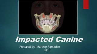
Impacted Canine
Prepared by :Marwan Ramadan
B.D.S
Impacted Canines
incidence :
 Impacted maxillary canines are quite common, and approximately 12%–15% of the population
present with impacted canines.
 They are localized palatally more often than labially.
 Twice common in female as it is in male
 Of all patient with impacted canine 8% have bilateral impaction
 the impacted canine presents five basic localizations (contralateral or ipsilateral and deep in the
bone) as follows:
1. Palatal localization
2. Palatal localization of crown and labial localization of root
3. Labial localization of crown and palatal localization of root
4. Labial localization
5. Ectopic positions
Etiology of impacted canine
 Several etiologic factors for canine impactions have been proposed: localized, systemic,
or genetic
Classification of maxillary Impacted Canines
According to axial inclination
Class I: Impacted canine located in the palate
 Horizontal
 Vertical
 Semi-Vertical
Class II: Impacted canine located in the buccal side
 Horizontal
 Vertical
 Semi-Vertical
Class III: Impacted canines located in both palatal as well as buccal alveolar bone.
Class IV: Impacted canines located vertically between incisors and premolars
Class V: Impacted canines located in edentulous maxilla
Class VI : when canine is placed in abnormal position, antral wall, infraorbital region
Classification of maxillary Impacted Canines
 According to According to Field and Ackerman(1935)
a. Labial position
(1) crown with intimate relationship with incisors
(2) crown well above apices of incisors
b. Palatal position
(1) Crown near surface in close relationship to root of incisors
(2) Crown deeply embedded in close relationship to apices of incisors
c. Intermediate position
(1) Crown between lateral incisor and first premolar root
(2) Crown above of these teeth with crown labially placed and root palatally placed,or vice versa
d. Unusual position
(1) In nasal or antral wall
(2) In infraorbital region
Classification of mandibular Impacted Canines
 CLASSIFICATION According to Field and Ackerman(1935)
a. Labial position
(1) Vertical
(2) Oblique
(3) horizontal
b. Unusual position
(1) At inferior border
(2) In mental protuberance
(3) Migrated to opposite side
Classification of mandibular Impacted Canines
Periapical radiograph
showing transmigrated and
erupted lower left canine in
the midline.
Classification of mandibular Impacted Canines
 CLASSIFICATION According to the depth
 Level A. The crown of the impacted canine tooth is at the cervical line of the adjacent teeth.
 Level B. The crown of the impacted canine tooth is between the cervical line and root apices of the
adjacent teeth.
 Level C. The crown of the impacted canines is beneath the root apices of the adjacent teeth
Sequelae of Canine Impaction
1. Labial or lingual malpositioning of the impacted tooth
2. Migration of the neighboring teeth and loss of arch length
3. Internal resorption
4. Dentigerous cyst formation
5. External root resorption of the impacted tooth, as well as the neighboring teeth,
6. Periodontal defects
7. Tooth transposition
8. Infection particularly with partial eruption
9. Referred pain and combinations of the above sequelae
Theories associated with palatally displaced
maxillary canines
 The guidance theory proposes that the canine erupts along the root
of the lateral incisor, which serves as a guide, and if the root of the
lateral incisor is absent or malformed, the canine will not erupt.
 The genetic theory includes other possibly associated dental
anomalies, such as missing, or small lateral incisors, also it may
associated with anomalies such as enamel hypoplasia,
infraocclusion of primary molars, and aplasia of second premolars.
Diagnosis and Localization:of Canine Impaction
 The diagnosis of canine impaction is based on both clinical and radiographic examinations
1. Delayed eruption of the permanent canine or prolonged retention of the deciduous
canine beyond 14–15 years of age
2. Absence of a normal labial canine bulge,
3. Presence of a palatal bulge in the canine region,
4. Delayed eruption, distal tipping, or migration (splaying) of the permanent lateral
incisor,
5. Loss of vitality and increased mobility of the permanent incisors
Clinical Evaluation
Diagnosis of Canine Impaction
Indicated in patients with unerupted and nonpalpable canine after the age of 11 years
Types
Intraoral
 Periapical and Occlusal
Extraoral
 OPG and lateral cephalogram
Digital imaging
 Ct scan and CBCT
 .
Radiographical Evaluation
Diagnosis of Canine Impaction
periapical film
 A single periapical film provides the clinician with a two-dimensional
representation of the dentition.
 It would relate the canine to the neighboring teeth both mesiodistally and
superoinferiorly
Diagnosis of Canine Impaction
 To evaluate the position of the
canine buccolingually
 Two periapical films are taken
of the same area, with the horizontal
angulation of the cone changed when
the second film is taken.
 If the object in question moves in the same direction as the cone, it is lingually positioned.
 If the object moves in the opposite direction, it is situated closer to the source of radiation and is
therefore buccally located
Tube-shift technique or Clark's rule or (SLOB) rule
Diagnosis of Canine Impaction
 The buccal object rule uses two radiographs taken with different vertical angulations of the
x-ray beam.
 An object located on the buccal side moves inferiorly with the beam
directed inferiorly,
 whereas an object located in a lingual or palatal position moves superiorly
Buccal object rule:
Diagnosis of Canine Impaction
Occlusal films
 help to determine the buccolingual position of the impacted canine in conjunction with
the periapical films, provided that the image of the impacted canine is not
superimposed on the other teeth
Radiographical Evaluation
Diagnosis of Canine Impaction
Extraoral films
1. lateral cephalograms
These can sometimes aid in the
determination of the position of
the impacted canine, particularly
its relationship to other facial
structures (e.g., the maxillary
sinus and the floor of the nose).
Radiographical Evaluation
A view of the anterior
section of the lateral skull
radiograph confirms the
palatal location of the fully
superimposed
impacted canines (arrow)
Diagnosis of Canine Impaction
Extraoral films
2. Panoramic films
 These are also used to localize impacted
teeth in all three planes of space
 as much the same as with two periapical films
in the tube-shift method, with the understanding
that the source of radiation comes from behind the
patient; thus, the movements are reversed for position.
 image size distortion; object further away from the
image receptor (film) will be depiceted more magnified
than objects closer to the film
Radiographical Evaluation
right maxillary canine impaction. The
image of the canine superimposes on
the middle of the root of the lateral
incisor
Diagnosis of Canine Impaction
Extraoral films
3. CT/CBCT
 Cone beam computed tomography (CBCT) can identify and locate the position of
impacted canines accurately.
 By using this imaging technique, dentists also can assess any damage to the roots
of adjacent teeth and the amount of bone surrounding each tooth.
 increased cost, time, radiation exposure, and medicolegal issues associated with
using CBCT limit its routine use
Radiographical Evaluation
Diagnosis of Canine Impaction
Extraoral films
Radiographical Evaluation
CT scan image showing impacted upper right canine
in the right maxillary sinus .
Ct scan Axial
view
Ct scan
coronal view
Diagnosis of Canine Impaction
Extraoral films
3. CBCT
Radiographical Evaluation
was designed to overcome some of the limitations of
conventional CT devices (eg high radiation dose to
produce the multiple images which are stacked to
produce a complete image
Three dimensional reconstructed image (CBCT) of
impacted maxillary canines
factors affecting treatment options
 Patient age
 General dental health and oral hygiene
 Whether space is available within the arch or if it can be established with
orthodontic manipulation
 Suitability of the first bicuspid to replace the canine
 Favorability of the position of the impacted canine and the likelihood of being able
to get it into position with orthodontic treatment
 Patient motivation for orthodontic treatment
 Medical contraindications for surgery
Treatment options
1. Interceptive
2. Surgical exposure with or without orthodontic traction
 Open surgical exposure with spontaneous eruption
 Open surgical exposure with packing only
 Open surgical exposure with packing and subsequent bonding of an auxillary.
 Closed surgical exposure and bonding of an attachment intraoperatively.(closed eruption
technique)
 Open surgical exposure and bonding of an attachment and repositioning of the covering
tissue (open eruption technique)
3. Surgical repositioning and alignment
4. Surgical Autotransplantation
5. Surgical removal
6.No treatment
1. Interceptive Treatment
 In Class I non crowded situations where the permanent maxillary canine is
impacted or erupting buccally or palatally, it may be the preventive treatment of
choice in patient 10-13 years old.
 However, intercept early if
 There is any root resorption
 3 not palpable in its normal position and radiographic examination confirms palatal ectopia
 When canines are impacted buccally, retained primary canine should be
extracted.
 However, interception does not guarantee correction or elimination of the
problem.
 If NO radiographic evidence of improvement seen one year after treatment,
then surgical exposure and orthodontic eruption, is indicated
2. Surgical Exposure with orthodontic traction
CONSIDERATION
 Patient must be willing to wear orthodontic appliance
 Patient must be motivated to maintain good OH
 Interceptive measures not suitable
 Position of malposition not too great to preclude treatment.
 The long axis of the 3 should not be too horizontal or oblique
 The optimal time for alignment is during adolescence
 3 must not be ankylosed
 The root of 3 should not be dilacerated
METHODS
A. surgical exposure with spontaneous eruption
B. surgical exposure with packing only
C. surgical exposure with packing and delayed bonding of an auxillary.
D. Open eruption technique .
E. Closed eruption technique
 For surgical exposure 3 surgical approach can be used
 1. Replacement flap technique
 2. Excisional exposure
 3. Apically position flap
 The goal is to choose a technique that exposes the canine within the a zone of keratinized
mucosa without involvement of the cementoenamel junction
A. surgical exposure with spontaneous eruption
 Usually has only soft tissue covering
 The technique involve excision of the gingivae over the canine with little exposure of the crown
and bone removal.
 This should be enough to allow eruption of the canine
unerupted maxillary left canine
The tooth was exposed and the
flap, which consisted of attached
gingiva, was apically
repositioned
At nine months post-surgery,
the tooth has erupted normally.
B. surgical exposure with packing
 The purpose of pack application is to prevent the gingival tissue from re-healing after exposure
 Indicated in deep impaction cases when spontaneous eruption is suspected
C. surgical exposure with packing and delayed bonding
of an auxillary
 Indicated in deep impaction cases when spontaneous eruption is failed
 of The packing is remove after a week postop and an attachment bonded with subsequent
traction using a fixed appliance.
 Adv
 Re-bonding can be done under direct vision
 Direct visualisation during movement
D. Open eruption technique
 In the “open eruption” technique, the crown of the impacted tooth is exposed with either an
opening cut into the overlying tissue without flap reflection, or a flap is reflected, a window
cut in it, and then the flap is repositioned
 simplest, most conservative and most direct manner to expose a tooth which is palpable
immediately under the oral mucosa
 attachment may then be bonded to the tooth and orthodontically encouraged eruption may
proceed without delay, to complete its alignment within a very short time.
A high buccal canine exposed
by circular incision of the
sulcus mucosa
Following alignment, the oral
mucosa is attached directly to
the gingiva
E. Closed eruption technique
 The alternative approach to surgical exposure, the closed eruption technique, has an attachment
placed at the time of the exposure and the tissues fully replaced and sutured to their former place,
to re-cover the impacted tooth
high labial canine has been exposed
with a full flap exposure, which
included the gingival margin of the
extracted deciduous canine
An attachment is bonded to the palatal aspect of the permanent
canine and its pigtail ligature is directed through the socket
vacated by the extracted deciduous tooth
The flap is sutured to its former place and vertical
traction will draw the tooth down, maintaining
alveolar bone on its labial side.
(a) The crown of an impacted canine is exposed using a wide flap, but with removal of minimal bone.
The unexposed crown lies between the roots of the central incisors, having traversed the midline
suture. (b) An attachment is bonded, while haemostasis is maintained by the surgeon. (c) The flap has
been divided to accommodate the ligature pigtail in its desired position, before being fully replaced and
sutured. (d) The labial spring auxiliary loop, seen in its passive position in (c), has been turned inwards
towards the palate and secured to the stainless steel ligature pigtail
Closed eruption
technique in palatally
impacted maxillary
canine
Surgical repositioning and alignment
 It suitable for tooth which are only mildly displaced.
 It involves de- rotation of the impacted canine within its socket
 It defer from transplantation because effort is made to avoid removal of the tooth from it socket
 The greater the displacement the poorer the prognosis as frequently the neurovascular bundle
are broken
Autotransplantation
 If patient is unwilling to wear orthodontic Appliance
 If the degree of malpositioning is too great for orthodontic alignment.
 Optimal time is when the root is about 50-75% formed
 Interceptive measure not appropriate or had failed
 Fixation at d recipient site is done using a preformed or vacuum splint which
covers the entire upper arch
 Splint is removed after 3-6wks and this is followed by bonded sectional fixed
appliance
Preoperative
Extraction of
the tooth from
impacted state
Intraoperative
after preparing
of the socket
Postoperative OBG showing alignment of
the tooth and splinting
Surgical removal
Indications
 Poor patient cooperation
 Patient decline treatment or is pleased with the appearance
 The tooth is lying in an unfavorable position (in horizontal position) and there is insufficient space
in the arch
 Presence of a pathology.
 Satisfactory occlusion and prognosis for treatment is poor.
 Evidence of early resorption of adjacent tooth.
 Good contact btw the 2 and 4,so as to substitute 4 for 3.
Methods of Surgical removal
Maxillary canine
 Extraction using labial approach
 Extraction using palatal approach
 Extraction of labially located canine with partial bone impaction
 Extraction of intra-alveolar impaction
 Ectopic Impacted Canine
Mandibular canine
 Extraction of mandibular canine with labial position
Flap design
 Buccal located canine: If the impacted canine is located buccally, we have three options depending on the
level of the tooth impaction.
1- Gingival crest incision can be made in the gingival sulcus.
2- If the impacted canine is high, the incision can be made horizontally above the papillae (flap with release
incision).
3- Vestibular incisions made at the level of the mucogingival junction should be made only when the impacted
canine is above the root apices.
 Palatal positioned canine: require long
incision to avoid damage to the
neurovascular bundles
(nasopalatine bundles &
the greater palatine bundle)
 Transversely located (in the middle of the ridge) we have to make 2 flaps one labially and the other one
palataly
Extraction of maxillary canine Using Labial Approach
OPG Impacted canine in right
and left side
Canine bulge on labial side trapezoidal flap
Reflection of the flap Bone removing Tooth sectioning
Extraction of maxillary canine Using Labial Approach
Removal of follicle
Bone smoothing
Removing of the crown Removing of the root
Surgical site after suturing
Extraction of maxillary canine Using palatal Approach
Bone removing Tooth sectioning
OPG Impacted canine in right
side
Palatal flap extend from
5 to 5
Reflection of the flap
Extraction of maxillary canine Using palatal Approach
Removing of crown Removing of root Re-approximation of
the flap
Surgical site after suturing
Extraction of Impacted Canine with Partial Bone Impaction
impacted maxillary canine
with a labial localization The ischemic protuberance
Trapezoidal incision
Extraction of Impacted Canine with Partial Bone Impaction
Creation of a groove between
the crown and bone, allowing
for positioning of the elevator
Removal of the tooth with elevator Bone smoothing
suturing
Extraction of Ectopic Impacted Canine
• The presence of ectopic impacted teeth is relatively
rare.
• Ectopic teeth are usually localized in the following
places:
1. underneath permanent teeth,
2. near the angle of the mandible,
3. inside the ramus,
4. near the mandibular notch,
5. the coronoid process,
6. the maxillary tuberosity,
7. the wall of the maxillary sinus,
8. the nasal cavity and,
9. rarely, near the orbit
Radiograph showing impacted canine with a labial
localization, which is in contact with the anterior wall
of the maxillary sinus
Extraction of Ectopic Impacted Canine
Incision in canine fossa region,
for sinus trephination using the
Caldwell–Luc approach
Reflection of the flap and
exposure of the anterior
wall of maxillary sinus Holes drilled through the bone
surface defining the border of bone
to be removed
Connecting holes to remove the
bone covering the impacted
tooth
Extraction of Ectopic Impacted Canine
Surgical field after suturing
a, Exposure and luxation of the impacted tooth using
straight elevator. b Removal of tooth using a hemostat
Extraction of mandibular canine: labially
positioned
Trapezoidal incision extending from the left lateral
incisor as far as the first premolar of the opposite side
of the mandible
Reflection of the flap
Extraction of mandibular canine: labially
positioned
Exposure of the crown of the impacted
tooth using a surgical bur
Luxation using the blade of the
elevator alternately on the
mesial and distal aspects of
crown of tooth
Surgical field after removal of
the impacted tooth and
odontomas
Extraction of mandibular canine: labially
positioned
Suturing
POSTOPERATIVE TREATMENT
 After Debridement and closure of incision the wound heals uneventfully
1. A post op radiograph is desirable
2. Cold application to face prevents disfiguring swelling and edema
3. Antibiotics are generally necessary especially if there is preexisting infection or the
antrum or nasal cavity has been opened.
4. Medication should be prescribed for pain

More Related Content

What's hot

deep bite management
deep bite managementdeep bite management
deep bite management
VilayatAli5
 
Class III Malocclusion
Class III MalocclusionClass III Malocclusion
Class III Malocclusion
Priyanka Vadhera
 
orthodontic management of impacted canine.
orthodontic management of impacted canine.orthodontic management of impacted canine.
orthodontic management of impacted canine.
Muhammad Shafad
 
Activator
ActivatorActivator
Activator
Ahmed Baattiah
 
Orthodontic Diagnosis
Orthodontic DiagnosisOrthodontic Diagnosis
Orthodontic Diagnosis
Muhammad Shafad
 
Class II malocclusion
Class II malocclusionClass II malocclusion
Class II malocclusion
Cing Sian Dal
 
Surgical orthodontics
Surgical orthodonticsSurgical orthodontics
Surgical orthodontics
Indian dental academy
 
Retention and Relapse in orthodontics
Retention and Relapse in orthodonticsRetention and Relapse in orthodontics
Retention and Relapse in orthodonticsEkta Chaudhary
 
Steiner analysis
Steiner analysisSteiner analysis
Steiner analysis
Nabil Rahman
 
Functional appliances
Functional appliancesFunctional appliances
Functional appliances
Dr Shahzad Hussain
 
Mangement of openbite in orthodontics
Mangement of openbite in orthodonticsMangement of openbite in orthodontics
Mangement of openbite in orthodontics
Ravikanth lakkakula
 
Mixed dentition analysis
Mixed dentition analysisMixed dentition analysis
Mixed dentition analysisRajesh Bariker
 
Canine impaction
Canine impactionCanine impaction
Canine impaction
Shashank Trivedi
 
Cephalometrics in orthodontics
Cephalometrics in orthodonticsCephalometrics in orthodontics
Cephalometrics in orthodontics
Dinesh Raj
 
Management of impacted canines
Management of impacted caninesManagement of impacted canines
Management of impacted canines
Miliya Parveen
 
Twin block
Twin block Twin block
Twin block
Dr Shilpa Dineshan
 
Cleft lip & palate management in orthodontics
Cleft lip & palate management in orthodonticsCleft lip & palate management in orthodontics
Cleft lip & palate management in orthodontics
Indian dental academy
 
Class ii malocclusion
Class ii malocclusionClass ii malocclusion
Class ii malocclusion
Abhidnya Madansure
 

What's hot (20)

deep bite management
deep bite managementdeep bite management
deep bite management
 
Class III Malocclusion
Class III MalocclusionClass III Malocclusion
Class III Malocclusion
 
orthodontic management of impacted canine.
orthodontic management of impacted canine.orthodontic management of impacted canine.
orthodontic management of impacted canine.
 
Orthognathic surgery
Orthognathic surgeryOrthognathic surgery
Orthognathic surgery
 
Activator
ActivatorActivator
Activator
 
Orthodontic Diagnosis
Orthodontic DiagnosisOrthodontic Diagnosis
Orthodontic Diagnosis
 
Class II malocclusion
Class II malocclusionClass II malocclusion
Class II malocclusion
 
Surgical orthodontics
Surgical orthodonticsSurgical orthodontics
Surgical orthodontics
 
Retention and Relapse in orthodontics
Retention and Relapse in orthodonticsRetention and Relapse in orthodontics
Retention and Relapse in orthodontics
 
Steiner analysis
Steiner analysisSteiner analysis
Steiner analysis
 
Functional appliances
Functional appliancesFunctional appliances
Functional appliances
 
Mangement of openbite in orthodontics
Mangement of openbite in orthodonticsMangement of openbite in orthodontics
Mangement of openbite in orthodontics
 
Mixed dentition analysis
Mixed dentition analysisMixed dentition analysis
Mixed dentition analysis
 
Canine impaction
Canine impactionCanine impaction
Canine impaction
 
Cephalometrics in orthodontics
Cephalometrics in orthodonticsCephalometrics in orthodontics
Cephalometrics in orthodontics
 
model-analysis
 model-analysis model-analysis
model-analysis
 
Management of impacted canines
Management of impacted caninesManagement of impacted canines
Management of impacted canines
 
Twin block
Twin block Twin block
Twin block
 
Cleft lip & palate management in orthodontics
Cleft lip & palate management in orthodonticsCleft lip & palate management in orthodontics
Cleft lip & palate management in orthodontics
 
Class ii malocclusion
Class ii malocclusionClass ii malocclusion
Class ii malocclusion
 

Similar to Impacted canine

CANINE IMPACTION 1.pptx
CANINE IMPACTION 1.pptxCANINE IMPACTION 1.pptx
CANINE IMPACTION 1.pptx
MalaM67
 
My lec 2
My lec 2My lec 2
impacted teeth
impacted teeth impacted teeth
Orthodontic management of impacted canines
Orthodontic management of impacted caninesOrthodontic management of impacted canines
Orthodontic management of impacted canines
Abdelrahman Mosaad
 
Canine impaction maxillary teeth part 2
Canine impaction   maxillary teeth part 2Canine impaction   maxillary teeth part 2
Canine impaction maxillary teeth part 2
VilvaKarthick
 
Orthodontic management of impactions
Orthodontic management of impactionsOrthodontic management of impactions
Orthodontic management of impactions
Abdelrahman Mosaad
 
Canine impaction
Canine impactionCanine impaction
Canine impaction
Jeff Zacharia
 
Management of impacted canine
Management of impacted canineManagement of impacted canine
Management of impacted canine
Indian dental academy
 
6 canine impaction .pptx
6 canine impaction .pptx6 canine impaction .pptx
6 canine impaction .pptx
DR DAVIS NADAKKAVUKARAN
 
Management of impacted canine (2)
Management of impacted canine (2)Management of impacted canine (2)
Management of impacted canine (2)
Indian dental academy
 
Maxillary canine and molar impaction
Maxillary canine and molar impactionMaxillary canine and molar impaction
Maxillary canine and molar impaction
KathirvelGopalakrish
 
localisation of impacted canine
localisation of impacted caninelocalisation of impacted canine
localisation of impacted canine
Royal medical services - JOS
 
craniofacial imaging-Recent advances
craniofacial imaging-Recent advances craniofacial imaging-Recent advances
craniofacial imaging-Recent advances
Tony Pious
 
Etiology of maxillary canine impaction
Etiology of maxillary canine impactionEtiology of maxillary canine impaction
Etiology of maxillary canine impaction
Dr. mahipal singh
 
Prevalence of impacted canines in Arab Population
Prevalence of impacted canines in Arab PopulationPrevalence of impacted canines in Arab Population
Prevalence of impacted canines in Arab Population
Abu-Hussein Muhamad
 
Maxillary canine impaction / oral surgery courses /certified fixed orthodon...
Maxillary canine impaction / oral surgery courses   /certified fixed orthodon...Maxillary canine impaction / oral surgery courses   /certified fixed orthodon...
Maxillary canine impaction / oral surgery courses /certified fixed orthodon...
Indian dental academy
 
Radiographic assessment in paediatric dentistry
Radiographic assessment in paediatric dentistryRadiographic assessment in paediatric dentistry
Radiographic assessment in paediatric dentistry
S. K.
 
Maxillary impacted canine management
Maxillary impacted canine managementMaxillary impacted canine management
Maxillary impacted canine management
Parag Deshmukh
 
Canine Impaction and Its Importance in Orthodontics
Canine Impaction and Its Importance in OrthodonticsCanine Impaction and Its Importance in Orthodontics
Canine Impaction and Its Importance in Orthodontics
Analhaq Shaikh
 

Similar to Impacted canine (20)

CANINE IMPACTION 1.pptx
CANINE IMPACTION 1.pptxCANINE IMPACTION 1.pptx
CANINE IMPACTION 1.pptx
 
My lec 2
My lec 2My lec 2
My lec 2
 
impacted teeth
impacted teeth impacted teeth
impacted teeth
 
Orthodontic management of impacted canines
Orthodontic management of impacted caninesOrthodontic management of impacted canines
Orthodontic management of impacted canines
 
Canine impaction maxillary teeth part 2
Canine impaction   maxillary teeth part 2Canine impaction   maxillary teeth part 2
Canine impaction maxillary teeth part 2
 
Orthodontic management of impactions
Orthodontic management of impactionsOrthodontic management of impactions
Orthodontic management of impactions
 
Canine impaction
Canine impactionCanine impaction
Canine impaction
 
Management of impacted canine
Management of impacted canineManagement of impacted canine
Management of impacted canine
 
6 canine impaction .pptx
6 canine impaction .pptx6 canine impaction .pptx
6 canine impaction .pptx
 
Management of impacted canine (2)
Management of impacted canine (2)Management of impacted canine (2)
Management of impacted canine (2)
 
Maxillary canine and molar impaction
Maxillary canine and molar impactionMaxillary canine and molar impaction
Maxillary canine and molar impaction
 
localisation of impacted canine
localisation of impacted caninelocalisation of impacted canine
localisation of impacted canine
 
craniofacial imaging-Recent advances
craniofacial imaging-Recent advances craniofacial imaging-Recent advances
craniofacial imaging-Recent advances
 
Etiology of maxillary canine impaction
Etiology of maxillary canine impactionEtiology of maxillary canine impaction
Etiology of maxillary canine impaction
 
Prevalence of impacted canines in Arab Population
Prevalence of impacted canines in Arab PopulationPrevalence of impacted canines in Arab Population
Prevalence of impacted canines in Arab Population
 
Maxillary canine impaction / oral surgery courses /certified fixed orthodon...
Maxillary canine impaction / oral surgery courses   /certified fixed orthodon...Maxillary canine impaction / oral surgery courses   /certified fixed orthodon...
Maxillary canine impaction / oral surgery courses /certified fixed orthodon...
 
Impacted teeth by DR luma
Impacted teeth by DR lumaImpacted teeth by DR luma
Impacted teeth by DR luma
 
Radiographic assessment in paediatric dentistry
Radiographic assessment in paediatric dentistryRadiographic assessment in paediatric dentistry
Radiographic assessment in paediatric dentistry
 
Maxillary impacted canine management
Maxillary impacted canine managementMaxillary impacted canine management
Maxillary impacted canine management
 
Canine Impaction and Its Importance in Orthodontics
Canine Impaction and Its Importance in OrthodonticsCanine Impaction and Its Importance in Orthodontics
Canine Impaction and Its Importance in Orthodontics
 

Recently uploaded

What Are Homeopathic Treatments for Migraines.pdf
What Are Homeopathic Treatments for Migraines.pdfWhat Are Homeopathic Treatments for Migraines.pdf
What Are Homeopathic Treatments for Migraines.pdf
Dharma Homoeopathy
 
GENERAL PHARMACOLOGY - INTRODUCTION DENTAL.ppt
GENERAL PHARMACOLOGY - INTRODUCTION DENTAL.pptGENERAL PHARMACOLOGY - INTRODUCTION DENTAL.ppt
GENERAL PHARMACOLOGY - INTRODUCTION DENTAL.ppt
Mangaiarkkarasi
 
Jaipur ❤cALL gIRLS 89O1183002 ❤ℂall Girls IN JaiPuR ESCORT SERVICE
Jaipur ❤cALL gIRLS 89O1183002 ❤ℂall Girls IN JaiPuR ESCORT SERVICEJaipur ❤cALL gIRLS 89O1183002 ❤ℂall Girls IN JaiPuR ESCORT SERVICE
Jaipur ❤cALL gIRLS 89O1183002 ❤ℂall Girls IN JaiPuR ESCORT SERVICE
ranishasharma67
 
Performance Standards for Antimicrobial Susceptibility Testing
Performance Standards for Antimicrobial Susceptibility TestingPerformance Standards for Antimicrobial Susceptibility Testing
Performance Standards for Antimicrobial Susceptibility Testing
Nguyễn Thị Vân Anh
 
QA Paediatric dentistry department, Hospital Melaka 2020
QA Paediatric dentistry department, Hospital Melaka 2020QA Paediatric dentistry department, Hospital Melaka 2020
QA Paediatric dentistry department, Hospital Melaka 2020
Azreen Aj
 
CHAPTER 1 SEMESTER V PREVENTIVE-PEDIATRICS.pdf
CHAPTER 1 SEMESTER V PREVENTIVE-PEDIATRICS.pdfCHAPTER 1 SEMESTER V PREVENTIVE-PEDIATRICS.pdf
CHAPTER 1 SEMESTER V PREVENTIVE-PEDIATRICS.pdf
Sachin Sharma
 
The Docs PPG - 30.05.2024.pptx..........
The Docs PPG - 30.05.2024.pptx..........The Docs PPG - 30.05.2024.pptx..........
The Docs PPG - 30.05.2024.pptx..........
TheDocs
 
Surgery-Mini-OSCE-All-Past-Years-Questions-Modified.
Surgery-Mini-OSCE-All-Past-Years-Questions-Modified.Surgery-Mini-OSCE-All-Past-Years-Questions-Modified.
Surgery-Mini-OSCE-All-Past-Years-Questions-Modified.
preciousstephanie75
 
Nursing Care of Client With Acute And Chronic Renal Failure.ppt
Nursing Care of Client With Acute And Chronic Renal Failure.pptNursing Care of Client With Acute And Chronic Renal Failure.ppt
Nursing Care of Client With Acute And Chronic Renal Failure.ppt
Rommel Luis III Israel
 
Contact Now 89011**83002 Dehradun ℂall Girls By Full Service ℂall Girl In De...
Contact Now  89011**83002 Dehradun ℂall Girls By Full Service ℂall Girl In De...Contact Now  89011**83002 Dehradun ℂall Girls By Full Service ℂall Girl In De...
Contact Now 89011**83002 Dehradun ℂall Girls By Full Service ℂall Girl In De...
aunty1x2
 
一比一原版纽约大学毕业证(NYU毕业证)成绩单留信认证
一比一原版纽约大学毕业证(NYU毕业证)成绩单留信认证一比一原版纽约大学毕业证(NYU毕业证)成绩单留信认证
一比一原版纽约大学毕业证(NYU毕业证)成绩单留信认证
o6ov5dqmf
 
GLOBAL WARMING BY PRIYA BHOJWANI @..pptx
GLOBAL WARMING BY PRIYA BHOJWANI @..pptxGLOBAL WARMING BY PRIYA BHOJWANI @..pptx
GLOBAL WARMING BY PRIYA BHOJWANI @..pptx
priyabhojwani1200
 
Roti bank chennai PPT [Autosaved].pptx1
Roti bank  chennai PPT [Autosaved].pptx1Roti bank  chennai PPT [Autosaved].pptx1
Roti bank chennai PPT [Autosaved].pptx1
roti bank
 
Dehradun ❤CALL Girls 8901183002 ❤ℂall Girls IN Dehradun ESCORT SERVICE❤
Dehradun ❤CALL Girls  8901183002 ❤ℂall  Girls IN Dehradun ESCORT SERVICE❤Dehradun ❤CALL Girls  8901183002 ❤ℂall  Girls IN Dehradun ESCORT SERVICE❤
Dehradun ❤CALL Girls 8901183002 ❤ℂall Girls IN Dehradun ESCORT SERVICE❤
aunty1x2
 
Antibiotic Stewardship by Anushri Srivastava.pptx
Antibiotic Stewardship by Anushri Srivastava.pptxAntibiotic Stewardship by Anushri Srivastava.pptx
Antibiotic Stewardship by Anushri Srivastava.pptx
AnushriSrivastav
 
How many patients does case series should have In comparison to case reports.pdf
How many patients does case series should have In comparison to case reports.pdfHow many patients does case series should have In comparison to case reports.pdf
How many patients does case series should have In comparison to case reports.pdf
pubrica101
 
HEAT WAVE presented by priya bhojwani..pptx
HEAT WAVE presented by priya bhojwani..pptxHEAT WAVE presented by priya bhojwani..pptx
HEAT WAVE presented by priya bhojwani..pptx
priyabhojwani1200
 
Introduction to Forensic Pathology course
Introduction to Forensic Pathology courseIntroduction to Forensic Pathology course
Introduction to Forensic Pathology course
fprxsqvnz5
 
Immunity to Veterinary parasitic infections power point presentation
Immunity to Veterinary parasitic infections power point presentationImmunity to Veterinary parasitic infections power point presentation
Immunity to Veterinary parasitic infections power point presentation
BeshedaWedajo
 
Haridwar ❤CALL Girls 🔝 89011★83002 🔝 ❤ℂall Girls IN Haridwar ESCORT SERVICE❤
Haridwar ❤CALL Girls 🔝 89011★83002 🔝 ❤ℂall Girls IN Haridwar ESCORT SERVICE❤Haridwar ❤CALL Girls 🔝 89011★83002 🔝 ❤ℂall Girls IN Haridwar ESCORT SERVICE❤
Haridwar ❤CALL Girls 🔝 89011★83002 🔝 ❤ℂall Girls IN Haridwar ESCORT SERVICE❤
ranishasharma67
 

Recently uploaded (20)

What Are Homeopathic Treatments for Migraines.pdf
What Are Homeopathic Treatments for Migraines.pdfWhat Are Homeopathic Treatments for Migraines.pdf
What Are Homeopathic Treatments for Migraines.pdf
 
GENERAL PHARMACOLOGY - INTRODUCTION DENTAL.ppt
GENERAL PHARMACOLOGY - INTRODUCTION DENTAL.pptGENERAL PHARMACOLOGY - INTRODUCTION DENTAL.ppt
GENERAL PHARMACOLOGY - INTRODUCTION DENTAL.ppt
 
Jaipur ❤cALL gIRLS 89O1183002 ❤ℂall Girls IN JaiPuR ESCORT SERVICE
Jaipur ❤cALL gIRLS 89O1183002 ❤ℂall Girls IN JaiPuR ESCORT SERVICEJaipur ❤cALL gIRLS 89O1183002 ❤ℂall Girls IN JaiPuR ESCORT SERVICE
Jaipur ❤cALL gIRLS 89O1183002 ❤ℂall Girls IN JaiPuR ESCORT SERVICE
 
Performance Standards for Antimicrobial Susceptibility Testing
Performance Standards for Antimicrobial Susceptibility TestingPerformance Standards for Antimicrobial Susceptibility Testing
Performance Standards for Antimicrobial Susceptibility Testing
 
QA Paediatric dentistry department, Hospital Melaka 2020
QA Paediatric dentistry department, Hospital Melaka 2020QA Paediatric dentistry department, Hospital Melaka 2020
QA Paediatric dentistry department, Hospital Melaka 2020
 
CHAPTER 1 SEMESTER V PREVENTIVE-PEDIATRICS.pdf
CHAPTER 1 SEMESTER V PREVENTIVE-PEDIATRICS.pdfCHAPTER 1 SEMESTER V PREVENTIVE-PEDIATRICS.pdf
CHAPTER 1 SEMESTER V PREVENTIVE-PEDIATRICS.pdf
 
The Docs PPG - 30.05.2024.pptx..........
The Docs PPG - 30.05.2024.pptx..........The Docs PPG - 30.05.2024.pptx..........
The Docs PPG - 30.05.2024.pptx..........
 
Surgery-Mini-OSCE-All-Past-Years-Questions-Modified.
Surgery-Mini-OSCE-All-Past-Years-Questions-Modified.Surgery-Mini-OSCE-All-Past-Years-Questions-Modified.
Surgery-Mini-OSCE-All-Past-Years-Questions-Modified.
 
Nursing Care of Client With Acute And Chronic Renal Failure.ppt
Nursing Care of Client With Acute And Chronic Renal Failure.pptNursing Care of Client With Acute And Chronic Renal Failure.ppt
Nursing Care of Client With Acute And Chronic Renal Failure.ppt
 
Contact Now 89011**83002 Dehradun ℂall Girls By Full Service ℂall Girl In De...
Contact Now  89011**83002 Dehradun ℂall Girls By Full Service ℂall Girl In De...Contact Now  89011**83002 Dehradun ℂall Girls By Full Service ℂall Girl In De...
Contact Now 89011**83002 Dehradun ℂall Girls By Full Service ℂall Girl In De...
 
一比一原版纽约大学毕业证(NYU毕业证)成绩单留信认证
一比一原版纽约大学毕业证(NYU毕业证)成绩单留信认证一比一原版纽约大学毕业证(NYU毕业证)成绩单留信认证
一比一原版纽约大学毕业证(NYU毕业证)成绩单留信认证
 
GLOBAL WARMING BY PRIYA BHOJWANI @..pptx
GLOBAL WARMING BY PRIYA BHOJWANI @..pptxGLOBAL WARMING BY PRIYA BHOJWANI @..pptx
GLOBAL WARMING BY PRIYA BHOJWANI @..pptx
 
Roti bank chennai PPT [Autosaved].pptx1
Roti bank  chennai PPT [Autosaved].pptx1Roti bank  chennai PPT [Autosaved].pptx1
Roti bank chennai PPT [Autosaved].pptx1
 
Dehradun ❤CALL Girls 8901183002 ❤ℂall Girls IN Dehradun ESCORT SERVICE❤
Dehradun ❤CALL Girls  8901183002 ❤ℂall  Girls IN Dehradun ESCORT SERVICE❤Dehradun ❤CALL Girls  8901183002 ❤ℂall  Girls IN Dehradun ESCORT SERVICE❤
Dehradun ❤CALL Girls 8901183002 ❤ℂall Girls IN Dehradun ESCORT SERVICE❤
 
Antibiotic Stewardship by Anushri Srivastava.pptx
Antibiotic Stewardship by Anushri Srivastava.pptxAntibiotic Stewardship by Anushri Srivastava.pptx
Antibiotic Stewardship by Anushri Srivastava.pptx
 
How many patients does case series should have In comparison to case reports.pdf
How many patients does case series should have In comparison to case reports.pdfHow many patients does case series should have In comparison to case reports.pdf
How many patients does case series should have In comparison to case reports.pdf
 
HEAT WAVE presented by priya bhojwani..pptx
HEAT WAVE presented by priya bhojwani..pptxHEAT WAVE presented by priya bhojwani..pptx
HEAT WAVE presented by priya bhojwani..pptx
 
Introduction to Forensic Pathology course
Introduction to Forensic Pathology courseIntroduction to Forensic Pathology course
Introduction to Forensic Pathology course
 
Immunity to Veterinary parasitic infections power point presentation
Immunity to Veterinary parasitic infections power point presentationImmunity to Veterinary parasitic infections power point presentation
Immunity to Veterinary parasitic infections power point presentation
 
Haridwar ❤CALL Girls 🔝 89011★83002 🔝 ❤ℂall Girls IN Haridwar ESCORT SERVICE❤
Haridwar ❤CALL Girls 🔝 89011★83002 🔝 ❤ℂall Girls IN Haridwar ESCORT SERVICE❤Haridwar ❤CALL Girls 🔝 89011★83002 🔝 ❤ℂall Girls IN Haridwar ESCORT SERVICE❤
Haridwar ❤CALL Girls 🔝 89011★83002 🔝 ❤ℂall Girls IN Haridwar ESCORT SERVICE❤
 

Impacted canine

  • 1. Impacted Canine Prepared by :Marwan Ramadan B.D.S
  • 2. Impacted Canines incidence :  Impacted maxillary canines are quite common, and approximately 12%–15% of the population present with impacted canines.  They are localized palatally more often than labially.  Twice common in female as it is in male  Of all patient with impacted canine 8% have bilateral impaction  the impacted canine presents five basic localizations (contralateral or ipsilateral and deep in the bone) as follows: 1. Palatal localization 2. Palatal localization of crown and labial localization of root 3. Labial localization of crown and palatal localization of root 4. Labial localization 5. Ectopic positions
  • 3. Etiology of impacted canine  Several etiologic factors for canine impactions have been proposed: localized, systemic, or genetic
  • 4. Classification of maxillary Impacted Canines According to axial inclination Class I: Impacted canine located in the palate  Horizontal  Vertical  Semi-Vertical Class II: Impacted canine located in the buccal side  Horizontal  Vertical  Semi-Vertical Class III: Impacted canines located in both palatal as well as buccal alveolar bone. Class IV: Impacted canines located vertically between incisors and premolars Class V: Impacted canines located in edentulous maxilla Class VI : when canine is placed in abnormal position, antral wall, infraorbital region
  • 5. Classification of maxillary Impacted Canines  According to According to Field and Ackerman(1935) a. Labial position (1) crown with intimate relationship with incisors (2) crown well above apices of incisors b. Palatal position (1) Crown near surface in close relationship to root of incisors (2) Crown deeply embedded in close relationship to apices of incisors c. Intermediate position (1) Crown between lateral incisor and first premolar root (2) Crown above of these teeth with crown labially placed and root palatally placed,or vice versa d. Unusual position (1) In nasal or antral wall (2) In infraorbital region
  • 6. Classification of mandibular Impacted Canines  CLASSIFICATION According to Field and Ackerman(1935) a. Labial position (1) Vertical (2) Oblique (3) horizontal b. Unusual position (1) At inferior border (2) In mental protuberance (3) Migrated to opposite side
  • 7. Classification of mandibular Impacted Canines Periapical radiograph showing transmigrated and erupted lower left canine in the midline.
  • 8. Classification of mandibular Impacted Canines  CLASSIFICATION According to the depth  Level A. The crown of the impacted canine tooth is at the cervical line of the adjacent teeth.  Level B. The crown of the impacted canine tooth is between the cervical line and root apices of the adjacent teeth.  Level C. The crown of the impacted canines is beneath the root apices of the adjacent teeth
  • 9. Sequelae of Canine Impaction 1. Labial or lingual malpositioning of the impacted tooth 2. Migration of the neighboring teeth and loss of arch length 3. Internal resorption 4. Dentigerous cyst formation 5. External root resorption of the impacted tooth, as well as the neighboring teeth, 6. Periodontal defects 7. Tooth transposition 8. Infection particularly with partial eruption 9. Referred pain and combinations of the above sequelae
  • 10. Theories associated with palatally displaced maxillary canines  The guidance theory proposes that the canine erupts along the root of the lateral incisor, which serves as a guide, and if the root of the lateral incisor is absent or malformed, the canine will not erupt.  The genetic theory includes other possibly associated dental anomalies, such as missing, or small lateral incisors, also it may associated with anomalies such as enamel hypoplasia, infraocclusion of primary molars, and aplasia of second premolars.
  • 11. Diagnosis and Localization:of Canine Impaction  The diagnosis of canine impaction is based on both clinical and radiographic examinations 1. Delayed eruption of the permanent canine or prolonged retention of the deciduous canine beyond 14–15 years of age 2. Absence of a normal labial canine bulge, 3. Presence of a palatal bulge in the canine region, 4. Delayed eruption, distal tipping, or migration (splaying) of the permanent lateral incisor, 5. Loss of vitality and increased mobility of the permanent incisors Clinical Evaluation
  • 12.
  • 13. Diagnosis of Canine Impaction Indicated in patients with unerupted and nonpalpable canine after the age of 11 years Types Intraoral  Periapical and Occlusal Extraoral  OPG and lateral cephalogram Digital imaging  Ct scan and CBCT  . Radiographical Evaluation
  • 14. Diagnosis of Canine Impaction periapical film  A single periapical film provides the clinician with a two-dimensional representation of the dentition.  It would relate the canine to the neighboring teeth both mesiodistally and superoinferiorly
  • 15. Diagnosis of Canine Impaction  To evaluate the position of the canine buccolingually  Two periapical films are taken of the same area, with the horizontal angulation of the cone changed when the second film is taken.  If the object in question moves in the same direction as the cone, it is lingually positioned.  If the object moves in the opposite direction, it is situated closer to the source of radiation and is therefore buccally located Tube-shift technique or Clark's rule or (SLOB) rule
  • 16. Diagnosis of Canine Impaction  The buccal object rule uses two radiographs taken with different vertical angulations of the x-ray beam.  An object located on the buccal side moves inferiorly with the beam directed inferiorly,  whereas an object located in a lingual or palatal position moves superiorly Buccal object rule:
  • 17. Diagnosis of Canine Impaction Occlusal films  help to determine the buccolingual position of the impacted canine in conjunction with the periapical films, provided that the image of the impacted canine is not superimposed on the other teeth Radiographical Evaluation
  • 18. Diagnosis of Canine Impaction Extraoral films 1. lateral cephalograms These can sometimes aid in the determination of the position of the impacted canine, particularly its relationship to other facial structures (e.g., the maxillary sinus and the floor of the nose). Radiographical Evaluation A view of the anterior section of the lateral skull radiograph confirms the palatal location of the fully superimposed impacted canines (arrow)
  • 19. Diagnosis of Canine Impaction Extraoral films 2. Panoramic films  These are also used to localize impacted teeth in all three planes of space  as much the same as with two periapical films in the tube-shift method, with the understanding that the source of radiation comes from behind the patient; thus, the movements are reversed for position.  image size distortion; object further away from the image receptor (film) will be depiceted more magnified than objects closer to the film Radiographical Evaluation right maxillary canine impaction. The image of the canine superimposes on the middle of the root of the lateral incisor
  • 20. Diagnosis of Canine Impaction Extraoral films 3. CT/CBCT  Cone beam computed tomography (CBCT) can identify and locate the position of impacted canines accurately.  By using this imaging technique, dentists also can assess any damage to the roots of adjacent teeth and the amount of bone surrounding each tooth.  increased cost, time, radiation exposure, and medicolegal issues associated with using CBCT limit its routine use Radiographical Evaluation
  • 21. Diagnosis of Canine Impaction Extraoral films Radiographical Evaluation CT scan image showing impacted upper right canine in the right maxillary sinus . Ct scan Axial view Ct scan coronal view
  • 22. Diagnosis of Canine Impaction Extraoral films 3. CBCT Radiographical Evaluation was designed to overcome some of the limitations of conventional CT devices (eg high radiation dose to produce the multiple images which are stacked to produce a complete image Three dimensional reconstructed image (CBCT) of impacted maxillary canines
  • 23. factors affecting treatment options  Patient age  General dental health and oral hygiene  Whether space is available within the arch or if it can be established with orthodontic manipulation  Suitability of the first bicuspid to replace the canine  Favorability of the position of the impacted canine and the likelihood of being able to get it into position with orthodontic treatment  Patient motivation for orthodontic treatment  Medical contraindications for surgery
  • 24. Treatment options 1. Interceptive 2. Surgical exposure with or without orthodontic traction  Open surgical exposure with spontaneous eruption  Open surgical exposure with packing only  Open surgical exposure with packing and subsequent bonding of an auxillary.  Closed surgical exposure and bonding of an attachment intraoperatively.(closed eruption technique)  Open surgical exposure and bonding of an attachment and repositioning of the covering tissue (open eruption technique) 3. Surgical repositioning and alignment 4. Surgical Autotransplantation 5. Surgical removal 6.No treatment
  • 25. 1. Interceptive Treatment  In Class I non crowded situations where the permanent maxillary canine is impacted or erupting buccally or palatally, it may be the preventive treatment of choice in patient 10-13 years old.  However, intercept early if  There is any root resorption  3 not palpable in its normal position and radiographic examination confirms palatal ectopia  When canines are impacted buccally, retained primary canine should be extracted.  However, interception does not guarantee correction or elimination of the problem.  If NO radiographic evidence of improvement seen one year after treatment, then surgical exposure and orthodontic eruption, is indicated
  • 26. 2. Surgical Exposure with orthodontic traction CONSIDERATION  Patient must be willing to wear orthodontic appliance  Patient must be motivated to maintain good OH  Interceptive measures not suitable  Position of malposition not too great to preclude treatment.  The long axis of the 3 should not be too horizontal or oblique  The optimal time for alignment is during adolescence  3 must not be ankylosed  The root of 3 should not be dilacerated
  • 27. METHODS A. surgical exposure with spontaneous eruption B. surgical exposure with packing only C. surgical exposure with packing and delayed bonding of an auxillary. D. Open eruption technique . E. Closed eruption technique  For surgical exposure 3 surgical approach can be used  1. Replacement flap technique  2. Excisional exposure  3. Apically position flap  The goal is to choose a technique that exposes the canine within the a zone of keratinized mucosa without involvement of the cementoenamel junction
  • 28. A. surgical exposure with spontaneous eruption  Usually has only soft tissue covering  The technique involve excision of the gingivae over the canine with little exposure of the crown and bone removal.  This should be enough to allow eruption of the canine unerupted maxillary left canine The tooth was exposed and the flap, which consisted of attached gingiva, was apically repositioned At nine months post-surgery, the tooth has erupted normally.
  • 29. B. surgical exposure with packing  The purpose of pack application is to prevent the gingival tissue from re-healing after exposure  Indicated in deep impaction cases when spontaneous eruption is suspected
  • 30. C. surgical exposure with packing and delayed bonding of an auxillary  Indicated in deep impaction cases when spontaneous eruption is failed  of The packing is remove after a week postop and an attachment bonded with subsequent traction using a fixed appliance.  Adv  Re-bonding can be done under direct vision  Direct visualisation during movement
  • 31.
  • 32. D. Open eruption technique  In the “open eruption” technique, the crown of the impacted tooth is exposed with either an opening cut into the overlying tissue without flap reflection, or a flap is reflected, a window cut in it, and then the flap is repositioned  simplest, most conservative and most direct manner to expose a tooth which is palpable immediately under the oral mucosa  attachment may then be bonded to the tooth and orthodontically encouraged eruption may proceed without delay, to complete its alignment within a very short time. A high buccal canine exposed by circular incision of the sulcus mucosa Following alignment, the oral mucosa is attached directly to the gingiva
  • 33. E. Closed eruption technique  The alternative approach to surgical exposure, the closed eruption technique, has an attachment placed at the time of the exposure and the tissues fully replaced and sutured to their former place, to re-cover the impacted tooth high labial canine has been exposed with a full flap exposure, which included the gingival margin of the extracted deciduous canine An attachment is bonded to the palatal aspect of the permanent canine and its pigtail ligature is directed through the socket vacated by the extracted deciduous tooth The flap is sutured to its former place and vertical traction will draw the tooth down, maintaining alveolar bone on its labial side.
  • 34. (a) The crown of an impacted canine is exposed using a wide flap, but with removal of minimal bone. The unexposed crown lies between the roots of the central incisors, having traversed the midline suture. (b) An attachment is bonded, while haemostasis is maintained by the surgeon. (c) The flap has been divided to accommodate the ligature pigtail in its desired position, before being fully replaced and sutured. (d) The labial spring auxiliary loop, seen in its passive position in (c), has been turned inwards towards the palate and secured to the stainless steel ligature pigtail Closed eruption technique in palatally impacted maxillary canine
  • 35. Surgical repositioning and alignment  It suitable for tooth which are only mildly displaced.  It involves de- rotation of the impacted canine within its socket  It defer from transplantation because effort is made to avoid removal of the tooth from it socket  The greater the displacement the poorer the prognosis as frequently the neurovascular bundle are broken
  • 36. Autotransplantation  If patient is unwilling to wear orthodontic Appliance  If the degree of malpositioning is too great for orthodontic alignment.  Optimal time is when the root is about 50-75% formed  Interceptive measure not appropriate or had failed  Fixation at d recipient site is done using a preformed or vacuum splint which covers the entire upper arch  Splint is removed after 3-6wks and this is followed by bonded sectional fixed appliance
  • 37. Preoperative Extraction of the tooth from impacted state Intraoperative after preparing of the socket Postoperative OBG showing alignment of the tooth and splinting
  • 38. Surgical removal Indications  Poor patient cooperation  Patient decline treatment or is pleased with the appearance  The tooth is lying in an unfavorable position (in horizontal position) and there is insufficient space in the arch  Presence of a pathology.  Satisfactory occlusion and prognosis for treatment is poor.  Evidence of early resorption of adjacent tooth.  Good contact btw the 2 and 4,so as to substitute 4 for 3.
  • 39. Methods of Surgical removal Maxillary canine  Extraction using labial approach  Extraction using palatal approach  Extraction of labially located canine with partial bone impaction  Extraction of intra-alveolar impaction  Ectopic Impacted Canine Mandibular canine  Extraction of mandibular canine with labial position
  • 40. Flap design  Buccal located canine: If the impacted canine is located buccally, we have three options depending on the level of the tooth impaction. 1- Gingival crest incision can be made in the gingival sulcus. 2- If the impacted canine is high, the incision can be made horizontally above the papillae (flap with release incision). 3- Vestibular incisions made at the level of the mucogingival junction should be made only when the impacted canine is above the root apices.  Palatal positioned canine: require long incision to avoid damage to the neurovascular bundles (nasopalatine bundles & the greater palatine bundle)  Transversely located (in the middle of the ridge) we have to make 2 flaps one labially and the other one palataly
  • 41. Extraction of maxillary canine Using Labial Approach OPG Impacted canine in right and left side Canine bulge on labial side trapezoidal flap Reflection of the flap Bone removing Tooth sectioning
  • 42. Extraction of maxillary canine Using Labial Approach Removal of follicle Bone smoothing Removing of the crown Removing of the root Surgical site after suturing
  • 43. Extraction of maxillary canine Using palatal Approach Bone removing Tooth sectioning OPG Impacted canine in right side Palatal flap extend from 5 to 5 Reflection of the flap
  • 44. Extraction of maxillary canine Using palatal Approach Removing of crown Removing of root Re-approximation of the flap Surgical site after suturing
  • 45. Extraction of Impacted Canine with Partial Bone Impaction impacted maxillary canine with a labial localization The ischemic protuberance Trapezoidal incision
  • 46. Extraction of Impacted Canine with Partial Bone Impaction Creation of a groove between the crown and bone, allowing for positioning of the elevator Removal of the tooth with elevator Bone smoothing suturing
  • 47. Extraction of Ectopic Impacted Canine • The presence of ectopic impacted teeth is relatively rare. • Ectopic teeth are usually localized in the following places: 1. underneath permanent teeth, 2. near the angle of the mandible, 3. inside the ramus, 4. near the mandibular notch, 5. the coronoid process, 6. the maxillary tuberosity, 7. the wall of the maxillary sinus, 8. the nasal cavity and, 9. rarely, near the orbit Radiograph showing impacted canine with a labial localization, which is in contact with the anterior wall of the maxillary sinus
  • 48. Extraction of Ectopic Impacted Canine Incision in canine fossa region, for sinus trephination using the Caldwell–Luc approach Reflection of the flap and exposure of the anterior wall of maxillary sinus Holes drilled through the bone surface defining the border of bone to be removed Connecting holes to remove the bone covering the impacted tooth
  • 49. Extraction of Ectopic Impacted Canine Surgical field after suturing a, Exposure and luxation of the impacted tooth using straight elevator. b Removal of tooth using a hemostat
  • 50. Extraction of mandibular canine: labially positioned Trapezoidal incision extending from the left lateral incisor as far as the first premolar of the opposite side of the mandible Reflection of the flap
  • 51. Extraction of mandibular canine: labially positioned Exposure of the crown of the impacted tooth using a surgical bur Luxation using the blade of the elevator alternately on the mesial and distal aspects of crown of tooth Surgical field after removal of the impacted tooth and odontomas
  • 52. Extraction of mandibular canine: labially positioned Suturing
  • 53. POSTOPERATIVE TREATMENT  After Debridement and closure of incision the wound heals uneventfully 1. A post op radiograph is desirable 2. Cold application to face prevents disfiguring swelling and edema 3. Antibiotics are generally necessary especially if there is preexisting infection or the antrum or nasal cavity has been opened. 4. Medication should be prescribed for pain