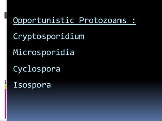
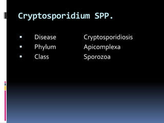
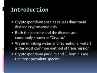
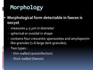
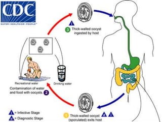
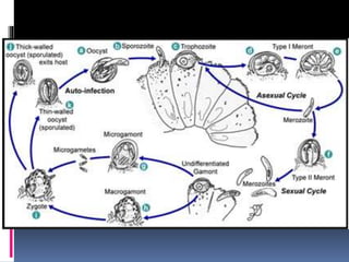
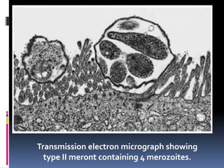
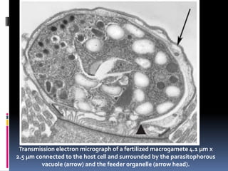
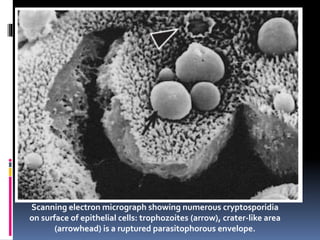
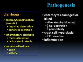
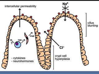
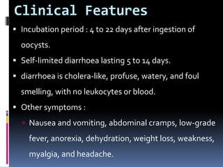
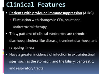
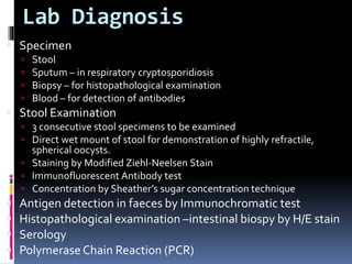
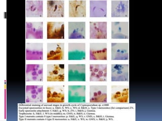
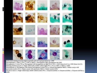
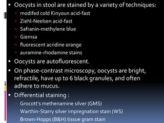
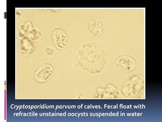
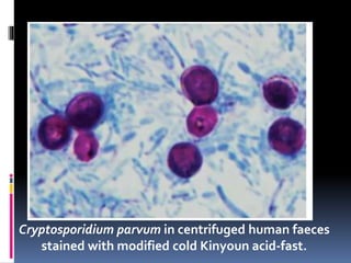
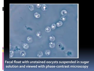
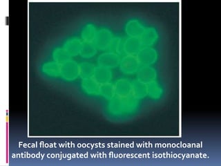
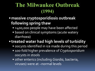
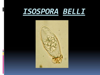
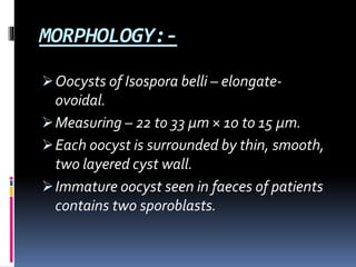
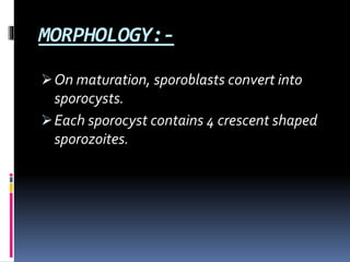
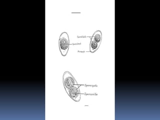
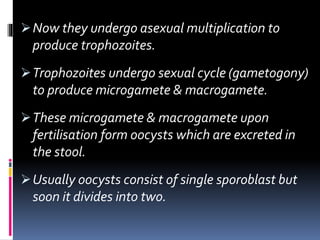
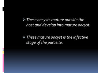
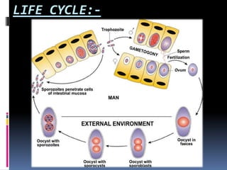
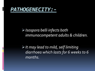
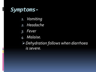
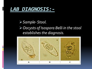
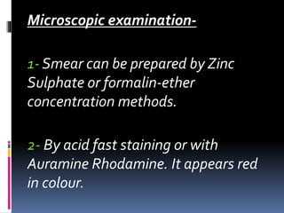
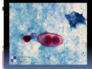
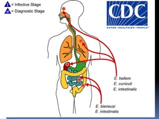
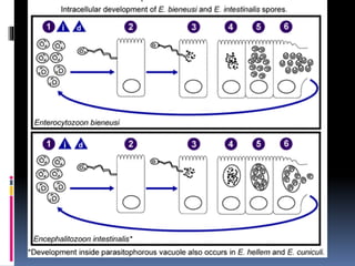
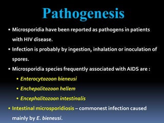
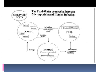
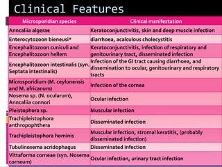
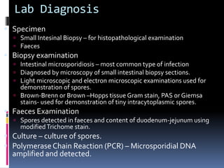
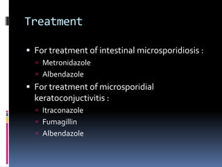
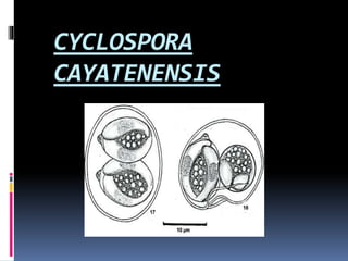
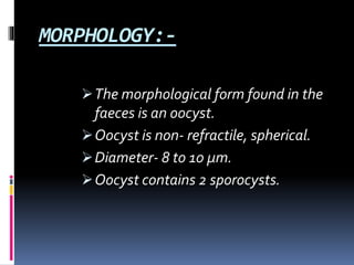
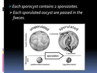
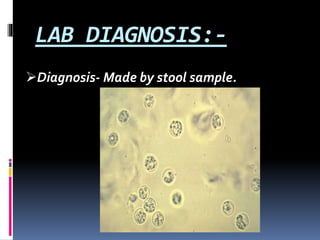
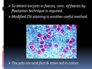
PRESENTED BY :
ABHISHEK KUMARYADAV
Opportunistic Protozoans :
Cryptosporidium
Microsporidia
Cyclospora
Isospora
Cryptosporidium SPP.
 Disease Cryptosporidiosis
 Phylum Apicomplexa
 Class Sporozoa
Introduction
 Cryptosporidium species causes diarrhoeal
disease cryptosporidiosis.
 Both the parasite and the disease are
commonly known as "Crypto.“
 Water (drinking water and recreational water)
is the most common method of transmission.
 Cryptosporidium parvum and C. hominis are
the most prevalent species.
Morphology
 Morphological form detectable in faeces is
oocyst
 measures 4-5 m in diameter
 spherical or ovoidal in shape
 contains four crescentic sporozoites and amylopectin
like granules (1-6 large dark granules).
 Two types :
 thin walled (autoinfection)
 thick walled (faeces)
LIFE CYCLE
Transmission electron micrograph showing
type II meront containing 4 merozoites.
Transmission electron micrograph of a fertilized macrogamete 4.1 µm x
2.5 µm connected to the host cell and surrounded by the parasitophorous
vacuole (arrow) and the feeder organelle (arrow head).
Scanning electron micrograph showing numerous cryptosporidia
on surface of epithelial cells: trophozoites (arrow), crater-like area
(arrowhead) is a ruptured parasitophorous envelope.
Scanning electron micrograph showing
type I meront releasing 8 merozoites
Transmission electron micrograph showing
macrogamont with nucleus and dark bodies
PATHOGENESIS
diarrhoea
• enterocyte malfunction
(osmotic)
• impaired absorption
• enhanced secretion
• inflammatory diarrhoea
• mucosal invasion
• leukocytes in stools
• secretory diarrhoea
• toxin
• watery
Pathogenesis
• enterocytes damaged or
killed
•villus atrophy (blunting)
• Na+ absorption
• permeability
• crypt cell hyperplasia
• Cl- secretion
• inflammation
Clinical Features
 Incubation period : 4 to 22 days after ingestion of
oocysts.
 Self-limited diarrhoea lasting 5 to 14 days.
 diarrhoea is cholera-like, profuse, watery, and foul
smelling, with no leukocytes or blood.
 Other symptoms :
 Nausea and vomiting, abdominal cramps, low-grade
fever, anorexia, dehydration, weight loss, weakness,
myalgia, and headache.
Clinical Features
 Patients with profound immunosuppression (AIDS) :
 Fluctuation with changes in CD4 count and
antiretroviral therapy.
 The 4 patterns of clinical syndromes are chronic
diarrhoea, cholera-like disease, transient diarrhoea, and
relapsing illness.
 Have a greater incidence of infection in extraintestinal
sites, such as the stomach, and the biliary, pancreatic,
and respiratory tracts.
Lab Diagnosis
 Specimen
 Stool
 Sputum – in respiratory cryptosporidiosis
 Biopsy – for histopathological examination
 Blood – for detection of antibodies
 Stool Examination
 3 consecutive stool specimens to be examined
 Direct wet mount of stool for demonstration of highly refractile,
spherical oocysts.
 Staining by Modified Ziehl-Neelsen Stain
 Immunofluorescent Antibody test
 Concentration by Sheather’s sugar concentration technique
 Antigen detection in faeces by Immunochromatic test
 Histopathological examination –intestinal biospy by H/E stain
 Serology
 Polymerase Chain Reaction (PCR)
 Oocysts in stool are stained by a variety of techniques:
 modifed cold Kinyoun acid-fast
 Ziehl-Neelsen acid-fast
 Safranin-methylene blue
 Giemsa
 fluorescent acridine orange
 auramine-rhodamine stains
 Oocysts are autofluorescent.
 On phase-contrast microscopy, oocysts are bright,
refractile, have up to 6 black granules, and often
adhere to mucus.
 Differential staining :
 Grocott’s methenamine silver (GMS)
 Warthin-Starry silver impregnation stain (WS)
 Brown-Hopps (B&H) tissue gram stain
Cryptosporidium parvum of calves. Fecal float with
refractile unstained oocysts suspended in water
Cryptosporidium parvum in centrifuged human faeces
stained with modified cold Kinyoun acid-fast.
Fecal float with unstained oocysts suspended in sugar
solution and viewed with phase-contrast microscopy
Fecal float with oocysts stained with monocloanal
antibody conjugated with fluorescent isothiocyanate.
Treatment
 There are several treatments for
cryptosporidium enteritis.
 Drugs such as nitazoxanide have been used in
children and adults.
 Other drugs that are sometimes used include:
 Atovaquone
 Metronidazole
Trimethoprim-sulfamethoxazole
The Milwaukee Outbreak
(1994)
•massive cryptosporidiosis outbreak
following spring thaw
• >400,000 people may have been affected
• based on clinical symptoms (acute watery
diarrhoea)
•treated water had high levels of turbidity
• oocysts identified in ice made during this period
• 100-fold higher prevalence of Cryptosporidium
oocysts in stools
• other enterics (including Giardia, bacteria,
viruses) were at ~normal levels
ISOSPORA BELLI
Sporozoan of human intestine.
Occurs throughout the world.
Most common in patients with
AIDS.
First described byVirchow in
1860 & named byWenyon in
1923.
It causes Isosporiasis which is a
human intestinal disease.
Isosporiasis:-
Infection causes acute, non-bloody
diarrhoea with crampy abdominal pain.
Can last for weeks & result in
malabsorption & weight loss.
MORPHOLOGY:-
Oocysts of Isospora belli – elongate-
ovoidal.
Measuring – 22 to 33 μm × 10 to 15 μm.
Each oocyst is surrounded by thin, smooth,
two layered cyst wall.
Immature oocyst seen in faeces of patients
contains two sporoblasts.
MORPHOLOGY:-
On maturation, sporoblasts convert into
sporocysts.
Each sporocyst contains 4 crescent shaped
sporozoites.
LIFE CYCLE
Life cycle of Isospora belli completes in one
host.
Humans acquire infection by ingestion of
food and water contaminated with faeces
containing sporulated (mature) oocysts.
8 sporozoites are released in the small
intestine & invade the epithelial cells of
distal duodenum & proximal jejunum.
Now they undergo asexual multiplication to
produce trophozoites.
Trophozoites undergo sexual cycle (gametogony)
to produce microgamete & macrogamete.
These microgamete & macrogamete upon
fertilisation form oocysts which are excreted in
the stool.
Usually oocysts consist of single sporoblast but
soon it divides into two.
These oocysts mature outside the
host and develop into mature oocyst.
These mature oocyst is the infective
stage of the parasite.
LIFE CYCLE:-
PATHOGENECITY:-
Isospora belli infects both
immunocompetent adults & children.
It may lead to mild, self limiting
diarrhoea which lasts for 6 weeks to 6
months.
Symptoms-
1. Vomiting
2. Headache
3. Fever
4. Malaise.
Dehydration follows when diarrhoea
is severe.
LAB DIAGNOSIS:-
Sample- Stool.
Oocysts of Isospora Belli in the stool
establishes the diagnosis.
Microscopic examination-
1- Smear can be prepared by Zinc
Sulphate or formalin-ether
concentration methods.
2- By acid fast staining or with
Auramine Rhodamine. It appears red
in colour.
3- Unstained oocysts are
autoflourescent.
They appear violet under UV light &
green under green or blue-violet light.
Lab Diagnosis Cont…..
POLYMERASE CHAIN REACTION
(PCR)
Highly sensitive and specific method.
TREATMENT:-
Cotrimoxazole is usually effective.
Microsporodia
 Phylum : Microspora
 Obligate, intracellular spore forming
protozoa.
 There are at least 15 microsporidian species
that have been identified as human
pathogens.
Nodular Cutaneous Microsporidiosis of the leg caused by
Encephalitozoon intestinalis in an HIV positive patient.
Morphology
 Unicellular, obligate intracellular parasites.
 In host cell, the parasite develops and multiply
(merogony-binary fission/schizogony-multiple
fission) and produces large number of spores
(sporogony).
 Spore is the infective stage, measures 0.5-0.2 m x
1-4 m; oval to cylindrical in shape; possess a thick
double layered wall.
Morphology
 Within cytoplasm, spore contains a coiled polar
tube which uncoils and thrusts forcefully into
the host cell and injects sporoplasm (infective
material).
 Spores :
 Stained with Gram’s stain, PAS, Giemsa stain or
modified trichome stain.
 Gram-positive and acid fast.
Electron micrograph of Anncaliia (Brachiola, Nosema) connori
spore in adrenal gland showing the coiled polar filament
(arrow) and two nuclei.
 Species and Genera :
 There are 15 genera which infect the humans :
 Anncaliia (formerly Brachiola) algerae
 A. connori
 A. vesicularum
 Encephalitozoon cuniculi
 E. hellem
 E. intestinalis
 Enterocytozoon bieneusi
 Microsporidium ceylonensis
 M. africanum
 Nosema ocularum
 Pleistophora sp.
 Trachipleistophora hominis
 T. anthropophthera
 Vittaforma corneae, and
 Tubulinosema acridophagus.
Spore of Enterocytozoon bieneusi demonstrating the
characteristic six turns of the polar tubule, which are
organized into two tiers of three turns each.
LIFE CYCLE
Pathogenesis
• Microsporidia have been reported as pathogens in patients
with HIV disease.
• Infection is probably by ingestion, inhalation or inoculation of
spores.
• Microsporidia species frequently associated with AIDS are :
• Enterocytozoon bieneusi
• Enchepalitozoon hellem
• Encephalitozoon intestinalis
• Intestinal microsporidiosis – commonest infection caused
mainly by E. bieneusi.
Clinical Features
Microsporidian species Clinical manifestation
Anncaliia algerae Keratoconjunctivitis, skin and deep muscle infection
Enterocytozoon bieneusi* diarrhoea, acalculous cholecystitis
Encephalitozoon cuniculi and
Encephalitozoon hellem
Keratoconjunctivitis, infection of respiratory and
genitourinary tract, disseminated infection
Encephalitozoon intestinalis (syn.
Septata intestinalis)
Infection of the GI tract causing diarrhoea, and
dissemination to ocular, genitourinary and respiratory
tracts
Microsporidium (M. ceylonensis
and M. africanum)
Infection of the cornea
Nosema sp. (N. ocularum),
Anncaliia connori
Ocular infection
Pleistophora sp. Muscular infection
Trachipleistophora
anthropophthera
Disseminated infection
Trachipleistophora hominis
Muscular infection, stromal keratitis, (probably
disseminated infection)
Tubulinosema acridophagus Disseminated infection
Vittaforma corneae (syn. Nosema
corneum)
Ocular infection, urinary tract infection
Lab Diagnosis
 Specimen
 Small Intesinal Biopsy – for histopathological examination
 Faeces
 Biopsy examination
 Intestinal microsporidiosis – most common type of infection
 Diagnosed by microscopy of small intestinal biopsy sections.
 Light microscopic and electron microscopic examinations used for
demonstration of spores.
 Brown-Brenn or Brown –Hopps tissue Gram stain, PAS or Giemsa
stains- used for demonstration of tiny intracytoplasmic spores.
 Faeces Examination
 Spores detected in faeces and content of duodenum-jejunum using
modifiedTrichome stain.
 Culture – culture of spores.
 Polymerase Chain Reaction (PCR) – Microsporidial DNA
amplified and detected.
Treatment
 For treatment of intestinal microsporidiosis :
 Metronidazole
 Albendazole
 For treatment of microsporidial
keratoconjuctivitis :
 Itraconazole
 Fumagillin
 Albendazole
CYCLOSPORA
CAYATENENSIS
It is a newly recognised protozoan
parasite.
Causes a disease named as
Cyclosporiasis in man particularly in
patients with AIDS.
First human case was reported in
Peru in 1995.
In recent years human cyclosporiasis
has emerged as an important
infection.
MORPHOLOGY:-
The morphological form found in the
faeces is an oocyst.
Oocyst is non- refractile, spherical.
Diameter- 8 to 10 μm.
Oocyst contains 2 sporocysts.
Each sporocyst contains 2 sporozoites.
Each sporulated oocyst are passed in the
faeces.
LIFE CYCLE:-
Source- Contaminated food & water
containing oocysts.
Host- Single host.
Has both sexual & asexual stage.
Man is infected by ingestion of food &
water contaminated with faeces.
The unsporulated oocyst in
faeces sporulates outside the
host.
Excystation of the sporocyst
releases 2 sporozoites which
infect the small intestine causing
diarrhoea.
PATHOGENESIS:-
Incubation period- 1 week.
Disease starts with acute watery diarrhoea
with nausea, loss of appetite, abdominal
pain, fever fatigue, weight loss.
Diarrhoea may be prolonged & associated
with muscle pain, vomiting, dehydration &
substantial weight loss.
Illness may last six weeks
before self-limiting.
If the disease is left
untreated, the illness may
relapse.
LAB DIAGNOSIS:-
Diagnosis- Made by stool sample.
To detect oocysts in faeces, conc. of faeces by
floatation technique is required.
Modified ZN staining is another useful method.
Oocysts are acid fast & stain red in colour.
TAKE HOME MESSAGE AND SUMMARY
 Acid fast Intestinal parasites
 Persistent diarrhoea – HIV/AIDS
 Cryptosporidium spp. – Oocyst
 Cyclospora spp. – Oocyst
 Isospora spp. – Oocyst
 Microsporidium Spp. - Spores
Opportunistic Protozoans - Microbiology

Opportunistic Protozoans - Microbiology

  • 1.
  • 2.
  • 3.
    Cryptosporidium SPP.  DiseaseCryptosporidiosis  Phylum Apicomplexa  Class Sporozoa
  • 4.
    Introduction  Cryptosporidium speciescauses diarrhoeal disease cryptosporidiosis.  Both the parasite and the disease are commonly known as "Crypto.“  Water (drinking water and recreational water) is the most common method of transmission.  Cryptosporidium parvum and C. hominis are the most prevalent species.
  • 5.
    Morphology  Morphological formdetectable in faeces is oocyst  measures 4-5 m in diameter  spherical or ovoidal in shape  contains four crescentic sporozoites and amylopectin like granules (1-6 large dark granules).  Two types :  thin walled (autoinfection)  thick walled (faeces)
  • 6.
  • 9.
    Transmission electron micrographshowing type II meront containing 4 merozoites.
  • 10.
    Transmission electron micrographof a fertilized macrogamete 4.1 µm x 2.5 µm connected to the host cell and surrounded by the parasitophorous vacuole (arrow) and the feeder organelle (arrow head).
  • 11.
    Scanning electron micrographshowing numerous cryptosporidia on surface of epithelial cells: trophozoites (arrow), crater-like area (arrowhead) is a ruptured parasitophorous envelope.
  • 12.
    Scanning electron micrographshowing type I meront releasing 8 merozoites
  • 13.
    Transmission electron micrographshowing macrogamont with nucleus and dark bodies
  • 14.
  • 15.
    diarrhoea • enterocyte malfunction (osmotic) •impaired absorption • enhanced secretion • inflammatory diarrhoea • mucosal invasion • leukocytes in stools • secretory diarrhoea • toxin • watery Pathogenesis • enterocytes damaged or killed •villus atrophy (blunting) • Na+ absorption • permeability • crypt cell hyperplasia • Cl- secretion • inflammation
  • 17.
    Clinical Features  Incubationperiod : 4 to 22 days after ingestion of oocysts.  Self-limited diarrhoea lasting 5 to 14 days.  diarrhoea is cholera-like, profuse, watery, and foul smelling, with no leukocytes or blood.  Other symptoms :  Nausea and vomiting, abdominal cramps, low-grade fever, anorexia, dehydration, weight loss, weakness, myalgia, and headache.
  • 18.
    Clinical Features  Patientswith profound immunosuppression (AIDS) :  Fluctuation with changes in CD4 count and antiretroviral therapy.  The 4 patterns of clinical syndromes are chronic diarrhoea, cholera-like disease, transient diarrhoea, and relapsing illness.  Have a greater incidence of infection in extraintestinal sites, such as the stomach, and the biliary, pancreatic, and respiratory tracts.
  • 19.
    Lab Diagnosis  Specimen Stool  Sputum – in respiratory cryptosporidiosis  Biopsy – for histopathological examination  Blood – for detection of antibodies  Stool Examination  3 consecutive stool specimens to be examined  Direct wet mount of stool for demonstration of highly refractile, spherical oocysts.  Staining by Modified Ziehl-Neelsen Stain  Immunofluorescent Antibody test  Concentration by Sheather’s sugar concentration technique  Antigen detection in faeces by Immunochromatic test  Histopathological examination –intestinal biospy by H/E stain  Serology  Polymerase Chain Reaction (PCR)
  • 22.
     Oocysts instool are stained by a variety of techniques:  modifed cold Kinyoun acid-fast  Ziehl-Neelsen acid-fast  Safranin-methylene blue  Giemsa  fluorescent acridine orange  auramine-rhodamine stains  Oocysts are autofluorescent.  On phase-contrast microscopy, oocysts are bright, refractile, have up to 6 black granules, and often adhere to mucus.  Differential staining :  Grocott’s methenamine silver (GMS)  Warthin-Starry silver impregnation stain (WS)  Brown-Hopps (B&H) tissue gram stain
  • 23.
    Cryptosporidium parvum ofcalves. Fecal float with refractile unstained oocysts suspended in water
  • 24.
    Cryptosporidium parvum incentrifuged human faeces stained with modified cold Kinyoun acid-fast.
  • 25.
    Fecal float withunstained oocysts suspended in sugar solution and viewed with phase-contrast microscopy
  • 26.
    Fecal float withoocysts stained with monocloanal antibody conjugated with fluorescent isothiocyanate.
  • 27.
    Treatment  There areseveral treatments for cryptosporidium enteritis.  Drugs such as nitazoxanide have been used in children and adults.  Other drugs that are sometimes used include:  Atovaquone  Metronidazole Trimethoprim-sulfamethoxazole
  • 28.
    The Milwaukee Outbreak (1994) •massivecryptosporidiosis outbreak following spring thaw • >400,000 people may have been affected • based on clinical symptoms (acute watery diarrhoea) •treated water had high levels of turbidity • oocysts identified in ice made during this period • 100-fold higher prevalence of Cryptosporidium oocysts in stools • other enterics (including Giardia, bacteria, viruses) were at ~normal levels
  • 29.
  • 30.
    Sporozoan of humanintestine. Occurs throughout the world. Most common in patients with AIDS. First described byVirchow in 1860 & named byWenyon in 1923. It causes Isosporiasis which is a human intestinal disease.
  • 31.
    Isosporiasis:- Infection causes acute,non-bloody diarrhoea with crampy abdominal pain. Can last for weeks & result in malabsorption & weight loss.
  • 32.
    MORPHOLOGY:- Oocysts of Isosporabelli – elongate- ovoidal. Measuring – 22 to 33 μm × 10 to 15 μm. Each oocyst is surrounded by thin, smooth, two layered cyst wall. Immature oocyst seen in faeces of patients contains two sporoblasts.
  • 33.
    MORPHOLOGY:- On maturation, sporoblastsconvert into sporocysts. Each sporocyst contains 4 crescent shaped sporozoites.
  • 35.
    LIFE CYCLE Life cycleof Isospora belli completes in one host. Humans acquire infection by ingestion of food and water contaminated with faeces containing sporulated (mature) oocysts. 8 sporozoites are released in the small intestine & invade the epithelial cells of distal duodenum & proximal jejunum.
  • 36.
    Now they undergoasexual multiplication to produce trophozoites. Trophozoites undergo sexual cycle (gametogony) to produce microgamete & macrogamete. These microgamete & macrogamete upon fertilisation form oocysts which are excreted in the stool. Usually oocysts consist of single sporoblast but soon it divides into two.
  • 37.
    These oocysts matureoutside the host and develop into mature oocyst. These mature oocyst is the infective stage of the parasite.
  • 38.
  • 39.
    PATHOGENECITY:- Isospora belli infectsboth immunocompetent adults & children. It may lead to mild, self limiting diarrhoea which lasts for 6 weeks to 6 months.
  • 40.
    Symptoms- 1. Vomiting 2. Headache 3.Fever 4. Malaise. Dehydration follows when diarrhoea is severe.
  • 41.
    LAB DIAGNOSIS:- Sample- Stool. Oocystsof Isospora Belli in the stool establishes the diagnosis.
  • 42.
    Microscopic examination- 1- Smearcan be prepared by Zinc Sulphate or formalin-ether concentration methods. 2- By acid fast staining or with Auramine Rhodamine. It appears red in colour.
  • 44.
    3- Unstained oocystsare autoflourescent. They appear violet under UV light & green under green or blue-violet light.
  • 45.
    Lab Diagnosis Cont….. POLYMERASECHAIN REACTION (PCR) Highly sensitive and specific method.
  • 46.
  • 47.
    Microsporodia  Phylum :Microspora  Obligate, intracellular spore forming protozoa.  There are at least 15 microsporidian species that have been identified as human pathogens.
  • 48.
    Nodular Cutaneous Microsporidiosisof the leg caused by Encephalitozoon intestinalis in an HIV positive patient.
  • 49.
    Morphology  Unicellular, obligateintracellular parasites.  In host cell, the parasite develops and multiply (merogony-binary fission/schizogony-multiple fission) and produces large number of spores (sporogony).  Spore is the infective stage, measures 0.5-0.2 m x 1-4 m; oval to cylindrical in shape; possess a thick double layered wall.
  • 50.
    Morphology  Within cytoplasm,spore contains a coiled polar tube which uncoils and thrusts forcefully into the host cell and injects sporoplasm (infective material).  Spores :  Stained with Gram’s stain, PAS, Giemsa stain or modified trichome stain.  Gram-positive and acid fast.
  • 52.
    Electron micrograph ofAnncaliia (Brachiola, Nosema) connori spore in adrenal gland showing the coiled polar filament (arrow) and two nuclei.
  • 53.
     Species andGenera :  There are 15 genera which infect the humans :  Anncaliia (formerly Brachiola) algerae  A. connori  A. vesicularum  Encephalitozoon cuniculi  E. hellem  E. intestinalis  Enterocytozoon bieneusi  Microsporidium ceylonensis  M. africanum  Nosema ocularum  Pleistophora sp.  Trachipleistophora hominis  T. anthropophthera  Vittaforma corneae, and  Tubulinosema acridophagus.
  • 54.
    Spore of Enterocytozoonbieneusi demonstrating the characteristic six turns of the polar tubule, which are organized into two tiers of three turns each.
  • 55.
  • 58.
    Pathogenesis • Microsporidia havebeen reported as pathogens in patients with HIV disease. • Infection is probably by ingestion, inhalation or inoculation of spores. • Microsporidia species frequently associated with AIDS are : • Enterocytozoon bieneusi • Enchepalitozoon hellem • Encephalitozoon intestinalis • Intestinal microsporidiosis – commonest infection caused mainly by E. bieneusi.
  • 60.
    Clinical Features Microsporidian speciesClinical manifestation Anncaliia algerae Keratoconjunctivitis, skin and deep muscle infection Enterocytozoon bieneusi* diarrhoea, acalculous cholecystitis Encephalitozoon cuniculi and Encephalitozoon hellem Keratoconjunctivitis, infection of respiratory and genitourinary tract, disseminated infection Encephalitozoon intestinalis (syn. Septata intestinalis) Infection of the GI tract causing diarrhoea, and dissemination to ocular, genitourinary and respiratory tracts Microsporidium (M. ceylonensis and M. africanum) Infection of the cornea Nosema sp. (N. ocularum), Anncaliia connori Ocular infection Pleistophora sp. Muscular infection Trachipleistophora anthropophthera Disseminated infection Trachipleistophora hominis Muscular infection, stromal keratitis, (probably disseminated infection) Tubulinosema acridophagus Disseminated infection Vittaforma corneae (syn. Nosema corneum) Ocular infection, urinary tract infection
  • 61.
    Lab Diagnosis  Specimen Small Intesinal Biopsy – for histopathological examination  Faeces  Biopsy examination  Intestinal microsporidiosis – most common type of infection  Diagnosed by microscopy of small intestinal biopsy sections.  Light microscopic and electron microscopic examinations used for demonstration of spores.  Brown-Brenn or Brown –Hopps tissue Gram stain, PAS or Giemsa stains- used for demonstration of tiny intracytoplasmic spores.  Faeces Examination  Spores detected in faeces and content of duodenum-jejunum using modifiedTrichome stain.  Culture – culture of spores.  Polymerase Chain Reaction (PCR) – Microsporidial DNA amplified and detected.
  • 62.
    Treatment  For treatmentof intestinal microsporidiosis :  Metronidazole  Albendazole  For treatment of microsporidial keratoconjuctivitis :  Itraconazole  Fumagillin  Albendazole
  • 63.
  • 64.
    It is anewly recognised protozoan parasite. Causes a disease named as Cyclosporiasis in man particularly in patients with AIDS. First human case was reported in Peru in 1995. In recent years human cyclosporiasis has emerged as an important infection.
  • 65.
    MORPHOLOGY:- The morphological formfound in the faeces is an oocyst. Oocyst is non- refractile, spherical. Diameter- 8 to 10 μm. Oocyst contains 2 sporocysts.
  • 66.
    Each sporocyst contains2 sporozoites. Each sporulated oocyst are passed in the faeces.
  • 67.
    LIFE CYCLE:- Source- Contaminatedfood & water containing oocysts. Host- Single host. Has both sexual & asexual stage. Man is infected by ingestion of food & water contaminated with faeces.
  • 68.
    The unsporulated oocystin faeces sporulates outside the host. Excystation of the sporocyst releases 2 sporozoites which infect the small intestine causing diarrhoea.
  • 70.
    PATHOGENESIS:- Incubation period- 1week. Disease starts with acute watery diarrhoea with nausea, loss of appetite, abdominal pain, fever fatigue, weight loss. Diarrhoea may be prolonged & associated with muscle pain, vomiting, dehydration & substantial weight loss.
  • 71.
    Illness may lastsix weeks before self-limiting. If the disease is left untreated, the illness may relapse.
  • 72.
  • 73.
    To detect oocystsin faeces, conc. of faeces by floatation technique is required. Modified ZN staining is another useful method. Oocysts are acid fast & stain red in colour.
  • 74.
    TAKE HOME MESSAGEAND SUMMARY  Acid fast Intestinal parasites  Persistent diarrhoea – HIV/AIDS  Cryptosporidium spp. – Oocyst  Cyclospora spp. – Oocyst  Isospora spp. – Oocyst  Microsporidium Spp. - Spores