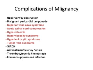
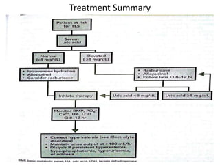
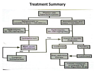
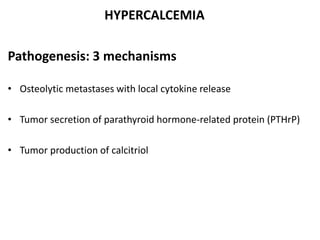
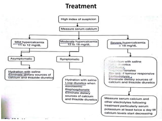
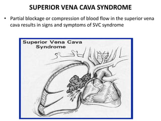
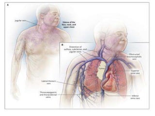
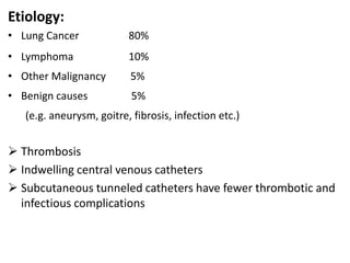
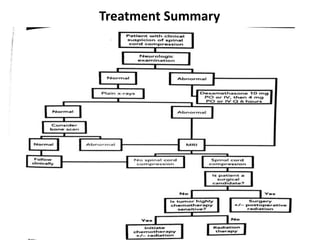
ONCOLOGICAL EMERGENCIES
DR. VISHAL KR. KANDHWAY
Max Superspeciality Hospital,
Patparganj
Complications of Mlignancy
- Upper airway obstruction
- Malignant pericardial tamponade
- Superior vena cava syndrome
- Acute spinal cord compression
- Hypercalcemia
- Hyperviscosity syndrome
- Hyperleukocytic syndrome
- Tumor lysis syndrome
- SIADH
- Adrenal insufficiency / crisis
- Thrombocytopenia / hemorrage
- Immunosuppression / infection
TLS
• Metabolic consequenses resulting from sudden release of
potassim, phosphates & purine metabolites from tumor cells
undergoing cell death.
Pathogenesis
• Chemotherapy is given to fast growing cells. This lysis causes a
shift of intracellular components into the circulation, affecting
the kidneys and causing dangerous electrolyte imbalances
Abnormalities
Hyperuricemia
Hyperkalemia
Hyprephophatemia
Hypocalcaemia
Raised LDH
Risk factors
Cancer type- ALL, Burkitts lymphoma
Following chemotherapy,radiation, chemoembolisation
Renal insufficiency
Dehydration
Signs & Symptoms
GI: nausea, vomiting, diarrhea
Cardiac: dysrhythmias
GU: flank pain, anuria, cloudy urine
Neuro: seizures, altered mentation, stupor
TREATMENT
HYDRATION
• High rate of fluids – 200-300ml/hr
• U.O. to be maintained @ 80-100ml/m2/h
• Urine specific gravity to be maintained <1.01
• Most effective strategy for preventing urate induced obstructive
uropathy
LOOP DIURETICS
• To maintain U.O.
• Contraindicated in hypovolemia or obstructive uropathy
URINE ALKALANIZATION
Controversial
Can lead formation of urinary xanthine crystals
May cause urinary obstruction
ALLOPURINOL
• Hypoxanthine analog that competitively inhibits xanthine oxidase
• Dose 600mg/day if uric acid <8mg/dl
• Start prior to chemo
• Caution in renal impairment
• Causes skin hypersensitivity reaction
RASBURICASE
• Oxidizes preformed uric acid to allantoin which is highly soluble in
urine
• Used when uric acid level >8mg/dl
• Dose-infusion 0.1-0.2mg/kg/day in 50 ml NS over 30 min for 5
days
• Adverse reaction- anaphylaxis, rash, hemolysis
Hyperkalemia
• Increase excretion: IVF, diuretics, Kaexylate
• Shift back into cells: IV insulin and dextrose
Hypocalemia- Only treat if extremely low.
HYPERPHOSPHATEMIA
•Phosphorus binder: aluminum hydroxide, Calcium carbonate,
sevelamer hydroxide, lanthanum carbonate
•Decrease oral intake
HEMODIALYSIS
• Volume overload
• Serum uric acid >10mg/dl
• Rapidly rising phosphorus
• Uncontrolled hyperkalemia
Treatment Summary
LEUCOSTASIS
• Syndrome associated with high number of immature
leukocytes(blasts) in peripheral circulation
• High & rapidly rising blast counts usually >100,000/ml
Associated malignancy
• AML especially myelomonocytic(M4) & monocytic(M5)
subtypes
• CML in blast crisis
• Rarely in ALL or CLL
Pathology:
• Occlusive intravascular aggregates of blasts blocks microcirculation
in multiple organs
Symptoms:
• Usually nonspecific
• SOB
• Headache
• Confusion
• Stupor
• Focal neurologoical deficit
TREATMENT
• Hydration
• Hydroxyurea 50-100mg/kg/day in 3 divided doses till blast count>
50,000/ml
• Allopurinol and/or rasburicase
• Leucopheresis till count< 50,000/ml
• Definitive treatment for cancer
Supportive Treatment:
RBC transfusion to be restricted
Correct DIC & thrombocytopenia
Broad spectrum antibiotics
Treatment Summary
HYPERCALCEMIA
Pathogenesis: 3 mechanisms
• Osteolytic metastases with local cytokine release
• Tumor secretion of parathyroid hormone-related protein (PTHrP)
• Tumor production of calcitriol
• Symptoms & Signs usually non-specific
GI:
• Constipation is most common
• Anorexia
• Vague abdominal pain
Renal dysfunction:
Nephrolithiasis
– More common in hyperparathyroidism
Nephrogenic diabetes insipidus
– Defect in concentrating ability
– Polyuria and polydipsia
Chronic renal failure
– Longstanding high calcium
Calcifcation, degeneration, and necrosis of tubules
Neuropsychiatric:
• Anxiety
• Depression
• Cognitive dysfunction
– Delerium
– Psychosis
– Hallucinations
– Somnolence
– Coma
CVS
• Short QT interval
• Supraventricualr arrhythmias
• Ventricular arrhythmias
Osteolytic metastases
Treatment
SUPERIOR VENA CAVA SYNDROME
• Partial blockage or compression of blood flow in the superior vena
cava results in signs and symptoms of SVC syndrome
Etiology:
• Lung Cancer 80%
• Lymphoma 10%
• Other Malignancy 5%
• Benign causes 5%
(e.g. aneurysm, goitre, fibrosis, infection etc.)
 Thrombosis
 Indwelling central venous catheters
 Subcutaneous tunneled catheters have fewer thrombotic and
infectious complications
Symptoms & Signs
As the obstruction develops venous collaterals are formed
Symptom onset depends on speed of SVC obstruction onset
Shortness of breath is the most common symptom
Facial swelling or head fullness
– exacerbated by bending forward or lying down
Cough
Arm edema
Cyanosis
Physical findings:
Venous distension
– neck
– chest wall
DIAGNOSIS
Radiographic studies:
• Most patients have an abnormal chest x-ray at presentation
• Most common findings are
– Mediastinal widening
– Pleural effusion
CT Chest
• Preferred choice
• IV contrast
– defines the level of obstruction
– Maps out collateral pathways
– Can identify underlying cause of obstruction
Venography:
• Bilateral upper arm venograpy
– superior to CT to define site of obstruction
– Does not define cause unless thrombosis is solely responsible
MRI:
Can be useful in patients with IV contrast allergies
Treatment
SPINAL CORD COMPRESSION
• Neoplastic invasion of the space between vertebrae and spinal
cord (epidural invasion)
– Usually from bone metastases
Causes
• Metastatic tumor from any primary site
• Prostate, breast, and lung carcinoma
– 15-20% of cases
• Renal cell, non-Hodgkin’s lymphoma, or myeloma
– 5-10% of cases
Spinal Locations:
• Thoracic spine: 60%
• Lumbosacral spine: 30%
• Cervical spine: 10%
Clinical Features
Pain:
• Usually first symptom
– 80-90% of the time
• Usually precedes other neurologic symptoms by several weeks
– Increases in intensity
• Severe local back pain
• Aggravated by recumbency
– Distension of venous plexus
• May become radicular
Motor:
• Weakness: 60-85%
• At or above conus medularis
– Extensors of the upper extremities
• Above the thoracic spine
– Affects flexors in the lower extremities
• Patients may be hyperreflexic below the lesion and have extensor
plantars
• Weakness tends to be symmetrical
• Progressive weakness is followed by loss of gait function then paralysis
• The severity of weakness is greatest with thoracic metastases
Sensory:
• Less common than motor findings
• Still present in majority of cases
• Ascending numbness and parathesias
Bowel & Bladder:
• Loss is late finding
• Autonomic neuropathy presents usually as urinary retension
– Rarely sole finding
Radiological investigations:
• CT myelography involves a lumbar puncture
– Contraindicated in brain metastases, thrombocytopenia, or
coagulopathy
– Can diagnose leptomeningeal metastases
• MRI
Images whole spine
High detail
Spares lumbar puncture
Patients in pain must lie still
MRI of epidural spinal cord compression in a women with past
history of breast cancer.
Pain management:
• Corticosteroids
– Decrease edema
• Opiates
– Needed to decrease pain for comfort and examination purposes
Radiation therapy:
• Relieves pain in most cases
• Post-neurological function usually determines response
• Response most associated with tumor type and radiosensitivity; eg.
lymphoma
Surgery:
• Changing role
• Recent controlled trial comparing aggressive surgery followed by
radiation vs. radiation alone
• Improvement in surgery + radiation therapy
Chemotherapy:
• Can be successful in chemosensitive tumors
– Hodgkin’s lymphoma
– Non-Hodgkin’s lymphoma
– Neuroblastoma
– Germ cell
– Breast cancer (hormonal manipulation)
– Prostate cancer (hormonal manipulation)
Treatment Summary
HYPERVISCOSITY SYNDROME
• Refers to the clinical sequelae of increased blood viscosity.
• Increased serum viscosity results from increased circulating serum
immunoglobulins and can be seen in diseases like multiple
myeloma.
• HVS can also result from increased cellular blood components in
hyperproliferative states such as leukemia, polycythemia &
myloproliferative disorders.
Clinical Presentation: consists of triad of mucosal bleeding, visual
changes & neurologic symptoms.
Pathophysiology:
• Viscosity is a property of liquid & is described as the resistance that
a liquid exibits to the flow of 1 layer over another.
• As serum proteins or cellular components increase, the blood
becomes more viscous. Vascular stasis & resultant hypoperfusion
then lead to clinical symptoms of HVS.
• HVS is associated most commonly with plasma cell dyscrasias & is
due to the large size of IgM.
Labs:
• Elevated serum viscosity (normal 1.4-1.8 centipoises)
• CBC- rouleaux formation
• WBC- 100,000/uL or greater
• TP & albumin
• Electrolytes: hypercalcemia, hyperphosphotemia & hyperkalemia
• Coagulation profile
• CXR, CT brain, MRI
Treatment:
• Plasmapheresis is the treatment of choice for initial management
& stablization.
• Other options are leukapheresis, plateletpheresis & phlebotomy
THANK YOU

Oncological emergencies

  • 1.
    ONCOLOGICAL EMERGENCIES DR. VISHALKR. KANDHWAY Max Superspeciality Hospital, Patparganj
  • 2.
    Complications of Mlignancy -Upper airway obstruction - Malignant pericardial tamponade - Superior vena cava syndrome - Acute spinal cord compression - Hypercalcemia - Hyperviscosity syndrome - Hyperleukocytic syndrome - Tumor lysis syndrome - SIADH - Adrenal insufficiency / crisis - Thrombocytopenia / hemorrage - Immunosuppression / infection
  • 3.
    TLS • Metabolic consequensesresulting from sudden release of potassim, phosphates & purine metabolites from tumor cells undergoing cell death.
  • 4.
    Pathogenesis • Chemotherapy isgiven to fast growing cells. This lysis causes a shift of intracellular components into the circulation, affecting the kidneys and causing dangerous electrolyte imbalances
  • 5.
  • 6.
    Risk factors Cancer type-ALL, Burkitts lymphoma Following chemotherapy,radiation, chemoembolisation Renal insufficiency Dehydration
  • 7.
    Signs & Symptoms GI:nausea, vomiting, diarrhea Cardiac: dysrhythmias GU: flank pain, anuria, cloudy urine Neuro: seizures, altered mentation, stupor
  • 8.
    TREATMENT HYDRATION • High rateof fluids – 200-300ml/hr • U.O. to be maintained @ 80-100ml/m2/h • Urine specific gravity to be maintained <1.01 • Most effective strategy for preventing urate induced obstructive uropathy LOOP DIURETICS • To maintain U.O. • Contraindicated in hypovolemia or obstructive uropathy
  • 9.
    URINE ALKALANIZATION Controversial Can leadformation of urinary xanthine crystals May cause urinary obstruction ALLOPURINOL • Hypoxanthine analog that competitively inhibits xanthine oxidase • Dose 600mg/day if uric acid <8mg/dl • Start prior to chemo • Caution in renal impairment • Causes skin hypersensitivity reaction
  • 10.
    RASBURICASE • Oxidizes preformeduric acid to allantoin which is highly soluble in urine • Used when uric acid level >8mg/dl • Dose-infusion 0.1-0.2mg/kg/day in 50 ml NS over 30 min for 5 days • Adverse reaction- anaphylaxis, rash, hemolysis Hyperkalemia • Increase excretion: IVF, diuretics, Kaexylate • Shift back into cells: IV insulin and dextrose Hypocalemia- Only treat if extremely low.
  • 11.
    HYPERPHOSPHATEMIA •Phosphorus binder: aluminumhydroxide, Calcium carbonate, sevelamer hydroxide, lanthanum carbonate •Decrease oral intake HEMODIALYSIS • Volume overload • Serum uric acid >10mg/dl • Rapidly rising phosphorus • Uncontrolled hyperkalemia
  • 12.
  • 13.
    LEUCOSTASIS • Syndrome associatedwith high number of immature leukocytes(blasts) in peripheral circulation • High & rapidly rising blast counts usually >100,000/ml Associated malignancy • AML especially myelomonocytic(M4) & monocytic(M5) subtypes • CML in blast crisis • Rarely in ALL or CLL
  • 14.
    Pathology: • Occlusive intravascularaggregates of blasts blocks microcirculation in multiple organs Symptoms: • Usually nonspecific • SOB • Headache • Confusion • Stupor • Focal neurologoical deficit
  • 15.
    TREATMENT • Hydration • Hydroxyurea50-100mg/kg/day in 3 divided doses till blast count> 50,000/ml • Allopurinol and/or rasburicase • Leucopheresis till count< 50,000/ml • Definitive treatment for cancer Supportive Treatment: RBC transfusion to be restricted Correct DIC & thrombocytopenia Broad spectrum antibiotics
  • 16.
  • 17.
    HYPERCALCEMIA Pathogenesis: 3 mechanisms •Osteolytic metastases with local cytokine release • Tumor secretion of parathyroid hormone-related protein (PTHrP) • Tumor production of calcitriol
  • 18.
    • Symptoms &Signs usually non-specific GI: • Constipation is most common • Anorexia • Vague abdominal pain
  • 19.
    Renal dysfunction: Nephrolithiasis – Morecommon in hyperparathyroidism Nephrogenic diabetes insipidus – Defect in concentrating ability – Polyuria and polydipsia Chronic renal failure – Longstanding high calcium Calcifcation, degeneration, and necrosis of tubules
  • 20.
    Neuropsychiatric: • Anxiety • Depression •Cognitive dysfunction – Delerium – Psychosis – Hallucinations – Somnolence – Coma CVS • Short QT interval • Supraventricualr arrhythmias • Ventricular arrhythmias
  • 21.
  • 22.
  • 23.
    SUPERIOR VENA CAVASYNDROME • Partial blockage or compression of blood flow in the superior vena cava results in signs and symptoms of SVC syndrome
  • 25.
    Etiology: • Lung Cancer80% • Lymphoma 10% • Other Malignancy 5% • Benign causes 5% (e.g. aneurysm, goitre, fibrosis, infection etc.)  Thrombosis  Indwelling central venous catheters  Subcutaneous tunneled catheters have fewer thrombotic and infectious complications
  • 26.
    Symptoms & Signs Asthe obstruction develops venous collaterals are formed Symptom onset depends on speed of SVC obstruction onset Shortness of breath is the most common symptom Facial swelling or head fullness – exacerbated by bending forward or lying down Cough Arm edema Cyanosis
  • 27.
  • 28.
    DIAGNOSIS Radiographic studies: • Mostpatients have an abnormal chest x-ray at presentation • Most common findings are – Mediastinal widening – Pleural effusion
  • 29.
    CT Chest • Preferredchoice • IV contrast – defines the level of obstruction – Maps out collateral pathways – Can identify underlying cause of obstruction
  • 30.
    Venography: • Bilateral upperarm venograpy – superior to CT to define site of obstruction – Does not define cause unless thrombosis is solely responsible
  • 31.
    MRI: Can be usefulin patients with IV contrast allergies
  • 32.
  • 33.
    SPINAL CORD COMPRESSION •Neoplastic invasion of the space between vertebrae and spinal cord (epidural invasion) – Usually from bone metastases Causes • Metastatic tumor from any primary site • Prostate, breast, and lung carcinoma – 15-20% of cases • Renal cell, non-Hodgkin’s lymphoma, or myeloma – 5-10% of cases
  • 34.
    Spinal Locations: • Thoracicspine: 60% • Lumbosacral spine: 30% • Cervical spine: 10%
  • 35.
    Clinical Features Pain: • Usuallyfirst symptom – 80-90% of the time • Usually precedes other neurologic symptoms by several weeks – Increases in intensity • Severe local back pain • Aggravated by recumbency – Distension of venous plexus • May become radicular
  • 36.
    Motor: • Weakness: 60-85% •At or above conus medularis – Extensors of the upper extremities • Above the thoracic spine – Affects flexors in the lower extremities • Patients may be hyperreflexic below the lesion and have extensor plantars • Weakness tends to be symmetrical • Progressive weakness is followed by loss of gait function then paralysis • The severity of weakness is greatest with thoracic metastases
  • 37.
    Sensory: • Less commonthan motor findings • Still present in majority of cases • Ascending numbness and parathesias
  • 38.
    Bowel & Bladder: •Loss is late finding • Autonomic neuropathy presents usually as urinary retension – Rarely sole finding
  • 39.
    Radiological investigations: • CTmyelography involves a lumbar puncture – Contraindicated in brain metastases, thrombocytopenia, or coagulopathy – Can diagnose leptomeningeal metastases • MRI Images whole spine High detail Spares lumbar puncture Patients in pain must lie still
  • 40.
    MRI of epiduralspinal cord compression in a women with past history of breast cancer.
  • 41.
    Pain management: • Corticosteroids –Decrease edema • Opiates – Needed to decrease pain for comfort and examination purposes
  • 42.
    Radiation therapy: • Relievespain in most cases • Post-neurological function usually determines response • Response most associated with tumor type and radiosensitivity; eg. lymphoma
  • 43.
    Surgery: • Changing role •Recent controlled trial comparing aggressive surgery followed by radiation vs. radiation alone • Improvement in surgery + radiation therapy
  • 44.
    Chemotherapy: • Can besuccessful in chemosensitive tumors – Hodgkin’s lymphoma – Non-Hodgkin’s lymphoma – Neuroblastoma – Germ cell – Breast cancer (hormonal manipulation) – Prostate cancer (hormonal manipulation)
  • 45.
  • 46.
    HYPERVISCOSITY SYNDROME • Refersto the clinical sequelae of increased blood viscosity. • Increased serum viscosity results from increased circulating serum immunoglobulins and can be seen in diseases like multiple myeloma. • HVS can also result from increased cellular blood components in hyperproliferative states such as leukemia, polycythemia & myloproliferative disorders.
  • 47.
    Clinical Presentation: consistsof triad of mucosal bleeding, visual changes & neurologic symptoms. Pathophysiology: • Viscosity is a property of liquid & is described as the resistance that a liquid exibits to the flow of 1 layer over another. • As serum proteins or cellular components increase, the blood becomes more viscous. Vascular stasis & resultant hypoperfusion then lead to clinical symptoms of HVS. • HVS is associated most commonly with plasma cell dyscrasias & is due to the large size of IgM.
  • 48.
    Labs: • Elevated serumviscosity (normal 1.4-1.8 centipoises) • CBC- rouleaux formation • WBC- 100,000/uL or greater • TP & albumin • Electrolytes: hypercalcemia, hyperphosphotemia & hyperkalemia • Coagulation profile • CXR, CT brain, MRI
  • 49.
    Treatment: • Plasmapheresis isthe treatment of choice for initial management & stablization. • Other options are leukapheresis, plateletpheresis & phlebotomy
  • 50.