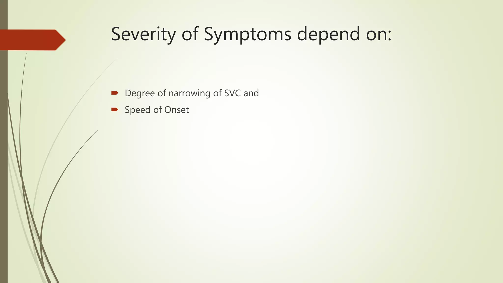
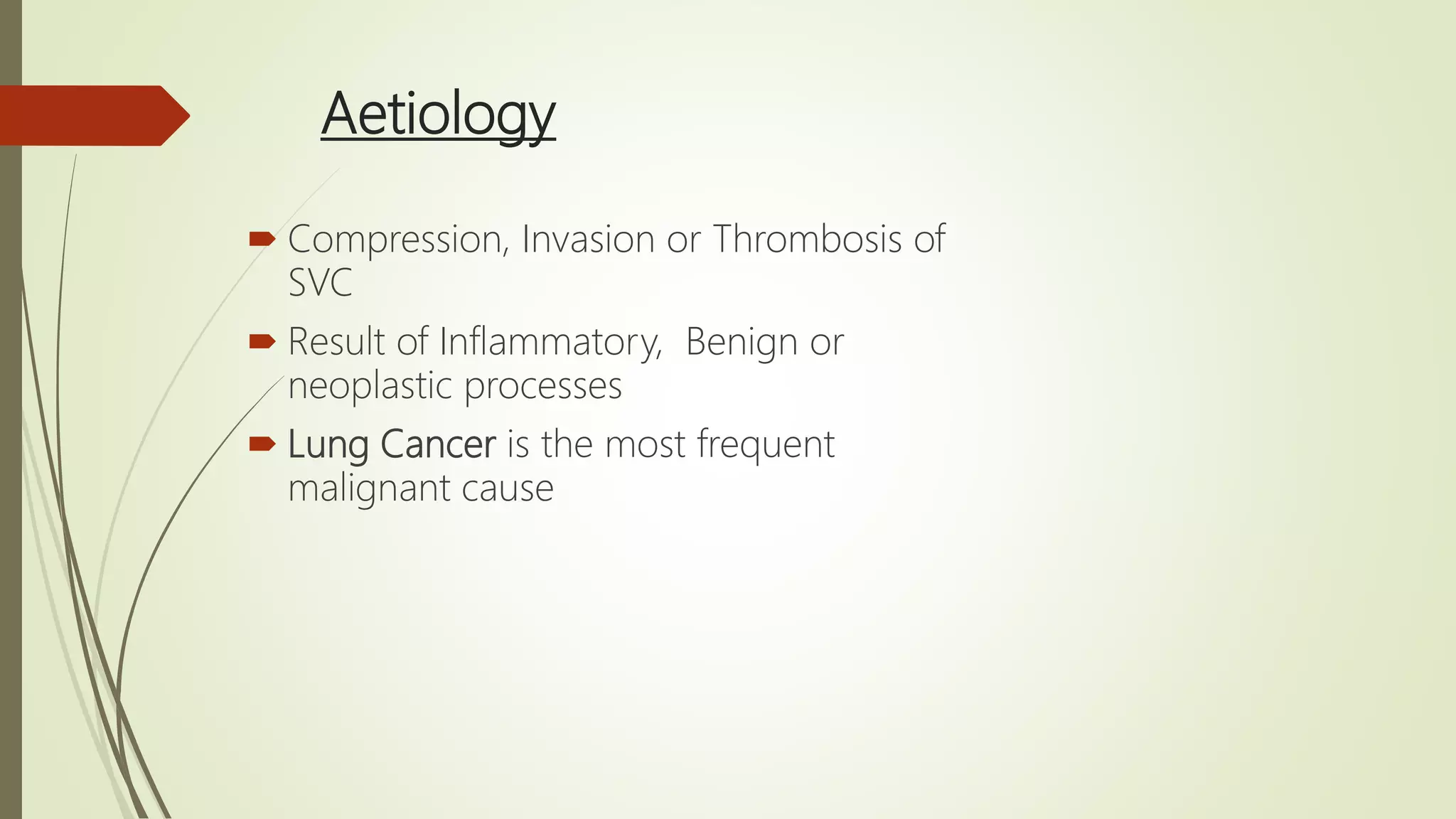
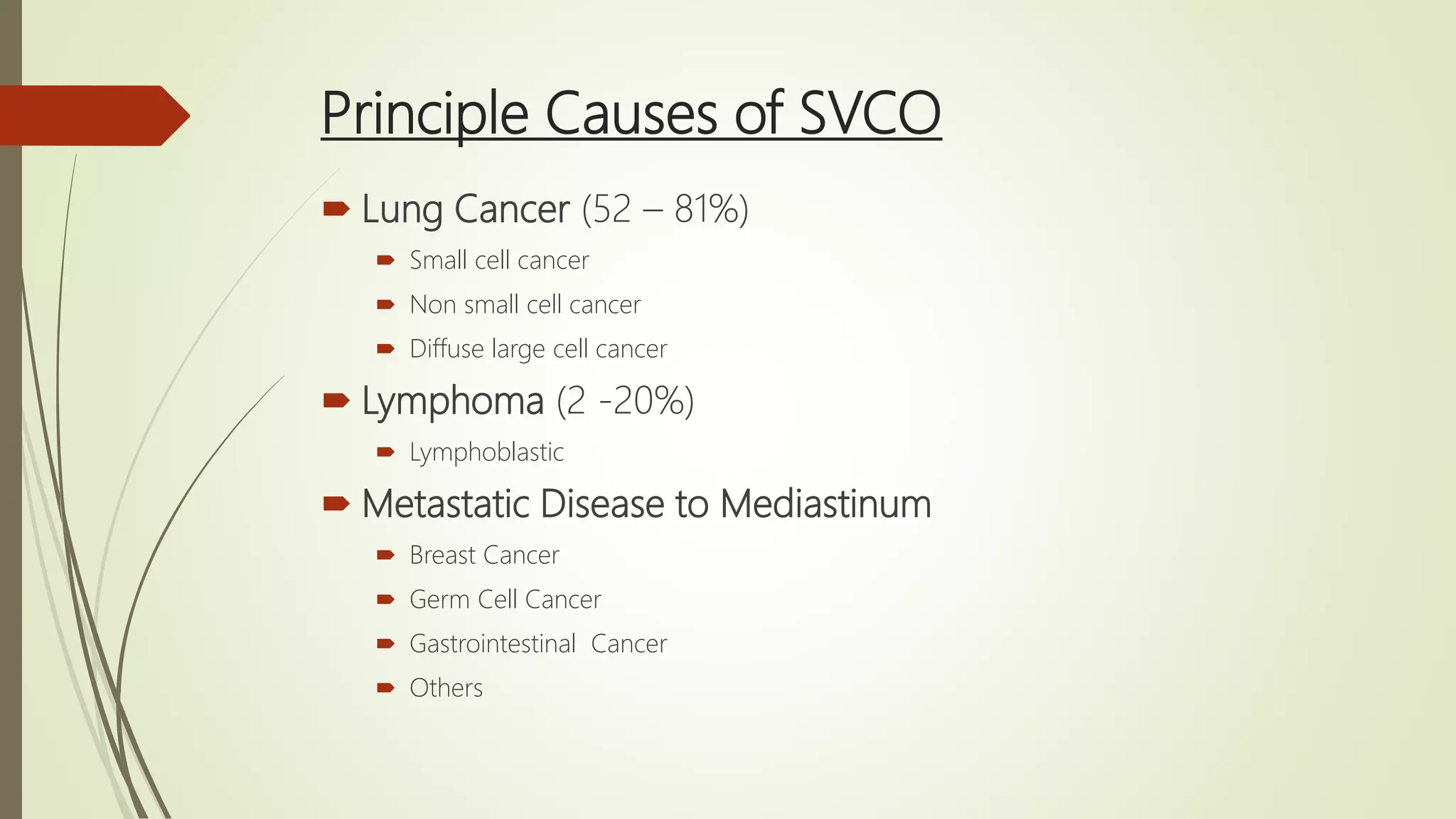
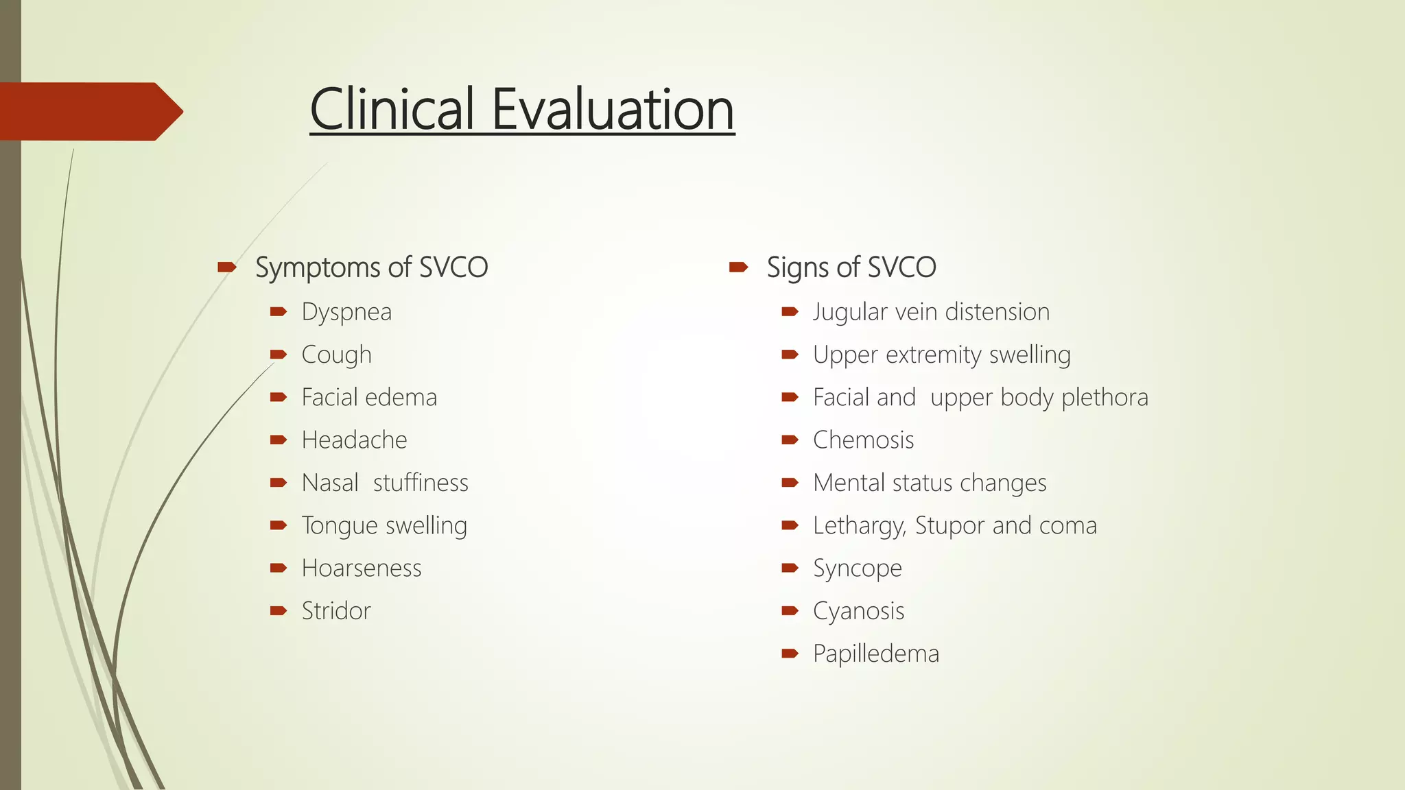
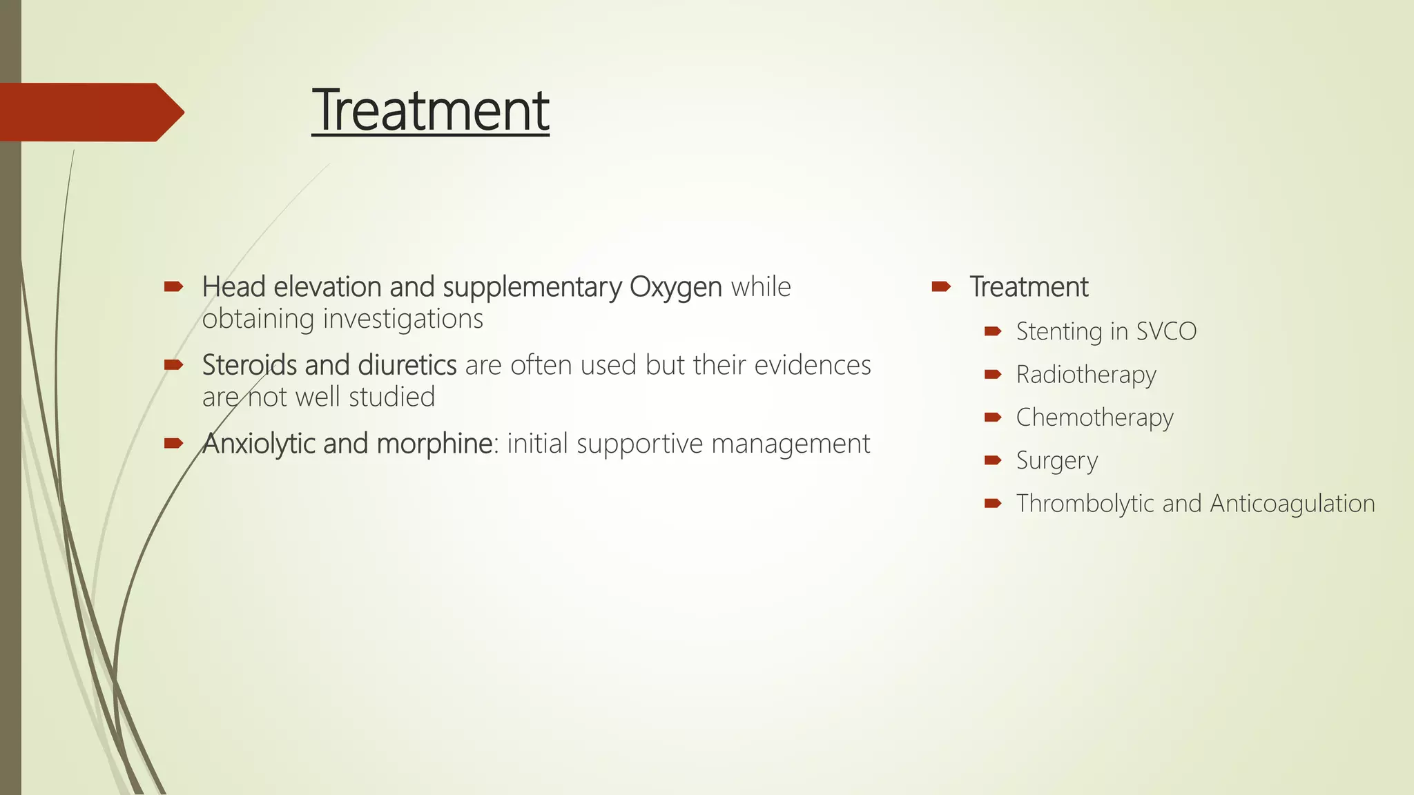
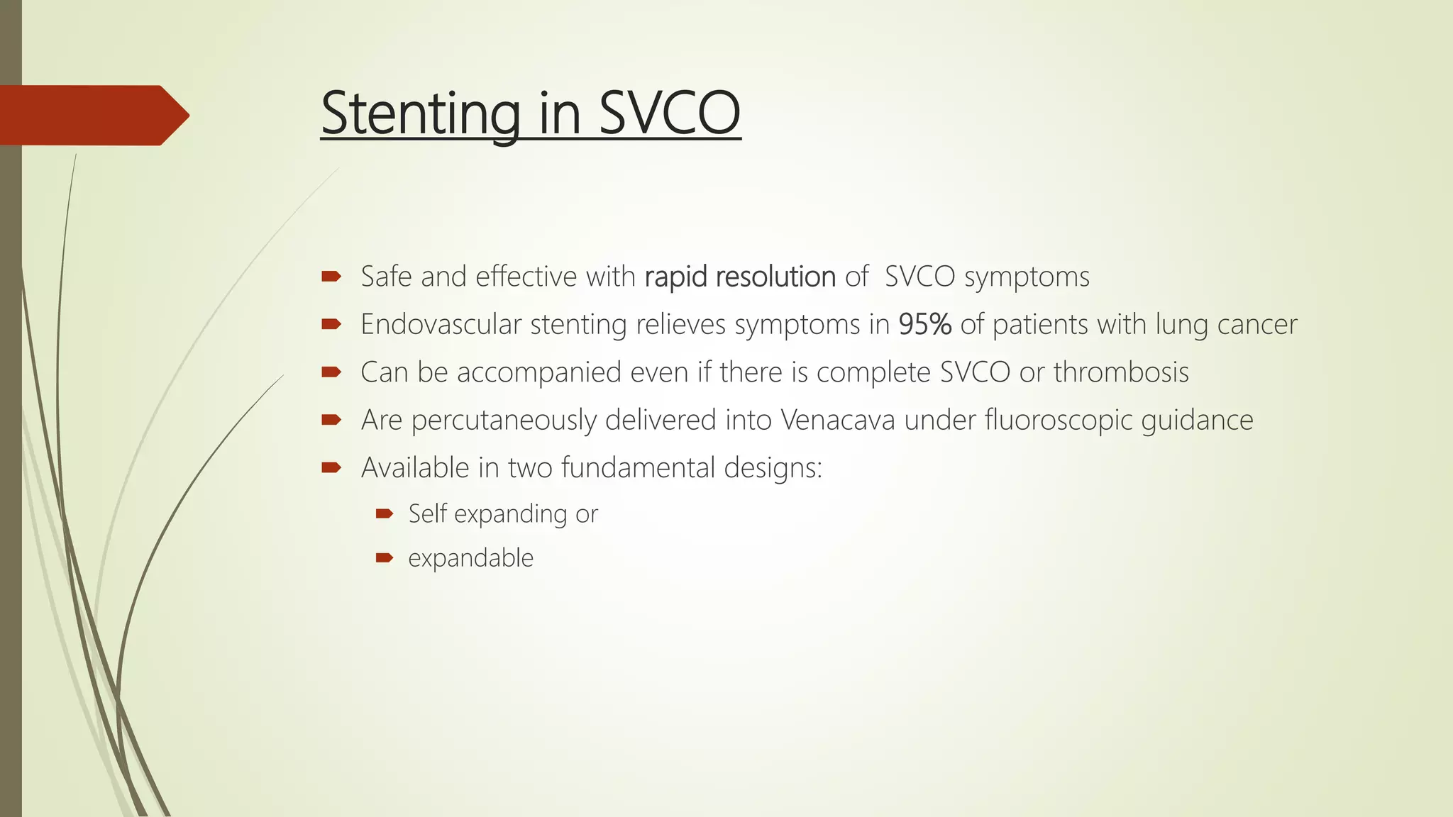
Superior Vena Cava Syndrome
(SCS)
Dr. Subhash Thakur
Clinical Oncologist, CMC, Bharatpur, Nepal
MD (PGIMER, Chandigarh)
Contents
 Introduction
 Physiology of SVC Obstruction
(SVCO)
 Aetiology
 Clinical Evaluation
 Investigations
 Treatment
Stenting in SVCO
Radiotherapy
Chemotherapy
Surgery
Thrombolytic and
Anticoagulation
 Summary
Introduction
 A range of signs and symptoms from compression of SVC or
associated greater veins (External Compression or Internal
Obstruction)
 Secondary to malignancies: 73-97%
 Rarely an emergency in absence of tracheal compression
 However, SVCO has an impact on prognosis
Median survival who receive treatment: 46 weeks
Median survival without treatment: 6 weeks
Physiology of Superior Vena Cava Obstruction
 Superior Venacava
Thin walled, compliant and easily
compressible vein
1/3rd of total venous return to heart
Head, neck and upper extremities
 SVC compression or Obstruction can
result in compromise of cardiac
output in acute setting, but within
few hours, collaterals develop
 Collateral vessels achieve steady state
blood return to the azygous vein or
inferior Venacava
Severity of Symptoms depend on:
 Degree of narrowing of SVC and
 Speed of Onset
Acute Setting
Elevated Venous pressure
Interstitial edema
Laryngeal and
Cerebral Edema
Aetiology
 Compression, Invasion or Thrombosis of
SVC
 Result of Inflammatory, Benign or
neoplastic processes
 Lung Cancer is the most frequent
malignant cause
Principle Causes of SVCO
 Lung Cancer (52 – 81%)
 Small cell cancer
 Non small cell cancer
 Diffuse large cell cancer
 Lymphoma (2 -20%)
 Lymphoblastic
 Metastatic Disease to Mediastinum
 Breast Cancer
 Germ Cell Cancer
 Gastrointestinal Cancer
 Others
 Primary Mediastinal Tumors
 Thymoma
 Sarcoma
 Melanoma
 Thymic Carcinoma
 Non-Malignant Causes
 Infectious diseases: syphilis, Tb, Histoplasmacytosis
 Central Thrombus and other iatrogenic causes
 Idiopathic fibrosing mediastinitis
 Congenital Heart Failure
 Goiter
Clinical Evaluation
 All patients with suspected SVCO should have thorough clinical history to assess
duration and speed of symptom onset
 History should also involve previous invasive procedures and malignancies
 Detail examination can rule common differentials like CHF and Cushing's
syndrome
 Careful examination of neurological system: subtle but life threatening due to
cerebral Edema
Clinical Evaluation
 Symptoms of SVCO
 Dyspnea
 Cough
 Facial edema
 Headache
 Nasal stuffiness
 Tongue swelling
 Hoarseness
 Stridor
 Signs of SVCO
 Jugular vein distension
 Upper extremity swelling
 Facial and upper body plethora
 Chemosis
 Mental status changes
 Lethargy, Stupor and coma
 Syncope
 Cyanosis
 Papilledema
Radiological Evaluation
 Chest X-ray
 Often abnormal
 Can identify superior
mediastinal masses or
mediastinal widening
 Hilar masses and pleural
effusion
 Contrast CT or MRI
 Gold standard
 Localize the level of SVCO and underlying pathology
 Tumor mass/size
 SVC diameter
 Length of stenosis/obstruction
 Evidence of SVCO thrombus
 Formation of collateral vessels
Tissue Biopsy
 Treatment is determined by underlying pathology
 Sub-acute setting, malignancy is suspected, tissue biopsy should be obtained
 Can be done via
 Bronchoscopy
 Endobronchial ultrasound
 Mediastinoscopy
 FNAC or excision biopsy
 CT guided biopsy
Treatment
 Treatment
 Stenting in SVCO
 Radiotherapy
 Chemotherapy
 Surgery
 Thrombolytic and Anticoagulation
 Head elevation and supplementary Oxygen while
obtaining investigations
 Steroids and diuretics are often used but their evidences
are not well studied
 Anxiolytic and morphine: initial supportive management
Stenting in SVCO
 Safe and effective with rapid resolution of SVCO symptoms
 Endovascular stenting relieves symptoms in 95% of patients with lung cancer
 Can be accompanied even if there is complete SVCO or thrombosis
 Are percutaneously delivered into Venacava under fluoroscopic guidance
 Available in two fundamental designs:
 Self expanding or
 expandable
 Immediate Endovascular Stenting
 Life threatening symptoms such as hemodynamic compromise, laryngeal edema or
cerebral edema
 Strongly recommended for patients with limited treatment approaches like
mesothelioma
 Questionable in chemo sensitive tumors like SCLC, Lymphoma and germ cell
tumors
Complications of stenting
 3 – 7%
 Infection
 Pulmonary emboli
 Pericardial tamponade
 Stent migration
 Perforation
 Bleeding
 Stent failure due to extrinsic
tumor compression, infiltration
of tumor through the stent or
thrombus
Radiotherapy
 An effective treatment modality for certain tumor types as an
 Initial intervention or
 Adjuvant treatment after stenting
 Subjective improvement is seen within 72 hours of initiation of therapy
 75% of malignancies associated SCS notice symptomatic improvement within 3 –
5 days, 90% in 1 week
 Objective response requires 1 – 3 weeks
 Dose: 30 Gray in 10 # or 50 Gray in 25 #
 For lymphomas, daily dose of 1.8 to 2 Gy is recommended and for lung cancers 2
to 3 Gy daily dose
 All locoregional diseases including hilar and supraclavicular region should be
treated with sufficient margin
Side effects of Radiotherapy
 Initial worsening of symptoms secondary to
 edema
 tumor necrosis with fever
 myelosuppression
 alopecia
 nausea, vomiting
 stomatitis
 esophagitis and
 infection
Failure of radiation therapy: Reasons
 Obstructive Thrombosis
 Tumor recurrence
 Radiation fibrosis
 Failure of development of collaterals secondary to fibrosis
 The mean post treatment survival is 6 to 7 months
Chemotherapy
 Treatment of Choice for
 Non Hodgkin lymphoma
 Germ Cell Tumors
 SCLC
 These tumors are exquisitely chemo sensitive
 Relief of symptoms: 80% of NHL, 77% in SCLC and 40% NSCL patients
 Symptoms usually improve within 1 – 2 weeks of treatment initiation
Targeted Therapy
 No data available till date
Surgery
 To bypass or resect tumors to decompress the venous system are effective in
selected patients
 However, invasive procedures in this predominantly palliative patients has very
limited role
 In patients with malignant Thymoma and Thymic carcinoma, surgery should be
evaluated as part of multimodal treatment strategy
Thrombolytics and Anticoagulation
 Benefit is unclear
 30 – 50 % of patients with SVCO have thrombosis at post mortem
 Experts recommendation: anticoagulation after thrombolysis to
 Prevent recurrence of thrombus and
 Reduce the incidence of pulmonary emboli
 Aspirin is often recommended after stent placement in absence of thrombus
Summary
 SCS is often clinically striking but rarely requires emergency intervention
 Treatment planning should be multidisciplinary
 Tissue biopsy is warranted to guide diagnosis and optimize treatment
 Life threatening symptoms or signs: intravascular stenting provide rapid relief
 In patients with malignancy, after stenting, radiotherapy/chemotherapy is
advised
 Chemo sensitive malignancies: Chemotherapy should be initiated

Superior Vena Cava Syndrome

  • 1.
    Superior Vena CavaSyndrome (SCS) Dr. Subhash Thakur Clinical Oncologist, CMC, Bharatpur, Nepal MD (PGIMER, Chandigarh)
  • 2.
    Contents  Introduction  Physiologyof SVC Obstruction (SVCO)  Aetiology  Clinical Evaluation  Investigations  Treatment Stenting in SVCO Radiotherapy Chemotherapy Surgery Thrombolytic and Anticoagulation  Summary
  • 3.
    Introduction  A rangeof signs and symptoms from compression of SVC or associated greater veins (External Compression or Internal Obstruction)  Secondary to malignancies: 73-97%
  • 4.
     Rarely anemergency in absence of tracheal compression  However, SVCO has an impact on prognosis Median survival who receive treatment: 46 weeks Median survival without treatment: 6 weeks
  • 5.
    Physiology of SuperiorVena Cava Obstruction  Superior Venacava Thin walled, compliant and easily compressible vein 1/3rd of total venous return to heart Head, neck and upper extremities
  • 6.
     SVC compressionor Obstruction can result in compromise of cardiac output in acute setting, but within few hours, collaterals develop  Collateral vessels achieve steady state blood return to the azygous vein or inferior Venacava
  • 7.
    Severity of Symptomsdepend on:  Degree of narrowing of SVC and  Speed of Onset
  • 8.
  • 9.
    Elevated Venous pressure Interstitialedema Laryngeal and Cerebral Edema
  • 11.
    Aetiology  Compression, Invasionor Thrombosis of SVC  Result of Inflammatory, Benign or neoplastic processes  Lung Cancer is the most frequent malignant cause
  • 12.
    Principle Causes ofSVCO  Lung Cancer (52 – 81%)  Small cell cancer  Non small cell cancer  Diffuse large cell cancer  Lymphoma (2 -20%)  Lymphoblastic  Metastatic Disease to Mediastinum  Breast Cancer  Germ Cell Cancer  Gastrointestinal Cancer  Others
  • 13.
     Primary MediastinalTumors  Thymoma  Sarcoma  Melanoma  Thymic Carcinoma  Non-Malignant Causes  Infectious diseases: syphilis, Tb, Histoplasmacytosis  Central Thrombus and other iatrogenic causes  Idiopathic fibrosing mediastinitis  Congenital Heart Failure  Goiter
  • 14.
    Clinical Evaluation  Allpatients with suspected SVCO should have thorough clinical history to assess duration and speed of symptom onset  History should also involve previous invasive procedures and malignancies  Detail examination can rule common differentials like CHF and Cushing's syndrome  Careful examination of neurological system: subtle but life threatening due to cerebral Edema
  • 15.
    Clinical Evaluation  Symptomsof SVCO  Dyspnea  Cough  Facial edema  Headache  Nasal stuffiness  Tongue swelling  Hoarseness  Stridor  Signs of SVCO  Jugular vein distension  Upper extremity swelling  Facial and upper body plethora  Chemosis  Mental status changes  Lethargy, Stupor and coma  Syncope  Cyanosis  Papilledema
  • 16.
    Radiological Evaluation  ChestX-ray  Often abnormal  Can identify superior mediastinal masses or mediastinal widening  Hilar masses and pleural effusion
  • 17.
     Contrast CTor MRI  Gold standard  Localize the level of SVCO and underlying pathology  Tumor mass/size  SVC diameter  Length of stenosis/obstruction  Evidence of SVCO thrombus  Formation of collateral vessels
  • 18.
    Tissue Biopsy  Treatmentis determined by underlying pathology  Sub-acute setting, malignancy is suspected, tissue biopsy should be obtained  Can be done via  Bronchoscopy  Endobronchial ultrasound  Mediastinoscopy  FNAC or excision biopsy  CT guided biopsy
  • 20.
    Treatment  Treatment  Stentingin SVCO  Radiotherapy  Chemotherapy  Surgery  Thrombolytic and Anticoagulation  Head elevation and supplementary Oxygen while obtaining investigations  Steroids and diuretics are often used but their evidences are not well studied  Anxiolytic and morphine: initial supportive management
  • 21.
    Stenting in SVCO Safe and effective with rapid resolution of SVCO symptoms  Endovascular stenting relieves symptoms in 95% of patients with lung cancer  Can be accompanied even if there is complete SVCO or thrombosis  Are percutaneously delivered into Venacava under fluoroscopic guidance  Available in two fundamental designs:  Self expanding or  expandable
  • 22.
     Immediate EndovascularStenting  Life threatening symptoms such as hemodynamic compromise, laryngeal edema or cerebral edema  Strongly recommended for patients with limited treatment approaches like mesothelioma  Questionable in chemo sensitive tumors like SCLC, Lymphoma and germ cell tumors
  • 23.
    Complications of stenting 3 – 7%  Infection  Pulmonary emboli  Pericardial tamponade  Stent migration  Perforation  Bleeding  Stent failure due to extrinsic tumor compression, infiltration of tumor through the stent or thrombus
  • 24.
    Radiotherapy  An effectivetreatment modality for certain tumor types as an  Initial intervention or  Adjuvant treatment after stenting  Subjective improvement is seen within 72 hours of initiation of therapy  75% of malignancies associated SCS notice symptomatic improvement within 3 – 5 days, 90% in 1 week  Objective response requires 1 – 3 weeks
  • 25.
     Dose: 30Gray in 10 # or 50 Gray in 25 #  For lymphomas, daily dose of 1.8 to 2 Gy is recommended and for lung cancers 2 to 3 Gy daily dose  All locoregional diseases including hilar and supraclavicular region should be treated with sufficient margin
  • 26.
    Side effects ofRadiotherapy  Initial worsening of symptoms secondary to  edema  tumor necrosis with fever  myelosuppression  alopecia  nausea, vomiting  stomatitis  esophagitis and  infection
  • 27.
    Failure of radiationtherapy: Reasons  Obstructive Thrombosis  Tumor recurrence  Radiation fibrosis  Failure of development of collaterals secondary to fibrosis  The mean post treatment survival is 6 to 7 months
  • 28.
    Chemotherapy  Treatment ofChoice for  Non Hodgkin lymphoma  Germ Cell Tumors  SCLC  These tumors are exquisitely chemo sensitive  Relief of symptoms: 80% of NHL, 77% in SCLC and 40% NSCL patients  Symptoms usually improve within 1 – 2 weeks of treatment initiation
  • 29.
    Targeted Therapy  Nodata available till date
  • 30.
    Surgery  To bypassor resect tumors to decompress the venous system are effective in selected patients  However, invasive procedures in this predominantly palliative patients has very limited role  In patients with malignant Thymoma and Thymic carcinoma, surgery should be evaluated as part of multimodal treatment strategy
  • 31.
    Thrombolytics and Anticoagulation Benefit is unclear  30 – 50 % of patients with SVCO have thrombosis at post mortem  Experts recommendation: anticoagulation after thrombolysis to  Prevent recurrence of thrombus and  Reduce the incidence of pulmonary emboli  Aspirin is often recommended after stent placement in absence of thrombus
  • 32.
    Summary  SCS isoften clinically striking but rarely requires emergency intervention  Treatment planning should be multidisciplinary  Tissue biopsy is warranted to guide diagnosis and optimize treatment  Life threatening symptoms or signs: intravascular stenting provide rapid relief  In patients with malignancy, after stenting, radiotherapy/chemotherapy is advised  Chemo sensitive malignancies: Chemotherapy should be initiated