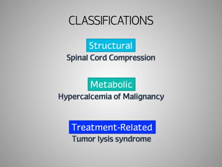
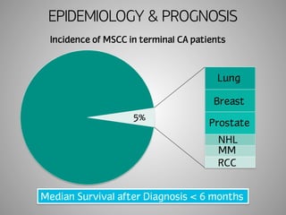
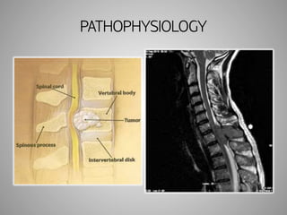
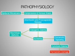
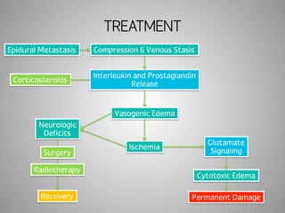
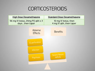
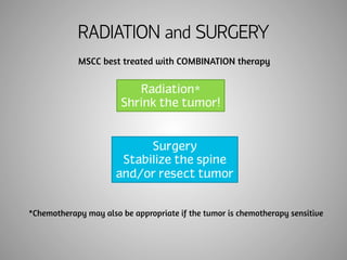
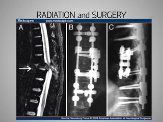
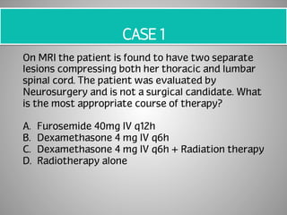
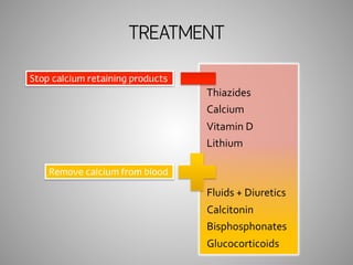
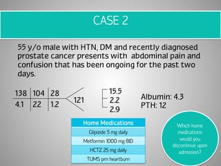
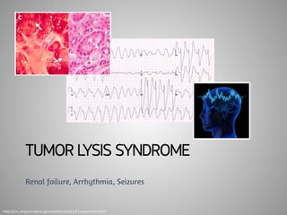
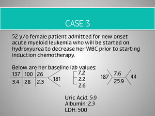
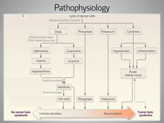
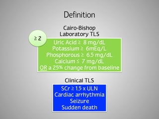
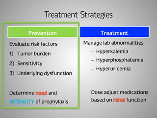
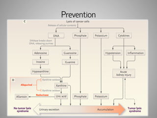
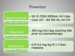
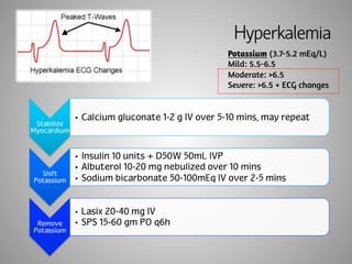
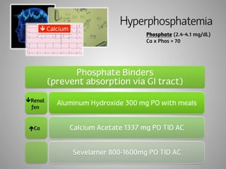
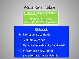
ONCOLOGIC EMERGENCIES 
Marti Larriva, PharmD 
PGY1 Pharmacy Practice Resident 
September 18, 2014
OBJECTIVES 
• Identify risks and benefits of MSCC therapy 
• Create a treatment plan for malignant 
hypercalcemia 
• Identify risk factors for TLS and synthesize 
appropriate prevention plan
Importance, Definitions, & Types 
OVERVIEW
ONCOLOGY EMERGENCY 
A clinical condition resulting from a 
metabolic, neurologic, cardiovascular, 
hematologic and/or infectious change 
caused by that 
to prevent 
loss of life or quality of life.
YOUR ROLE 
Awareness 
Prevention 
Monitoring 
Treatment
CLASSIFICATIONS 
Structural 
Metabolic 
Treatment-Related
MALIGNANT SPINAL CORD 
COMPRESSION (MSCC) 
Back pain, paralysis, loss of bowel/bladder control
CASE 1 
46 y/o female with stage IV breast cancer and no 
other significant PMH is admitted to your general 
medicine team s/p fall with cc: back pain, 
numbness/tingling and weakness in both lower 
extremities.
EPIDEMIOLOGY & PROGNOSIS 
Incidence of MSCC in terminal CA patients 
5% 
Lung 
Breast 
Prostate 
NHL 
MM 
RCC 
Median Survival after Diagnosis < 6 months
PATHOPHYSIOLOGY
PATHOPHYSIOLOGY 
Epidural Metastasis Compression & Venous Stasis 
Interleukin and Prostaglandin 
Release 
Vasogenic Edema 
Ischemia 
Neurologic 
Deficits 
Glutamate 
Signaling 
Cytotoxic Edema 
Permanent Damage
TREATMENT 
Epidural Metastasis Compression & Venous Stasis 
Interleukin and Prostaglandin 
Release 
Vasogenic Edema 
Ischemia 
Corticosteroids 
Neurologic 
Deficits 
Glutamate 
Signaling 
Cytotoxic Edema 
Surgery 
Radiotherapy 
Recovery Permanent Damage
CORTICOSTEROIDS 
High Dose Dexamethasone Standard Dose Dexamethasone 
96 mg IV bolus, 24mg PO q6h x 3 
days , then taper 
10 mg IV bolus, then 
4 mg IV q6h, then taper 
Adverse 
Effects Benefits 
GI perforation 
Infection 
Psychosis 
Severe Neuro 
deficits
RADIATION and SURGERY 
MSCC best treated with COMBINATION therapy 
Radiation* 
Shrink the tumor! 
Surgery 
Stabilize the spine 
and/or resect tumor 
*Chemotherapy may also be appropriate if the tumor is chemotherapy sensitive
RADIATION and SURGERY
CASE 1 
46 y/o female with stage IV breast cancer and no 
other significant PMH is admitted to your general 
medicine team s/p fall with cc: back pain, 
numbness/tingling and weakness in both lower 
extremities.
CASE 1 
On MRI the patient is found to have two separate 
lesions compressing both her thoracic and lumbar 
spinal cord. The patient was evaluated by 
Neurosurgery and is not a surgical candidate. What 
is the most appropriate course of therapy? 
A. Furosemide 40mg IV q12h 
B. Dexamethasone 4 mg IV q6h 
C. Dexamethasone 4 mg IV q6h + Radiation therapy 
D. Radiotherapy alone
HYPERCALCEMIA 
OF MALIGNANCY 
Bones, stones, groans, and psychological undertones
CASE 2 
55 y/o male with HTN, DM and recently diagnosed 
prostate cancer presents with abdominal pain and 
confusion that has been ongoing for the past two 
days. 
138 104 28 
4.1 22 1.2 
121 
15.5 
2.2 
2.9 
Albumin: 4.3 
PTH: 12 
Home Medications 
Glipizide 5 mg daily 
Metformin 1000 mg BID 
HCTZ 25 mg daily 
TUMS prn heartburn
EPIDEMIOLOGY & PROGNOSIS 
Patients experiencing hypercalcemia during 
disease course 
1/3 
Breast 
Prostate 
Renal Cell 
MM 
T Cell Leukemia 
T Cell Lymphoma 
Patients hospitalized for 
hypercalcemia 
Survived 
30 days 
Died after 
30 days
PATHOPHYSIOLOGY 
PTHrP Vitamin D 
Bone 
Metastasis 
Released 
from 
tumor 
Osteoclast 
stimulation 
Absorption 
Resorption 
Bone 
store 
release 
80% 
of 
cases
Parathyroid hormone related peptide
Degrees of hypercalcemia 
Mild 
10.5-11.9mg/dL 
Moderate 
12-13.9 mg/dL 
Severe 
≥ 14 mg/dL 
Confusion 
Lethargy 
Coma 
N/V 
Anorexia 
Polydipsia 
Polyuria 
Constipation 
Weight Loss 
Bone Pain 
Fatigue 
Corrected Calcium = Calcium + 0.8 (4-Albumin)
TREATMENT 
Stop calcium retaining products 
Thiazides 
Calcium 
Vitamin 
D 
Lithium 
Fluids 
+ 
Diuretics 
Calcitonin 
Bisphosphonates 
Glucocorticoids 
Remove calcium from blood
TREATMENT 
Hydration (6-12h*) 
NS 300-500cc/hr until euvolemic Caution: HF, Renal failure* 
Diuresis (6-12h) 
Lasix 20-40 mg IV q12-24h Avoid Thiazides 
Calcitonin (12-24h) 
Calcitonin 4-8IU/kg IM/IV q12h Tachyphylaxis often occurs, IN ineffective 
Bisphosphonates (48-72h) 
Glucocorticoids (3-5 days) 
Hydrocortisone 100 mg IV q6h 
Prednisone 60 mg PO daily 
Usually limited to lymphomas 
Zoledronic Acid 4 mg IV 
Pamidronate 60-90 mg IV 
Caution: poor dentition (ONJ) or reduced CrCl 
PTH
Monitoring 
Therapeutic Efficacy Therapeutic Toxicity 
Potassium 
Magnesium 
Phosphate 
Serum Creatinine 
Urine output (I/O) 
Calcium 
Albumin 
Ionized calcium
CASE 2 
55 y/o male with HTN, DM and recently diagnosed 
prostate cancer presents with abdominal pain and 
confusion that has been ongoing for the past two 
days. 
138 104 28 
4.1 22 1.2 
121 
15.5 
2.2 
2.9 
Albumin: 4.3 
PTH: 12 
Home Medications 
Glipizide 5 mg daily 
Metformin 1000 mg BID 
HCTZ 25 mg daily 
TUMS prn heartburn 
Which home 
medications 
would you 
discontinue upon 
admission?
CASE 2 
The patient has already been started on NS running 
at 300mL/hr, what would be the next step in 
treatment and what would you monitor? 
A. Hydrocortisone 100 mg IV q6h; Monitor Ca, K, Mg 
B. Furosemide 40 mg IV q24h; Monitor Ca, K, Mg, SCr 
C. Zoledronic Acid 4 mg IV x 1; Monitor Phos, Ca 
D. Dialysis; Monitor Ca 
E. B and C
TUMOR LYSIS SYNDROME 
Renal failure, Arrhythmia, Seizures 
http://circ.ahajournals.org/content/116/1/e2/F3.expansion.html
CASE 3 
52 y/o female patient admitted for new onset 
acute myeloid leukemia who will be started on 
hydroxyurea to decrease her WBC prior to starting 
induction chemotherapy. 
Below are her baseline lab values: 
137 100 26 
181 
3.4 28 2.3 
7.2 
2.2 
2.6 
Uric Acid: 5.9 
Albumin: 2.3 
LDH: 500 
7.6 
25.9 
187 44
Epidemiology & Prognosis 
Cancer 
Mass 
Bulky 
or 
metastatic 
Organ 
involvement 
Lysis 
Potential 
Proliferation 
Rate 
Cell 
Sensitivity 
Chemo 
Intensity 
Patient 
Factors 
Renal 
Impairment 
Hypotension/ 
Volume 
depletion 
Nephrotoxins 
TLS é 
Risk of 
death during 
treatment 
and é LOS
Pathophysiology
TLS Induced Renal Failure
Definition 
Cairo-Bishop 
Laboratory TLS 
Uric Acid ≥ 
8 mg/dL 
Potassium ≥ 
6mEq/L 
Phosphorous ≥ 
6.5 mg/dL 
Calcium ≤ 
7 mg/dL 
OR a 25% change from baseline 
Clinical TLS 
SCr ≥ 1.5 x ULN 
Cardiac arrhythmia 
Seizure 
Sudden death 
≥ 2
Treatment Strategies 
Prevention 
Evaluate risk factors 
1) Tumor burden 
2) Sensitivity 
3) Underlying dysfunction 
Determine and 
of prophylaxis 
Treatment 
Manage lab abnormalities 
– Hyperkalemia 
– Hyperphosphatemia 
– Hyperuricemia 
Dose adjust medications 
based on function
Prevention
Prevention 
• NS IV 2500-3000mL/m2/day 
• Goal: UO = 80-100 mL/m2/hr 
Hyperhydration 
• 300 mg/m2/day starting 48h 
prior to chemotherapy 
Caution: 
Heart 
or 
Renal 
failure 
Allopurinol 
• 0.15-0.2 mg/kg IV x 1 then 
reassess 
No 
need 
for 
renal 
dose 
adjust 
Rasburicase 
Ensure 
proper 
G6PD 
function
Risk Stratification 
Low Intermediate High 
Solid 
Tumors* 
Multiple 
Myeloma 
*Exception: 
Germ cell 
cancer OR 
small cell 
lung cancer 
Leukemia/Lymphoma 
Less aggressive 
Indolent 
Burkitt’s 
Lymphoma 
Elevated LDH>2x ULN 
Elevated WBC>25
Prevention 
Low Intermediate High 
Monitor 
Hyper-hydration 
prn Normal 
Hydration 
Allopurinol 
Monitor 
*prn loop diuretic 
Hyper-hydration 
Allopurinol 
OR 
Rasburicase 
*prn loop diuretic 
Monitor
Treatment Strategies 
Prevention 
Evaluate risk factors 
1) Tumor burden 
2) Sensitivity 
3) Underlying dysfunction 
Determine and 
of prophylaxis 
Treatment 
Manage lab abnormalities 
– Hyperkalemia 
– Hyperphosphatemia 
– Hyperuricemia 
Dose adjust medications 
based on function
Hyperkalemia 
Potassium (3.7-5.2 mEq/L) 
Mild: 5.5-6.5 
Moderate: >6.5 
Severe: >6.5 + ECG changes 
Stabilize 
Myocardium 
• Calcium gluconate 1-2 g IV over 5-10 mins, may repeat 
Shift 
Potassium 
• Insulin 10 units + D50W 50mL IVP 
• Albuterol 10-20 mg nebulized over 10 mins 
• Sodium bicarbonate 50-100mEq IV over 2-5 mins 
Remove 
Potassium 
• Lasix 20-40 mg IV 
• SPS 15-60 gm PO q6h
Hyperphosphatemia 
Phosphate (2.4-4.1 mg/dL) 
Ca x Phos > 70 
Phosphate Binders 
(prevent absorption via GI tract) 
Aluminum Hydroxide 300 mg PO with meals 
Calcium Acetate 1337 mg PO TID AC 
Sevelamer 800-1600mg PO TID AC 
ê Calcium 
êRenal 
fxn 
éCa
Hyperuricemia 
Uric Acid 
Low <4 
Intermediate: 4-8 
High >8 
Flush out 
uric acid 
• NS 2500-3000mL/m2/day 
• Goal: UO = 80-100mL/m2/hr 
Metabolize 
Uric Acid 
• Rasburicase 0.2 mg/kg IV x 1, 
redose based upon UA levels
Acute Renal Failure 
First line: Hyperhydration 
NS IV 3L/m2/day 
Goal UO 80-100mL/m2/hr 
Dialysis if: 
1) No response to fluids 
2) Volume overload 
3) Hyperkalemia despite treatment 
4) Phosphate > 10.2mg/dL + 
symptomatic hypocalcemia
CASE 3 
52 y/o female patient admitted for new onset 
acute myeloid leukemia who will be started on 
hydroxyurea to decrease her WBC prior to starting 
induction chemotherapy. 
Below are her baseline lab values: 
137 100 26 
181 
3.4 28 2.3 
7.2 
2.2 
2.6 
Uric Acid: 5.9 
Albumin: 2.3 
LDH: 500 
7.6 
25.9 
187 44 
Ht: 149.9 cm 
Wt: 99.9 kg
CASE 3 
Based upon her baseline lab values and risk for TLS 
what preventative regimen should she be placed 
on? 
A. Intermediate Risk (hyperhydration + allopurinol) 
B. Low Risk (normal hydration) 
C. High Risk (hyperhydration + rasburicase OR 
allopurinol) 
Why did 
you choose 
this 
answer?
Take Home Points 
1) MSCC should be treated initially with steroids 
(dexamethasone), but with 
radiotherapy and surgery is more effective overall. 
2) HOM is best treated with to 
decrease calcium in the acute setting, but 
are first line to obtain 
normocalcemia over the coming weeks and should 
be started concomitantly. 
3) Severity and occurrence TLS can be reduced through 
adequate and treatment 
prior to chemotherapy.
Other Oncologic Emergencies 
Structural 
Pericardial Effusion 
Superior Vena Cava Syndrome 
Increased ICP 
Metabolic 
SIADH 
Treatment-Related 
Neutropenic Fever 
Hypersensitivity 
Hematologic 
Hyperviscosity
Questions? 
marti.larriva@ucsf.edu
REFERENCES 
1. 
George 
R, 
Jeba 
J, 
Ramkumar 
G 
et 
al. 
Interventions 
for 
the 
treatment 
of 
metastatic 
extradural 
spinal 
cord 
compression 
in 
adults. 
Cochrane 
Database 
Syst 
Rev. 
2008;(4):CD006716. 
doi(4):CD006716. 
2. 
Heimdal 
K, 
Hirschberg 
H, 
Slettebo 
H 
et 
al. 
High 
incidence 
of 
serious 
side 
effects 
of 
high-­‐dose 
dexamethasone 
treatment 
in 
patients 
with 
epidural 
spinal 
cord 
compression. 
J 
Neurooncol. 
1992;12(2):141-­‐4. 
3. 
Howard 
SC, 
Jones 
DP, 
Pui 
CH. 
The 
tumor 
lysis 
syndrome. 
N 
Engl 
J 
Med. 
2011;364(19):1844-­‐54. 
4. 
Kraft 
MD, 
Btaiche 
IF, 
Sacks 
GS 
et 
al. 
Treatment 
of 
electrolyte 
disorders 
in 
adult 
patients 
in 
the 
intensive 
care 
unit. 
Am 
J 
Health 
Syst 
Pharm. 
2005;62(16):1663-­‐82. 
5. 
Lewis 
MA, 
Hendrickson 
AW, 
Moynihan 
TJ. 
Oncologic 
emergencies: 
Pathophysiology, 
presentation, 
diagnosis, 
and 
treatment. 
CA 
Cancer 
J 
Clin. 
2011. 
6. 
Loblaw 
DA, 
Mitera 
G, 
Ford 
M 
et 
al. 
A 
2011 
updated 
systematic 
review 
and 
clinical 
practice 
guideline 
for 
the 
management 
of 
malignant 
extradural 
spinal 
cord 
compression. 
Int 
J 
Radiat 
Oncol 
Biol 
Phys. 
2012;84(2):312-­‐7. 
7. 
McCurdy 
MT, 
Shanholtz 
CB. 
Oncologic 
emergencies. 
Crit 
Care 
Med. 
2012;40(7):2212-­‐22. 
8. 
Mundy 
GR, 
Edwards 
JR. 
PTH-­‐related 
peptide 
(PTHrP) 
in 
hypercalcemia. 
J 
Am 
Soc 
Nephrol. 
2008;19(4):672-­‐5. 
9. 
Prasad 
D, 
Schiff 
D. 
Malignant 
spinal-­‐cord 
compression. 
Lancet 
Oncol. 
2005;6(1):15-­‐24. 
10. 
Samphao 
S, 
Eremin 
JM, 
Eremin 
O. 
Oncological 
emergencies: 
Clinical 
importance 
and 
principles 
of 
management. 
Eur 
J 
Cancer 
Care 
(Engl). 
2010;19(6):707-­‐13.

Oncologic emergencies

  • 1.
    ONCOLOGIC EMERGENCIES MartiLarriva, PharmD PGY1 Pharmacy Practice Resident September 18, 2014
  • 2.
    OBJECTIVES • Identifyrisks and benefits of MSCC therapy • Create a treatment plan for malignant hypercalcemia • Identify risk factors for TLS and synthesize appropriate prevention plan
  • 3.
  • 5.
    ONCOLOGY EMERGENCY Aclinical condition resulting from a metabolic, neurologic, cardiovascular, hematologic and/or infectious change caused by that to prevent loss of life or quality of life.
  • 6.
    YOUR ROLE Awareness Prevention Monitoring Treatment
  • 7.
  • 8.
    MALIGNANT SPINAL CORD COMPRESSION (MSCC) Back pain, paralysis, loss of bowel/bladder control
  • 9.
    CASE 1 46y/o female with stage IV breast cancer and no other significant PMH is admitted to your general medicine team s/p fall with cc: back pain, numbness/tingling and weakness in both lower extremities.
  • 10.
    EPIDEMIOLOGY & PROGNOSIS Incidence of MSCC in terminal CA patients 5% Lung Breast Prostate NHL MM RCC Median Survival after Diagnosis < 6 months
  • 11.
  • 12.
    PATHOPHYSIOLOGY Epidural MetastasisCompression & Venous Stasis Interleukin and Prostaglandin Release Vasogenic Edema Ischemia Neurologic Deficits Glutamate Signaling Cytotoxic Edema Permanent Damage
  • 13.
    TREATMENT Epidural MetastasisCompression & Venous Stasis Interleukin and Prostaglandin Release Vasogenic Edema Ischemia Corticosteroids Neurologic Deficits Glutamate Signaling Cytotoxic Edema Surgery Radiotherapy Recovery Permanent Damage
  • 14.
    CORTICOSTEROIDS High DoseDexamethasone Standard Dose Dexamethasone 96 mg IV bolus, 24mg PO q6h x 3 days , then taper 10 mg IV bolus, then 4 mg IV q6h, then taper Adverse Effects Benefits GI perforation Infection Psychosis Severe Neuro deficits
  • 15.
    RADIATION and SURGERY MSCC best treated with COMBINATION therapy Radiation* Shrink the tumor! Surgery Stabilize the spine and/or resect tumor *Chemotherapy may also be appropriate if the tumor is chemotherapy sensitive
  • 16.
  • 17.
    CASE 1 46y/o female with stage IV breast cancer and no other significant PMH is admitted to your general medicine team s/p fall with cc: back pain, numbness/tingling and weakness in both lower extremities.
  • 18.
    CASE 1 OnMRI the patient is found to have two separate lesions compressing both her thoracic and lumbar spinal cord. The patient was evaluated by Neurosurgery and is not a surgical candidate. What is the most appropriate course of therapy? A. Furosemide 40mg IV q12h B. Dexamethasone 4 mg IV q6h C. Dexamethasone 4 mg IV q6h + Radiation therapy D. Radiotherapy alone
  • 19.
    HYPERCALCEMIA OF MALIGNANCY Bones, stones, groans, and psychological undertones
  • 20.
    CASE 2 55y/o male with HTN, DM and recently diagnosed prostate cancer presents with abdominal pain and confusion that has been ongoing for the past two days. 138 104 28 4.1 22 1.2 121 15.5 2.2 2.9 Albumin: 4.3 PTH: 12 Home Medications Glipizide 5 mg daily Metformin 1000 mg BID HCTZ 25 mg daily TUMS prn heartburn
  • 21.
    EPIDEMIOLOGY & PROGNOSIS Patients experiencing hypercalcemia during disease course 1/3 Breast Prostate Renal Cell MM T Cell Leukemia T Cell Lymphoma Patients hospitalized for hypercalcemia Survived 30 days Died after 30 days
  • 22.
    PATHOPHYSIOLOGY PTHrP VitaminD Bone Metastasis Released from tumor Osteoclast stimulation Absorption Resorption Bone store release 80% of cases
  • 23.
  • 24.
    Degrees of hypercalcemia Mild 10.5-11.9mg/dL Moderate 12-13.9 mg/dL Severe ≥ 14 mg/dL Confusion Lethargy Coma N/V Anorexia Polydipsia Polyuria Constipation Weight Loss Bone Pain Fatigue Corrected Calcium = Calcium + 0.8 (4-Albumin)
  • 25.
    TREATMENT Stop calciumretaining products Thiazides Calcium Vitamin D Lithium Fluids + Diuretics Calcitonin Bisphosphonates Glucocorticoids Remove calcium from blood
  • 26.
    TREATMENT Hydration (6-12h*) NS 300-500cc/hr until euvolemic Caution: HF, Renal failure* Diuresis (6-12h) Lasix 20-40 mg IV q12-24h Avoid Thiazides Calcitonin (12-24h) Calcitonin 4-8IU/kg IM/IV q12h Tachyphylaxis often occurs, IN ineffective Bisphosphonates (48-72h) Glucocorticoids (3-5 days) Hydrocortisone 100 mg IV q6h Prednisone 60 mg PO daily Usually limited to lymphomas Zoledronic Acid 4 mg IV Pamidronate 60-90 mg IV Caution: poor dentition (ONJ) or reduced CrCl PTH
  • 27.
    Monitoring Therapeutic EfficacyTherapeutic Toxicity Potassium Magnesium Phosphate Serum Creatinine Urine output (I/O) Calcium Albumin Ionized calcium
  • 28.
    CASE 2 55y/o male with HTN, DM and recently diagnosed prostate cancer presents with abdominal pain and confusion that has been ongoing for the past two days. 138 104 28 4.1 22 1.2 121 15.5 2.2 2.9 Albumin: 4.3 PTH: 12 Home Medications Glipizide 5 mg daily Metformin 1000 mg BID HCTZ 25 mg daily TUMS prn heartburn Which home medications would you discontinue upon admission?
  • 29.
    CASE 2 Thepatient has already been started on NS running at 300mL/hr, what would be the next step in treatment and what would you monitor? A. Hydrocortisone 100 mg IV q6h; Monitor Ca, K, Mg B. Furosemide 40 mg IV q24h; Monitor Ca, K, Mg, SCr C. Zoledronic Acid 4 mg IV x 1; Monitor Phos, Ca D. Dialysis; Monitor Ca E. B and C
  • 30.
    TUMOR LYSIS SYNDROME Renal failure, Arrhythmia, Seizures http://circ.ahajournals.org/content/116/1/e2/F3.expansion.html
  • 31.
    CASE 3 52y/o female patient admitted for new onset acute myeloid leukemia who will be started on hydroxyurea to decrease her WBC prior to starting induction chemotherapy. Below are her baseline lab values: 137 100 26 181 3.4 28 2.3 7.2 2.2 2.6 Uric Acid: 5.9 Albumin: 2.3 LDH: 500 7.6 25.9 187 44
  • 32.
    Epidemiology & Prognosis Cancer Mass Bulky or metastatic Organ involvement Lysis Potential Proliferation Rate Cell Sensitivity Chemo Intensity Patient Factors Renal Impairment Hypotension/ Volume depletion Nephrotoxins TLS é Risk of death during treatment and é LOS
  • 33.
  • 34.
  • 35.
    Definition Cairo-Bishop LaboratoryTLS Uric Acid ≥ 8 mg/dL Potassium ≥ 6mEq/L Phosphorous ≥ 6.5 mg/dL Calcium ≤ 7 mg/dL OR a 25% change from baseline Clinical TLS SCr ≥ 1.5 x ULN Cardiac arrhythmia Seizure Sudden death ≥ 2
  • 36.
    Treatment Strategies Prevention Evaluate risk factors 1) Tumor burden 2) Sensitivity 3) Underlying dysfunction Determine and of prophylaxis Treatment Manage lab abnormalities – Hyperkalemia – Hyperphosphatemia – Hyperuricemia Dose adjust medications based on function
  • 37.
  • 38.
    Prevention • NSIV 2500-3000mL/m2/day • Goal: UO = 80-100 mL/m2/hr Hyperhydration • 300 mg/m2/day starting 48h prior to chemotherapy Caution: Heart or Renal failure Allopurinol • 0.15-0.2 mg/kg IV x 1 then reassess No need for renal dose adjust Rasburicase Ensure proper G6PD function
  • 39.
    Risk Stratification LowIntermediate High Solid Tumors* Multiple Myeloma *Exception: Germ cell cancer OR small cell lung cancer Leukemia/Lymphoma Less aggressive Indolent Burkitt’s Lymphoma Elevated LDH>2x ULN Elevated WBC>25
  • 40.
    Prevention Low IntermediateHigh Monitor Hyper-hydration prn Normal Hydration Allopurinol Monitor *prn loop diuretic Hyper-hydration Allopurinol OR Rasburicase *prn loop diuretic Monitor
  • 41.
    Treatment Strategies Prevention Evaluate risk factors 1) Tumor burden 2) Sensitivity 3) Underlying dysfunction Determine and of prophylaxis Treatment Manage lab abnormalities – Hyperkalemia – Hyperphosphatemia – Hyperuricemia Dose adjust medications based on function
  • 42.
    Hyperkalemia Potassium (3.7-5.2mEq/L) Mild: 5.5-6.5 Moderate: >6.5 Severe: >6.5 + ECG changes Stabilize Myocardium • Calcium gluconate 1-2 g IV over 5-10 mins, may repeat Shift Potassium • Insulin 10 units + D50W 50mL IVP • Albuterol 10-20 mg nebulized over 10 mins • Sodium bicarbonate 50-100mEq IV over 2-5 mins Remove Potassium • Lasix 20-40 mg IV • SPS 15-60 gm PO q6h
  • 43.
    Hyperphosphatemia Phosphate (2.4-4.1mg/dL) Ca x Phos > 70 Phosphate Binders (prevent absorption via GI tract) Aluminum Hydroxide 300 mg PO with meals Calcium Acetate 1337 mg PO TID AC Sevelamer 800-1600mg PO TID AC ê Calcium êRenal fxn éCa
  • 44.
    Hyperuricemia Uric Acid Low <4 Intermediate: 4-8 High >8 Flush out uric acid • NS 2500-3000mL/m2/day • Goal: UO = 80-100mL/m2/hr Metabolize Uric Acid • Rasburicase 0.2 mg/kg IV x 1, redose based upon UA levels
  • 45.
    Acute Renal Failure First line: Hyperhydration NS IV 3L/m2/day Goal UO 80-100mL/m2/hr Dialysis if: 1) No response to fluids 2) Volume overload 3) Hyperkalemia despite treatment 4) Phosphate > 10.2mg/dL + symptomatic hypocalcemia
  • 46.
    CASE 3 52y/o female patient admitted for new onset acute myeloid leukemia who will be started on hydroxyurea to decrease her WBC prior to starting induction chemotherapy. Below are her baseline lab values: 137 100 26 181 3.4 28 2.3 7.2 2.2 2.6 Uric Acid: 5.9 Albumin: 2.3 LDH: 500 7.6 25.9 187 44 Ht: 149.9 cm Wt: 99.9 kg
  • 47.
    CASE 3 Basedupon her baseline lab values and risk for TLS what preventative regimen should she be placed on? A. Intermediate Risk (hyperhydration + allopurinol) B. Low Risk (normal hydration) C. High Risk (hyperhydration + rasburicase OR allopurinol) Why did you choose this answer?
  • 48.
    Take Home Points 1) MSCC should be treated initially with steroids (dexamethasone), but with radiotherapy and surgery is more effective overall. 2) HOM is best treated with to decrease calcium in the acute setting, but are first line to obtain normocalcemia over the coming weeks and should be started concomitantly. 3) Severity and occurrence TLS can be reduced through adequate and treatment prior to chemotherapy.
  • 49.
    Other Oncologic Emergencies Structural Pericardial Effusion Superior Vena Cava Syndrome Increased ICP Metabolic SIADH Treatment-Related Neutropenic Fever Hypersensitivity Hematologic Hyperviscosity
  • 50.
  • 51.
    REFERENCES 1. George R, Jeba J, Ramkumar G et al. Interventions for the treatment of metastatic extradural spinal cord compression in adults. Cochrane Database Syst Rev. 2008;(4):CD006716. doi(4):CD006716. 2. Heimdal K, Hirschberg H, Slettebo H et al. High incidence of serious side effects of high-­‐dose dexamethasone treatment in patients with epidural spinal cord compression. J Neurooncol. 1992;12(2):141-­‐4. 3. Howard SC, Jones DP, Pui CH. The tumor lysis syndrome. N Engl J Med. 2011;364(19):1844-­‐54. 4. Kraft MD, Btaiche IF, Sacks GS et al. Treatment of electrolyte disorders in adult patients in the intensive care unit. Am J Health Syst Pharm. 2005;62(16):1663-­‐82. 5. Lewis MA, Hendrickson AW, Moynihan TJ. Oncologic emergencies: Pathophysiology, presentation, diagnosis, and treatment. CA Cancer J Clin. 2011. 6. Loblaw DA, Mitera G, Ford M et al. A 2011 updated systematic review and clinical practice guideline for the management of malignant extradural spinal cord compression. Int J Radiat Oncol Biol Phys. 2012;84(2):312-­‐7. 7. McCurdy MT, Shanholtz CB. Oncologic emergencies. Crit Care Med. 2012;40(7):2212-­‐22. 8. Mundy GR, Edwards JR. PTH-­‐related peptide (PTHrP) in hypercalcemia. J Am Soc Nephrol. 2008;19(4):672-­‐5. 9. Prasad D, Schiff D. Malignant spinal-­‐cord compression. Lancet Oncol. 2005;6(1):15-­‐24. 10. Samphao S, Eremin JM, Eremin O. Oncological emergencies: Clinical importance and principles of management. Eur J Cancer Care (Engl). 2010;19(6):707-­‐13.

Editor's Notes

  • #5 Cancer 2nd leading cause of death after HD Many oncologic emergencies can contribute to this number if left untreated
  • #6 Defined as… Condition resulting from various changes caused by CA or its treatment REQUIRES intervention to prevent loss of life OR quality of life ---Many of the interventions we will talk about today are focused on improving quality of life
  • #7 May not be going into oncology… does not mean you will not see oncology patients! Already seen 2, and they were not on my onc rotation…will share Aware – knowing these emergencies exist will help you to recognize when action is needed Prevention – oftentimes we can use medications to prevent emergencies from occurring, knowing what these are and when they are appropriate will be important Monitoring for effectiveness of prevention or occurrence of emergencies commonly associated with certain cancers Treatment – pharmacist need to know treatments, when to use what when and at what dose
  • #8 Structural – result of tumor pushing on something it is not supposed to be pushing on, focus spinal cord compression Metabolic – changes in electrolytes as a result of cancer, focus HOM Treatment-related – self explanatory, focus tumor lysis syndrome
  • #9 S/S malignant spinal cord compression include back pain, paralysis – usually paraplegia (presenting as numbness weakness initially), loss of bowel/bladder control (including retention) Progression to irreversible paraplegia can be complete within hours to days, delays in patient recognition of symptoms can delay treatment and alter the patient’s outcomes
  • #10 Time-lapse of disease progression Tues PTA pt awoke with R leg weakness Weds notices L leg weakness Thurs fell while walking in the house Thursday evening - Admitted
  • #11 5% in terminal CA patients in the last two years of life. 3 most common cancers (breast, lung, and prostate) each making up about 15-20 percent and NHL, MM, and RCC making up 5-10% of cases. Usually complication of known cancer, but between 8 and 34% can have MSCC as a first symptom of CA. Outcome/ability to ambulate after diagnosis is highly dependent upon ability to ambulate on diagnosis -> Early diagnosis is best Median survival after diagnosis < 6mo, but prompt treatment palliates pain and prevents paralysis McCurdy
  • #12 Malignant spinal cord compression can result from the metastatic spread of any type of tumor to the bony tissue: Spinous process posteriorly or vertebral body anteriorly can have disease This tumor can compress the thecal sac putting pressure on the SC Destruction of bone by tumor can further complicate the issue with retropulsion of the boney fragments into the epidural space Right – MRI (gold standard) shows disease in spinous process pushing on the epidural space
  • #13 Hematogenous spread forms epidural mets Compression leads to inflammatory response – IL and PG release Swelling at the site results from this signaling pathway which can contribute to ischemia both edema and ischemia can worsen neurologic deficits prolonged ischemia can result in glutamate signaling which is associated with cytotoxic edema and permanent damage Prasad/Schiff
  • #14 Treatment is MULTImodal radiotherapy, spinal decompression, and steroids. Study (Patchell, Lancet 2005): given dexamethasone with surgery and radiation vs dex + radiation alone. Found that those that received all 3 had higher percentage of ambulatory patients (84% vs 57%) and longer duration of ambulation (median 122 days vs 13 days).
  • #15 Dexamethasone no mineralocorticoid activity – advantage = no sodium/water retention also CNS penetration! Patients with no neuro deficits, but radiographic SCC do not need steroids Initial studies looking at the use of corticosteroids in addition to radiotherapy used high doses and showed improved duration of ambulation after radiotherapy Later studies looking at lower doses vs. higher doses of corticosteroids showed there was no difference in response to therapy, but more adverse effects consistent with steroid: GI perforation d/t decreased prostaglandin mediated production of the mucosal protective layer Increased infection – increased WBC, but neutrophils unable to travel to site of infection d/t decreased adhesion molecule expression. Also, glucocorticoids reduce lymphocytes (mainly T cells) and prevent phagocytosis. Psychosis – disturbed perception of reality (hallucinations, delusions, though disorg) Taper over 10-14 days; high dose taper over 10, standard over 2 weeks (Loblaw, McCurdy)
  • #16 Radiation will help to decrease the size of the tumor Surgery may be necessary to stabilize the spinal column or resect the tumor
  • #17 Spinal cord injury in 38 y/o male with HL who demonstrated subluxation of T9 into the epidural space requiring stabilization by titanium
  • #18 Time-lapse of disease progression Tues PTA pt awoke with R leg weakness Weds notices L leg weakness Thurs fell while walking in the house Thursday evening - Admitted
  • #22 Epidemiologically about 1/3 of patients with cancer will experience hypercalcemia during their disease course Of this 1/3 the most common types of cancer experiencing hypercalcemia are listed here – some were already mentioned as causing bone metastases (breast, prostate, and multiple myeloma). Others can also cause hypercalcemia The prognosis of patients hospitalized for hypercalcemia is poor with only half surviving after 30 days. Median survival after diagnosis is 3-4 months. Although treatment may not prolong life extensively it can be palliative or allow for definitive therapy
  • #23 3 major mechanisms for hypercalcemia in cancer patients Bone metastasis is one of the more straight forward causes of hypercalcemia where the tumor met stimulates bone break down by activating osteoclasts – note NOT all bone mets are created equal, breast cancer and multiple myeloma are well known for this, but prostate cancer although it does metastasize to the bone, it does not stimulate osteoclasts. Tumor-bone interaction vs. bone met. The other 2 mechanisms are a result of direct release of calcium modulating agents by the tumor itself. Vitamin D release by the tumor leads to systemic increases in absorption of Ca from gut, resorption of Ca from kidney and release of stores from bone. The most common cause of hypercalcemia of malignancy is parathyroid hormone related peptide. It is similar to endogenous PTH, but does not alter absorption of Ca. Lets look a bit more closely at its mechanism.
  • #24 Endogenous PTH acts on 2 organs ways: on the kidney to increase calcium reabsorption and synthesize vitamin D (increases calcium absorption) bone resorption
  • #25 Normal Calcium levels – 8.8-10.4 General guidelines for degrees listed: mild and moderate levels are associated more with GI symptoms bone pain (lytic lesions) *Polydipsia and polyuria, effort to urinate out calcium (osmotic diuresis) severe levels are associated with psych symptoms Always correct for albumin in these patients. Calcium is bound to albumin in the blood and total calcium may appear lower in low albumin states, but in reality there may be more free calcium in these patients that is not being accounted for and a patient with high calcium may actually appear to have normal calcium levels.
  • #26 Treatment is pretty straightforward: remove agents causing or contributing to high calcium *thiazides – retention of calcium at the level of the kidney due to disruption of ion gradients *Vit D – increased absorption, resorption, and bone store release *Lithium (long term for bipolar) – increases endogenous PTH Remove calcium from body *combination of modalities listed here, lets talk about them in detail
  • #27 Hydration can reduce calcium by increasing the rate of urination and preventing retention of calcium, this can reduce Ca by 20-40% within the first 6-12 hours of treatment. Generally hydration does not return patients to normocalcemia on its own and additional agents are needed. Lasix prevents resorption of calcium from the renal tubules and increases diuresis of calcium overall, this should only be used after repletion of fluid volume in these patients Calcitonin can be used in conjunction with other therapies to counteract the effects of PTH at the level of the gut, kidney and bone not only method of treatment b/c tachyphylaxis develops often Must use IV/IM forms Bisphosphonates are really the mainstay and long term solution for HOM* Emphasize that although they take longer to work they can be started at the same time as hydration/diuresis/bisphosphonates reductions in calcium usually take at least 2 days, but normocalcemia can be seen starting a week after treatment Wait 7 days before retreating to see full effect Zoledronic acid has been shown to be more effective, but cannot be used in patients with CrCl <30 and Pamidronate can potentially be administered over a slower rate Patients with poor dentition are at an increased risk of developing ONJ, a rare but severe side effect of bisphosphonate therapy Glucocorticoids help to reduce osteoclast stimulation by inflammatory mediators released in patients with lymphomas - usually reserved for these patients only
  • #28 UO – make sure fluids and diuretics are working to increase urinary excretion of calcium K and Mg 2/2 treatment with lasix Phosphate – 2/2 treatment with bisphosphonates which will alter the ability of the body to free up bone stores of phosphate Serum Cr – overdiuresis, underhydration prior to diuresis
  • #32 CrCl ~ 27 with Adjusted weight of 68kg
  • #34 Lysis results in the release of all on the intracellular contents DNA – broken down into nucleic acids, phosphate, and sugars Purine nucleic acids, adenosine and guanine get broken down into uric acid by xanthine oxidase The degree of elevation of the uric acid can vary with renal function, the better it is the less likely it will accumulate. Worse kidney function leads to accumulation. Phosphate released from DNA and from intracellular stores of ATP (higher in cancer cells) accumulates in renal dysfunction K – primary intracellular cation accumulate in renal dysfunction Cytokines – decreased perfusion to the kidney Kidney injury contributes to TLS, but TLS can also worsen kidney function. - uric acid, calcium+phosphate, and xanthine can form crystals
  • #35 A: A scanning electron micrograph shows large uric acid crystals (arrowhead), which served as seeds for the formation of calcium oxalate crystals (arrows). C: Autopsy of a 4-year-old boy who had high-grade non-Hodgkin’s lymphoma and died of acute tumor lysis syndrome. Linear yellow streaks of precipitated uric acid in the renal medulla are shown in the left panel (arrows); a single tubule containing a uric acid crystal (arrowhead) is shown in the right panel. Howard et al.
  • #36 Tumor lysis can be defined by laboratory values or by clinical signs of end organ dysfunction Specific cut-offs in lab values are delineated by the Cairo-Bishop criteria shown here. Not only are these specific cut-offs considered, but a 25% increase from baseline can be considered TLS. Note that these changes must occur within 3 days prior to or 7 days after chemotherapy. Not all TLS occurs after Clinical TLS is more of a diagnosis based upon the end effects of these electrolyte abnormalities: SCr bumps from uric acid, calcium phosphate, and potentially xanthine crystal damage. Arrhythmias as a result of hyperkalemia and hypocalcemia and can result in sudden death Hypocalcemia as a result of hyperphos can lead to seizure
  • #38 Prevention of tumor lysis: preventing kidney dysfunction – hydration, hydration, hydration flushing out K, Phos – hydration, hydration, hydration use of agents to prevent uric acid accumulation or treat it once its occurred Allopurinol inhibits xanthine oxidase, preventing FORMATION of uric acid – but will no get rid of it if it is already there! Rasburicase = ase, enzyme! Breaks down uric acid into a more soluble form, ie allantoin, easily renally excreted.
  • #39 Hyperhydration Normal hydration ½ this rate Can give diuretics to improve tolerance of hyperhydration Allopurinol No need for allopurinol adjustment when used for this indication due to short duration (usually <7 days) Most-feared side effects of allopurinol not dose-related (TEN/SJS), dose-related side effects rare and mild Degree of xanthine oxidase inhibition is dose-related Rasburicase G6PD – hemolytic anemia, methemoglobinemia fixed dosing sample on ice after taken
  • #40 LDH ULN ~200
  • #43 Often the earliest and most serious complication of TLS fluids to maintain UO >100mL/hr Loop diuretics
  • #44 Hypocalcemia that is asymptomatic should not be treated, but arrhythmias, seizures, or tetany should be treated with calcium gluconate 1g under continuous ECG monitoring - least amount of calcium should be given to decrease the chances of calcium phosphate crystal formation Elevated phos can be difficult to control without dialysis, only option is to prevent further intake of phosphate by using binders
  • #45 No allopurinol – won’t lower high uric acid levels!
  • #47 CrCl ~ 27 with Adjusted weight of 68kg
  • #48 WBC> 25 LDH > 400 baseline renal dysfunction intermediate uric acid level at baseline
  • #49  ----- Meeting Notes (8/19/14 18:46) ----- IV HOM.
  • #50 Hyperviscosity