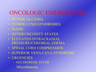
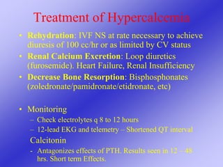
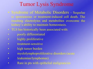
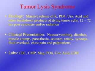
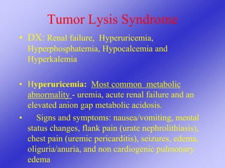
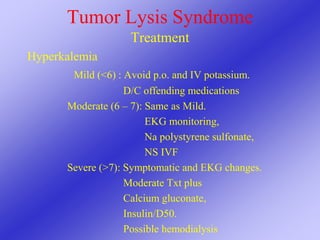
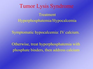
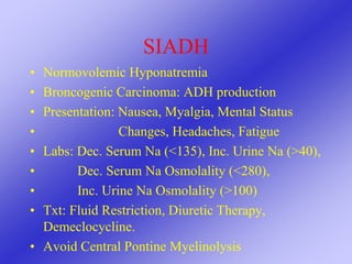
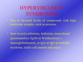
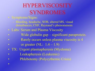
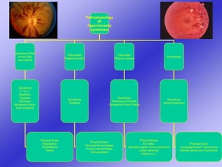
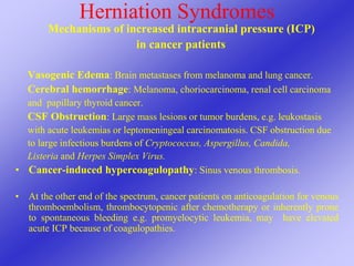
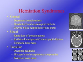
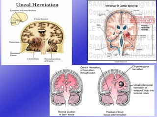
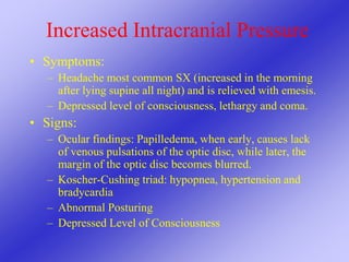
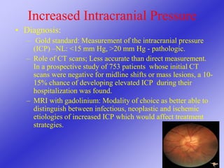
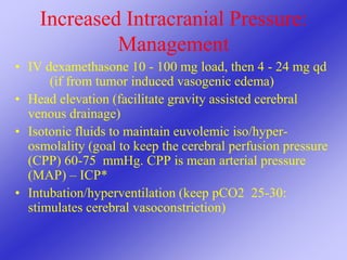
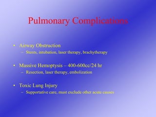
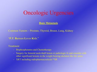
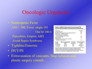
ONCOLOGIC
EMERGENCIES
Terry Gruchow MHS PA-C
Internal Hospital Medicine
Moffitt Cancer Center
Tampa, Florida
March 10, 2018
Oncologic Emergencies
I have no financial relationships, commercial
interests or conflicts to disclose
EMERGENCIES
• Crisis
• Unforeseen combination of circumstances
• Condition requiring immediate action
ONCOLOGIC EMERGENCIES
• HYPERCALCEMIA
• TUMOR LYSIS SYNDROMES
• SIADH
• HYPERVISCOSITY STATES
• ELEVATED INTRACRANIAL
PRESSURE/CEREBRAL EDEMA
• SPINAL CORD COMPRESSION
• SUPERIOR VENA CAVA SYNDROME
• URGENCIES
– NEUTROPENIC FEVER
Miscellaneous
Hypercalcemia of Malignancy
Humoral Hypercalcemia (PTHrP)- Bone
resorbing activity. Interacts with renal PTH receptor -
Stimulate renal calcium resorption
- Squamous Cell Carcinoma
- Renal Cell Carcinoma
- Ovarian and Breast Cancer
1,25 (OH)2 Vit. D Production – Increased
osteoclastic bone resorption. Intestinal absorption of
calcium
- Myeloma
- Lymphoma
Hypercalcemia of Malignancy
Local Osteolytic Hypercalcemia
– Breast cancer
– Multiple myeloma
– Lymphoma/leukemia
– Lung cancer
– Prostate cancer
Clinical Manifestations of
Hypercalcemia
• Total Calcium > 12 mg/dL
• Rapidity of increase
• MOANS, STONES and GROANS
– Altered mental status, renal calculi,
constipation/cramps, n/v, seizures
• RENAL: Dehydration, polyuria/dipsia
• CV: bradycardia, arrhythmia (Shortened QT)
• Muscle weakness, lethargy, psychosis
Treatment of Hypercalcemia
• MILD Hypercalcemia
– Avoid inactivity and ambulate
– Avoid dehydration
• Severe Hypercalcemia
– Rehydrate
– Increase renal calcium excretion
– Decrease Bone Resorption
Treatment of Hypercalcemia
• Rehydration: IVF NS at rate necessary to achieve
diuresis of 100 cc/hr or as limited by CV status
• Renal Calcium Excretion: Loop diuretics
(furosemide). Heart Failure, Renal Insufficiency
• Decrease Bone Resorption: Bisphosphonates
(zoledronate/pamidronate/etidronate, etc)
• Monitoring
– Check electrolytes q 8 to 12 hours
– 12-lead EKG and telemetry – Shortened QT interval
Calcitonin
- Antagonizes effects of PTH. Results seen in 12 – 48
hrs. Short term Effects.
Tumor Lysis Syndrome
• Syndrome of Metabolic Disorders – Sequelae
to spontaneous or treatment-induced cell death. The
resulting electrolytes and metabolites overcome the
kidney’s ability to maintain homeostasis.
• TLS has historically been associated with:
• poorly differentiated
• highly proliferative
• treatment-sensitive
• high tumor burden
• myelolymphoproliferative disorders (acute
• leukemias/lymphomas)
• Rare in pts with epithelial malignancies
Tumor Lysis Syndrome
• Etiology: Massive release of K, PO4, Uric Acid and
other breakdown products of dying tumor cells, 12 – 72
hrs post cytotoxic and/or radiation
• Clinical Presentation: Nausea/vomiting, diarrhea,
muscle cramps, paresthesia, seizures, tetany, syncope,
fluid overload, chest pain and palpitations.
• Labs: CBC, CMP, Mag, PO4, Uric Acid, LDH
Tumor Lysis Syndrome
• DX: Renal failure, Hyperuricemia,
Hyperphosphatemia, Hypocalcemia and
Hyperkalemia
• Hyperuricemia: Most common metabolic
abnormality - uremia, acute renal failure and an
elevated anion gap metabolic acidosis.
• Signs and symptoms: nausea/vomiting, mental
status changes, flank pain (urate nephrolithiasis),
chest pain (uremic pericarditis), seizures, edema,
oliguria/anuria, and non cardiogenic pulmonary
edema
Tumor Lysis Syndrome:
Hyperphosphatemia
Manifestations: Symptomatic hypocalcemia. ARF,
Arrhythmias
Hypocalcemia
• Manifestations: Muscle cramps, Paresthesias, Tetany,
Mental status changes, Seizures, Hypotension and QT
prolongation.
• Trousseau’s and Chvostek’s - dramatic when present but of
unknown sensitivity and specificity.
Hyperkalemia
Presentation: Muscle weakness, Ekg Changes
Tumor Lysis Syndrome
• Laboratory TLS: 2 or more of  Uric acid
• Potassium
• Phosphate
• Calcium
• Clinical TLS: Lab TLS plus  renal failure,
• arrhythmias,
• seizures,
• sudden death
Treatment of Tumor Lysis
Syndrome
“Prevention is better than cure”
Allopurinol prophylaxis with isotonic saline prior
to antitumor treatment
• Treat individual electrolyte abnormalities
• Treat Renal failure or cardiac arrhythmias
Tumor Lysis Syndrome
Treatment
Hyperuricemia:
Hydration/Diuresis – NS IVF and Diuretics
(Lasix or Mannitol)
Decrease Uric Acid Production –
Allopurinol
Increase Uric Acid Destruction –
Rasburicase
Use of Allopurinol vs Rasburicase - Based on
WBC, LDH, Uric Acid and
Tumor Type and Extent
Tumor Lysis Syndrome
Treatment
Hyperkalemia
Mild (<6) : Avoid p.o. and IV potassium.
D/C offending medications
Moderate (6 – 7): Same as Mild.
EKG monitoring,
Na polystyrene sulfonate,
NS IVF
Severe (>7): Symptomatic and EKG changes.
Moderate Txt plus
Calcium gluconate,
Insulin/D50.
Possible hemodialysis
Tumor Lysis Syndrome
Treatment
Hyperphosphatemia/Hypocalcemia
Symptomatic hypocalcemia: IV calcium.
Otherwise, treat hyperphosphatemia with
phosphate binders, then address calcium
SIADH
• Normovolemic Hyponatremia
• Broncogenic Carcinoma: ADH production
• Presentation: Nausea, Myalgia, Mental Status
• Changes, Headaches, Fatigue
• Labs: Dec. Serum Na (<135), Inc. Urine Na (>40),
• Dec. Serum Na Osmolality (<280),
• Inc. Urine Na Osmolality (>100)
• Txt: Fluid Restriction, Diuretic Therapy,
Demeclocycline.
• Avoid Central Pontine Myelinolysis
HYPERVISCOSITY
SYNDROMES
• Due to elevated levels of compounds with high
molecular weights, such as proteins.
• Seen in polycythemias, leukemia, monoclonal
gammopathies (IgM) in Waldenstrom’s
macroglobulinemia, or IgA or IgG in multiple
myeloma, sickle cell anemia and sepsis.
HYPERVISCOSITY
SYNDROMES
• Symptoms/Signs:
– Bleeding, headache, SOB, altered MS, visual
disturbances, CHF, Raynaud’s phenomenom
• Labs: Serum and Plasma Viscosity
• Wide globulin gap – significant paraprotein.
• Rarely occurs unless plasma viscosity is 4
• or greater (NL: 1.4 – 1.9)
• TX: Urgent plasmapheresis (Myeloma)
• Leukapheresis (Leukemic)
• Phlebotomy (Polycythemic Crisis)
•
Pathophysiology
Of
Hyperviscosity
syndromes
Decreased Flow
In the CNS
vasculature
Decreased
Platelet function
Expanded
Plasma volume
Thrombosis
Symptoms
5 “D’s”
Deafness
Diplopia
Dizziness
Decreased vision
Dull headache
Symptoms
Epistaxis
Symptoms
Shortness of breath
Congestive Heart Failure
Symptoms
Visual Inpairment
Physical Exam
Nystagmus
Papilledema
Ataxia
Physical Exam
Mucosal hemorrhages
Fundus hemorrhages
And exudates
Physical Exam
S3, rales,
elevated jugular venous pressure
Lower extremity
Edema e.t.c.
Physical Exam
“sausaqge-linked” retinal veins
Central retinal vein thrombosis
Herniation Syndromes
Mechanisms of increased intracranial pressure (ICP)
in cancer patients
Vasogenic Edema: Brain metastases from melanoma and lung cancer.
Cerebral hemorrhage: Melanoma, choriocarcinoma, renal cell carcinoma
and papillary thyroid cancer.
CSF Obstruction: Large mass lesions or tumor burdens, e.g. leukostasis
with acute leukemias or leptomeningeal carcinomatosis. CSF obstruction due
to large infectious burdens of Cryptococcus, Aspergillus, Candida,
Listeria and Herpes Simplex Virus.
• Cancer-induced hypercoagulopathy: Sinus venous thrombosis.
• At the other end of the spectrum, cancer patients on anticoagulation for venous
thromboembolism, thrombocytopenic after chemotherapy or inherently prone
to spontaneous bleeding e.g. promyelocytic leukemia, may have elevated
acute ICP because of coagulopathies.
Herniation Syndromes
• Central
– Decreased consciousness
– Headache/Focal neurological deficits
– Cheyne Stoke respirations/fixed pupil
• Uncal
– Rapid loss of consciousness
– Ipsilateral hemiparesis/Lateral pupil dilation
– Temporal lobe mass
• Tonsillar
– Occipital headache
– Hiccups/emesis/respiratory compromise
– Posterior fossa mass
Increased Intracranial Pressure
• Symptoms:
– Headache most common SX (increased in the morning
after lying supine all night) and is relieved with emesis.
– Depressed level of consciousness, lethargy and coma.
• Signs:
– Ocular findings: Papilledema, when early, causes lack
of venous pulsations of the optic disc, while later, the
margin of the optic disc becomes blurred.
– Koscher-Cushing triad: hypopnea, hypertension and
bradycardia
– Abnormal Posturing
– Depressed Level of Consciousness
Increased Intracranial Pressure
• Diagnosis:
– Gold standard: Measurement of the intracranial pressure
(ICP) –NL: <15 mm Hg, >20 mm Hg - pathologic.
– Role of CT scans; Less accurate than direct measurement.
In a prospective study of 753 patients whose initial CT
scans were negative for midline shifts or mass lesions, a 10-
15% chance of developing elevated ICP during their
hospitalization was found.
– MRI with gadolinium: Modality of choice as better able to
distinguish between infectious, neoplastic and ischemic
etiologies of increased ICP which would affect treatment
strategies.
Increased Intracranial Pressure:
Management
• IV dexamethasone 10 - 100 mg load, then 4 - 24 mg qd
(if from tumor induced vasogenic edema)
• Head elevation (facilitate gravity assisted cerebral
venous drainage)
• Isotonic fluids to maintain euvolemic iso/hyper-
osmolality (goal to keep the cerebral perfusion pressure
(CPP) 60-75 mmHg. CPP is mean arterial pressure
(MAP) – ICP*
• Intubation/hyperventilation (keep pCO2 25-30:
stimulates cerebral vasoconstriction)
Increased Intracranial Pressure:
Management
• IV Mannitol in unstable patients to promote
osmotic diuresis
– (dose of 20-25% @ 0.75-1.0 g/kg IV initially,
then 0.25-0.5g/kg every 3-6 h is recommended
by most experts. This is to be discontinued if
the serum osmolality exceeds 300)
• Neuro-surgical decompression (herniation)
• Supportive Care
Spinal Cord Compression
• Infection
• Hematoma
• Herniated disc
• Vertebral fractures
• Myelopathy
• Metastasis (prostate, thyroid, breast, lung, RCC,
NHL,,MM). Thoracic (70%), LS (20%), Cervical
(10%). P T Barnum Loves Kids
• Leptomeningial disease
Spinal Cord Compression
• Symptoms  Back pain, not relieved by lying
down
•  Weakness/sensory paresthesia
•  Thoracic: Weak/paresthesia is
• ascending
• Gait disorder
• Sphincter late
•  Lumbar: Sphincter early
Spinal Cord Compression
• Suspect the diagnosis
• Aggressive evaluation with MRI (CT
myelogram). Entire Spine
• Neurosurgery: Tissue dx, Resection of
Tumor, Vertebral Fracture, Vertebral
Stabilization. Initial Consult
• XRT consultation
• IV Dexamethasone
• Pain control
Cardiac Tamponade/Pericardial
Injury
• Secondary to metastasis: Thoracic
• Radiation injury
• Infection
• Uremia
• Hypothyroidism
Cardiac Tamponade
• Beck’s Triad
• Hypotension  Decline in cardiac output
• Muffled Heart Sounds
• Distended Neck Veins
• Dyspnea  Most common
• Cough, chest pain, generalized weakness
• Tachycardia, peripheral edema, Kussmaul’s sign,
pulsus paradoxus (abnormally large decrease in
systolic blood pressure (>10 mmHg) on
inspiration)
Cardiac Tamponade
• Workup  EKG: Sinus tachycardia
• : Low voltage
• : Electrical alternans
•  ECHO: Right atrial collapse
•  CXR
•  Pericardial Bx
Cardiac Tamponade
• Management  Asymptomatic with mild
effusion: No emergent treatment
• Symptomatic: Drain fluid.
Pericardiocentesis or Surgical
pericardiectomy
Superior Vena Cava Syndrome
• Thoracic Malignancy 85 to 95% of cases, CVC
• DX: SOB, facial/neck/UE swelling, chest pain,
headaches. Bending forward or lying flat worse
• PE: thoracic vein distention, edema of face,
tachypnea
• Workup: CXR, CT, Histologic dx to determine
therapy
• TX: Treat Underlying disease, stenting, surgery,
thrombolytics
GU Obstruction
• Causes: prostate CA, Bladder CA, Retroperitoneal tumors
like sarcoma and metastasis
• Dx: CT scan, ultrasound, MRI in some cases elevated
creatinine, anuria
• Treatment:
– Relative obstruction – TURP/TURBT, stents percutaneous
nephrostomy tubes
– Treat infections, fluid imbalances, etc.
Leukostasis
• Acute Leukemias  High blast counts
• Presentations: Resp. distress, confusion,
CNS bleed
• Clinical dx  Heart failure, infection,
• Imaging: CXR  nonspecific diffuse
infiltrate
• Txt: Leukapheresis. Hydrea as adjunct.
Pulmonary Complications
• Airway Obstruction
– Stents, intubation, laser therapy, brachytherapy
• Massive Hemoptysis – 400-600cc/24 hr
– Resection, laser therapy, embolization
• Toxic Lung Injury
– Supportative care, must exclude other acute causes
Oncologic Urgencies
Bony Metastasis
Common Tumors – Prostate, Thyroid, Breast, Lung, Kidney
“P.T. Barnum Loves Kids.”
Treatment
– Bisphosphonates and Chemotherapy
– Surgery for femoral neck/shaft lesion or pathologic fx and consider with
other significant lesions in the weight bearing skeleton like the spine.
– XRT including radiopharmaceuticals 89SR
Oncologic Urgencies
• Neutropenic Fever
ANC< 500, Fever: single 101
One hr 100.4
Panculture, Empiric ABX
Avoid Sepsis Syndrome
• Typhlitis/Enteritis
• DVT/PE
• Extravasation of vesicants. Stop infusion and
plastic surgery consult.
Oncologic Emergencies
Thank you for your attendance and attention

Oncologic Emergencies & Symptom Management

  • 1.
    ONCOLOGIC EMERGENCIES Terry Gruchow MHSPA-C Internal Hospital Medicine Moffitt Cancer Center Tampa, Florida March 10, 2018
  • 2.
    Oncologic Emergencies I haveno financial relationships, commercial interests or conflicts to disclose
  • 3.
    EMERGENCIES • Crisis • Unforeseencombination of circumstances • Condition requiring immediate action
  • 4.
    ONCOLOGIC EMERGENCIES • HYPERCALCEMIA •TUMOR LYSIS SYNDROMES • SIADH • HYPERVISCOSITY STATES • ELEVATED INTRACRANIAL PRESSURE/CEREBRAL EDEMA • SPINAL CORD COMPRESSION • SUPERIOR VENA CAVA SYNDROME • URGENCIES – NEUTROPENIC FEVER Miscellaneous
  • 5.
    Hypercalcemia of Malignancy HumoralHypercalcemia (PTHrP)- Bone resorbing activity. Interacts with renal PTH receptor - Stimulate renal calcium resorption - Squamous Cell Carcinoma - Renal Cell Carcinoma - Ovarian and Breast Cancer 1,25 (OH)2 Vit. D Production – Increased osteoclastic bone resorption. Intestinal absorption of calcium - Myeloma - Lymphoma
  • 6.
    Hypercalcemia of Malignancy LocalOsteolytic Hypercalcemia – Breast cancer – Multiple myeloma – Lymphoma/leukemia – Lung cancer – Prostate cancer
  • 7.
    Clinical Manifestations of Hypercalcemia •Total Calcium > 12 mg/dL • Rapidity of increase • MOANS, STONES and GROANS – Altered mental status, renal calculi, constipation/cramps, n/v, seizures • RENAL: Dehydration, polyuria/dipsia • CV: bradycardia, arrhythmia (Shortened QT) • Muscle weakness, lethargy, psychosis
  • 8.
    Treatment of Hypercalcemia •MILD Hypercalcemia – Avoid inactivity and ambulate – Avoid dehydration • Severe Hypercalcemia – Rehydrate – Increase renal calcium excretion – Decrease Bone Resorption
  • 9.
    Treatment of Hypercalcemia •Rehydration: IVF NS at rate necessary to achieve diuresis of 100 cc/hr or as limited by CV status • Renal Calcium Excretion: Loop diuretics (furosemide). Heart Failure, Renal Insufficiency • Decrease Bone Resorption: Bisphosphonates (zoledronate/pamidronate/etidronate, etc) • Monitoring – Check electrolytes q 8 to 12 hours – 12-lead EKG and telemetry – Shortened QT interval Calcitonin - Antagonizes effects of PTH. Results seen in 12 – 48 hrs. Short term Effects.
  • 10.
    Tumor Lysis Syndrome •Syndrome of Metabolic Disorders – Sequelae to spontaneous or treatment-induced cell death. The resulting electrolytes and metabolites overcome the kidney’s ability to maintain homeostasis. • TLS has historically been associated with: • poorly differentiated • highly proliferative • treatment-sensitive • high tumor burden • myelolymphoproliferative disorders (acute • leukemias/lymphomas) • Rare in pts with epithelial malignancies
  • 11.
    Tumor Lysis Syndrome •Etiology: Massive release of K, PO4, Uric Acid and other breakdown products of dying tumor cells, 12 – 72 hrs post cytotoxic and/or radiation • Clinical Presentation: Nausea/vomiting, diarrhea, muscle cramps, paresthesia, seizures, tetany, syncope, fluid overload, chest pain and palpitations. • Labs: CBC, CMP, Mag, PO4, Uric Acid, LDH
  • 12.
    Tumor Lysis Syndrome •DX: Renal failure, Hyperuricemia, Hyperphosphatemia, Hypocalcemia and Hyperkalemia • Hyperuricemia: Most common metabolic abnormality - uremia, acute renal failure and an elevated anion gap metabolic acidosis. • Signs and symptoms: nausea/vomiting, mental status changes, flank pain (urate nephrolithiasis), chest pain (uremic pericarditis), seizures, edema, oliguria/anuria, and non cardiogenic pulmonary edema
  • 13.
    Tumor Lysis Syndrome: Hyperphosphatemia Manifestations:Symptomatic hypocalcemia. ARF, Arrhythmias Hypocalcemia • Manifestations: Muscle cramps, Paresthesias, Tetany, Mental status changes, Seizures, Hypotension and QT prolongation. • Trousseau’s and Chvostek’s - dramatic when present but of unknown sensitivity and specificity. Hyperkalemia Presentation: Muscle weakness, Ekg Changes
  • 14.
    Tumor Lysis Syndrome •Laboratory TLS: 2 or more of  Uric acid • Potassium • Phosphate • Calcium • Clinical TLS: Lab TLS plus  renal failure, • arrhythmias, • seizures, • sudden death
  • 15.
    Treatment of TumorLysis Syndrome “Prevention is better than cure” Allopurinol prophylaxis with isotonic saline prior to antitumor treatment • Treat individual electrolyte abnormalities • Treat Renal failure or cardiac arrhythmias
  • 16.
    Tumor Lysis Syndrome Treatment Hyperuricemia: Hydration/Diuresis– NS IVF and Diuretics (Lasix or Mannitol) Decrease Uric Acid Production – Allopurinol Increase Uric Acid Destruction – Rasburicase Use of Allopurinol vs Rasburicase - Based on WBC, LDH, Uric Acid and Tumor Type and Extent
  • 17.
    Tumor Lysis Syndrome Treatment Hyperkalemia Mild(<6) : Avoid p.o. and IV potassium. D/C offending medications Moderate (6 – 7): Same as Mild. EKG monitoring, Na polystyrene sulfonate, NS IVF Severe (>7): Symptomatic and EKG changes. Moderate Txt plus Calcium gluconate, Insulin/D50. Possible hemodialysis
  • 18.
    Tumor Lysis Syndrome Treatment Hyperphosphatemia/Hypocalcemia Symptomatichypocalcemia: IV calcium. Otherwise, treat hyperphosphatemia with phosphate binders, then address calcium
  • 19.
    SIADH • Normovolemic Hyponatremia •Broncogenic Carcinoma: ADH production • Presentation: Nausea, Myalgia, Mental Status • Changes, Headaches, Fatigue • Labs: Dec. Serum Na (<135), Inc. Urine Na (>40), • Dec. Serum Na Osmolality (<280), • Inc. Urine Na Osmolality (>100) • Txt: Fluid Restriction, Diuretic Therapy, Demeclocycline. • Avoid Central Pontine Myelinolysis
  • 20.
    HYPERVISCOSITY SYNDROMES • Due toelevated levels of compounds with high molecular weights, such as proteins. • Seen in polycythemias, leukemia, monoclonal gammopathies (IgM) in Waldenstrom’s macroglobulinemia, or IgA or IgG in multiple myeloma, sickle cell anemia and sepsis.
  • 21.
    HYPERVISCOSITY SYNDROMES • Symptoms/Signs: – Bleeding,headache, SOB, altered MS, visual disturbances, CHF, Raynaud’s phenomenom • Labs: Serum and Plasma Viscosity • Wide globulin gap – significant paraprotein. • Rarely occurs unless plasma viscosity is 4 • or greater (NL: 1.4 – 1.9) • TX: Urgent plasmapheresis (Myeloma) • Leukapheresis (Leukemic) • Phlebotomy (Polycythemic Crisis) •
  • 22.
    Pathophysiology Of Hyperviscosity syndromes Decreased Flow In theCNS vasculature Decreased Platelet function Expanded Plasma volume Thrombosis Symptoms 5 “D’s” Deafness Diplopia Dizziness Decreased vision Dull headache Symptoms Epistaxis Symptoms Shortness of breath Congestive Heart Failure Symptoms Visual Inpairment Physical Exam Nystagmus Papilledema Ataxia Physical Exam Mucosal hemorrhages Fundus hemorrhages And exudates Physical Exam S3, rales, elevated jugular venous pressure Lower extremity Edema e.t.c. Physical Exam “sausaqge-linked” retinal veins Central retinal vein thrombosis
  • 23.
    Herniation Syndromes Mechanisms ofincreased intracranial pressure (ICP) in cancer patients Vasogenic Edema: Brain metastases from melanoma and lung cancer. Cerebral hemorrhage: Melanoma, choriocarcinoma, renal cell carcinoma and papillary thyroid cancer. CSF Obstruction: Large mass lesions or tumor burdens, e.g. leukostasis with acute leukemias or leptomeningeal carcinomatosis. CSF obstruction due to large infectious burdens of Cryptococcus, Aspergillus, Candida, Listeria and Herpes Simplex Virus. • Cancer-induced hypercoagulopathy: Sinus venous thrombosis. • At the other end of the spectrum, cancer patients on anticoagulation for venous thromboembolism, thrombocytopenic after chemotherapy or inherently prone to spontaneous bleeding e.g. promyelocytic leukemia, may have elevated acute ICP because of coagulopathies.
  • 24.
    Herniation Syndromes • Central –Decreased consciousness – Headache/Focal neurological deficits – Cheyne Stoke respirations/fixed pupil • Uncal – Rapid loss of consciousness – Ipsilateral hemiparesis/Lateral pupil dilation – Temporal lobe mass • Tonsillar – Occipital headache – Hiccups/emesis/respiratory compromise – Posterior fossa mass
  • 26.
    Increased Intracranial Pressure •Symptoms: – Headache most common SX (increased in the morning after lying supine all night) and is relieved with emesis. – Depressed level of consciousness, lethargy and coma. • Signs: – Ocular findings: Papilledema, when early, causes lack of venous pulsations of the optic disc, while later, the margin of the optic disc becomes blurred. – Koscher-Cushing triad: hypopnea, hypertension and bradycardia – Abnormal Posturing – Depressed Level of Consciousness
  • 27.
    Increased Intracranial Pressure •Diagnosis: – Gold standard: Measurement of the intracranial pressure (ICP) –NL: <15 mm Hg, >20 mm Hg - pathologic. – Role of CT scans; Less accurate than direct measurement. In a prospective study of 753 patients whose initial CT scans were negative for midline shifts or mass lesions, a 10- 15% chance of developing elevated ICP during their hospitalization was found. – MRI with gadolinium: Modality of choice as better able to distinguish between infectious, neoplastic and ischemic etiologies of increased ICP which would affect treatment strategies.
  • 28.
    Increased Intracranial Pressure: Management •IV dexamethasone 10 - 100 mg load, then 4 - 24 mg qd (if from tumor induced vasogenic edema) • Head elevation (facilitate gravity assisted cerebral venous drainage) • Isotonic fluids to maintain euvolemic iso/hyper- osmolality (goal to keep the cerebral perfusion pressure (CPP) 60-75 mmHg. CPP is mean arterial pressure (MAP) – ICP* • Intubation/hyperventilation (keep pCO2 25-30: stimulates cerebral vasoconstriction)
  • 29.
    Increased Intracranial Pressure: Management •IV Mannitol in unstable patients to promote osmotic diuresis – (dose of 20-25% @ 0.75-1.0 g/kg IV initially, then 0.25-0.5g/kg every 3-6 h is recommended by most experts. This is to be discontinued if the serum osmolality exceeds 300) • Neuro-surgical decompression (herniation) • Supportive Care
  • 30.
    Spinal Cord Compression •Infection • Hematoma • Herniated disc • Vertebral fractures • Myelopathy • Metastasis (prostate, thyroid, breast, lung, RCC, NHL,,MM). Thoracic (70%), LS (20%), Cervical (10%). P T Barnum Loves Kids • Leptomeningial disease
  • 31.
    Spinal Cord Compression •Symptoms  Back pain, not relieved by lying down •  Weakness/sensory paresthesia •  Thoracic: Weak/paresthesia is • ascending • Gait disorder • Sphincter late •  Lumbar: Sphincter early
  • 32.
    Spinal Cord Compression •Suspect the diagnosis • Aggressive evaluation with MRI (CT myelogram). Entire Spine • Neurosurgery: Tissue dx, Resection of Tumor, Vertebral Fracture, Vertebral Stabilization. Initial Consult • XRT consultation • IV Dexamethasone • Pain control
  • 33.
    Cardiac Tamponade/Pericardial Injury • Secondaryto metastasis: Thoracic • Radiation injury • Infection • Uremia • Hypothyroidism
  • 34.
    Cardiac Tamponade • Beck’sTriad • Hypotension  Decline in cardiac output • Muffled Heart Sounds • Distended Neck Veins • Dyspnea  Most common • Cough, chest pain, generalized weakness • Tachycardia, peripheral edema, Kussmaul’s sign, pulsus paradoxus (abnormally large decrease in systolic blood pressure (>10 mmHg) on inspiration)
  • 35.
    Cardiac Tamponade • Workup EKG: Sinus tachycardia • : Low voltage • : Electrical alternans •  ECHO: Right atrial collapse •  CXR •  Pericardial Bx
  • 36.
    Cardiac Tamponade • Management Asymptomatic with mild effusion: No emergent treatment • Symptomatic: Drain fluid. Pericardiocentesis or Surgical pericardiectomy
  • 37.
    Superior Vena CavaSyndrome • Thoracic Malignancy 85 to 95% of cases, CVC • DX: SOB, facial/neck/UE swelling, chest pain, headaches. Bending forward or lying flat worse • PE: thoracic vein distention, edema of face, tachypnea • Workup: CXR, CT, Histologic dx to determine therapy • TX: Treat Underlying disease, stenting, surgery, thrombolytics
  • 39.
    GU Obstruction • Causes:prostate CA, Bladder CA, Retroperitoneal tumors like sarcoma and metastasis • Dx: CT scan, ultrasound, MRI in some cases elevated creatinine, anuria • Treatment: – Relative obstruction – TURP/TURBT, stents percutaneous nephrostomy tubes – Treat infections, fluid imbalances, etc.
  • 40.
    Leukostasis • Acute Leukemias High blast counts • Presentations: Resp. distress, confusion, CNS bleed • Clinical dx  Heart failure, infection, • Imaging: CXR  nonspecific diffuse infiltrate • Txt: Leukapheresis. Hydrea as adjunct.
  • 41.
    Pulmonary Complications • AirwayObstruction – Stents, intubation, laser therapy, brachytherapy • Massive Hemoptysis – 400-600cc/24 hr – Resection, laser therapy, embolization • Toxic Lung Injury – Supportative care, must exclude other acute causes
  • 42.
    Oncologic Urgencies Bony Metastasis CommonTumors – Prostate, Thyroid, Breast, Lung, Kidney “P.T. Barnum Loves Kids.” Treatment – Bisphosphonates and Chemotherapy – Surgery for femoral neck/shaft lesion or pathologic fx and consider with other significant lesions in the weight bearing skeleton like the spine. – XRT including radiopharmaceuticals 89SR
  • 43.
    Oncologic Urgencies • NeutropenicFever ANC< 500, Fever: single 101 One hr 100.4 Panculture, Empiric ABX Avoid Sepsis Syndrome • Typhlitis/Enteritis • DVT/PE • Extravasation of vesicants. Stop infusion and plastic surgery consult.
  • 44.
    Oncologic Emergencies Thank youfor your attendance and attention