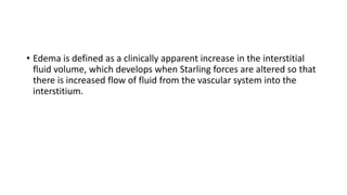
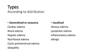
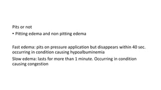
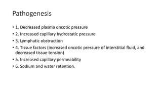
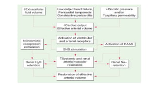
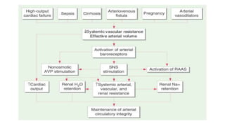
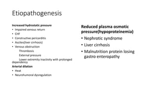
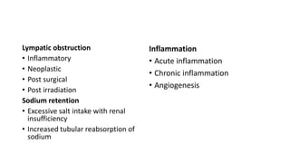
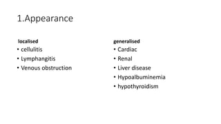
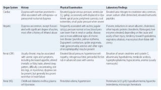
OEDEMA
Akshai George Paul
• Edema is defined as a clinically apparent increase in the interstitial
fluid volume, which develops when Starling forces are altered so that
there is increased flow of fluid from the vascular system into the
interstitium.
Types
According to distribution
• Generalised or anasarca
Cardiac edema
Renal edema
Hepatic edema
Nutritional edema
Cyclic premenstrual edema
idiopathic
• Localised
Venous edema
Lymphatic edema
Inflammatory edema
allergic
Pits or not
• Pitting edema and non pitting edema
Fast edema: pits on pressure application but disappears within 40 sec.
occurring in condition causing hypoalbuminemia
Slow edema: lasts for more than 1 minute. Occurring in condition
causing congestion
• Renal edema
nephrotic edema
nephritic edema
• Cardiac edema
• Pulmonary edema
• Cerebral edema
vasogenic edema
cytotoxic edema
interstitial edema
• Hepatic edema
• Nutritional edema
• Myxodema
Pathogenesis
• 1. Decreased plasma oncotic pressure
• 2. Increased capillary hydrostatic pressure
• 3. Lymphatic obstruction
• 4. Tissue factors (increased oncotic pressure of interstitial fluid, and
decreased tissue tension)
• 5. Increased capillary permeability
• 6. Sodium and water retention.
Etiopathogenesis
Increased hydrostatic pressure
• Impaired venous return
• CHF
• Constructive pericarditis
• Ascites(liver cirrhosis)
• Venous obstruction
Thrombosis
External pressure
Lower extremity inactivity with prolonged
dependency
Arterial dilation
• Heat
• Neurohumoral dysregulation
Reduced plasma osmotic
pressure(hypoproteinemia)
• Nephrotic syndrome
• Liver cirrhosis
• Malnutrition protein losing
gastro enteropathy
Lympatic obstruction
• Inflammatory
• Neoplastic
• Post surgical
• Post irradiation
Sodium retention
• Excessive salt intake with renal
insufficiency
• Increased tubular reabsorption of
sodium
Inflammation
• Acute inflammation
• Chronic inflammation
• Angiogenesis
DRUGS ASSOCIATED WITH EDEMA FORMATION
• Non steroidal anti-inflammatory drugs
• Antihypertensive agents
• Direct arterial/arteriolar vasodilators
Hydralazine
clonidine
Methyldopa
Guanethidine
Minoxidil
• calcium channel antagonists
• a-Adrenergic antagonists
• Thiazolidinediones
• Steroid hormones
Glucocorticoids
Anabolic steroids
Estrogens
Progestins
• Cyclosporine
• Growth hormone
• Immunotherapies
Interleukin2
OKT3 monoclonal antibody
Grading of oedema
Approach to oedema
History
1.Appearance
localised
• cellulitis
• Lymphangitis
• Venous obstruction
generalised
• Cardiac
• Renal
• Liver disease
• Hypoalbuminemia
• hypothyroidism
2.Onset
sudden
• a/c nephritis
• a/c anaphylaxis
insidious
3.First site of appearance
Periorbital area - renal cause
Dependant part - cardiac oedema
• leg
• sacrum
4.Other symptoms
Hypothyroidism
• Constipation
• Cold intolerance
• Feeling sleepy
Are there any features of nutritional deficiency
Starvation/malnutrition
hypoproteinemia
Filariasis/cellulitis/lymphangitis
• Recurrent attack of fever and rigor
• History of fever and signs of inflammation
5.Associated features
• Oliguria and smoky urine - nephritis
• Orthopnoea and PND - cardiac cause
• Urticaria and manifestation of allergy - angiodema
• GI symptom - cirrhosis of liver - ascites
• Chest pain and cough/dyspnoea - mediastinal obstruction due to tumour
• Signs of inflammation over area - inflammatory cause
6.Past, present and family history
• Past history - cardiac renal or liver disease
• Family history of oedema –Milroy’s oedema
• Drug history- nifedipine, estrogen, steroids, NSAIDS
• Is the patient pregnant-unilateral pedal oedema
• Relationship to menstrual period-cyclical oedema(periodical)
• History of surgery and prolonged travel-DVT
Examination of patient
General examination
Build and nourishment
• Poorly nourished if, oedema is due to any nutritional deficiency
Pallor
• Cardiac/GI causes
Icterus
• Cirrhosis of liver
Cyanosis and clubbing
• Cardiac oedema
Lymphadenopathy
• Filariasis
How to demonstrate oedema clinically
Ambulant patient
• Over medial malleolus or 5cm above it with right thumb apply
pressure for minimum 30 seconds
• Almost always look for dimple after applying pressure otherwise we
can miss a case of minimal oedema
Bedridden patient
• Apply pressure over sacrum with right thumb for 30 seconds and look
for dimple
Examination of patient with cardiac oedema
Inspection
• Dyspnoeic
• Engorged or pulsatile veins in neck
palpation
• Pitting oedema
• Soft tender hepatomegaly
• Apex outside MCL – cardiomegaly
Auscultation
• May be associated with RV gallop rhythm
Examination of patient with renal oedema
Inspection
• Periorbital oedema
• Swelling of scrotal sac
Palpation
• Pitting oedema
Percussion and auscultation are less significant
Examination of patient with ascites
Inspection
• Generalised swelling of abdomen, flanks usually full, umbilicus
everted
Percussion
• Puddle sign
• Fluid thrill
• Shifting dullness
Evolution of oedema in different systemic disease
• Congestive cardiac failure; legs –face – ascites
• Nephrotic syndrome; face(periorbital puffiness) – legs – ascites(with
scrotal swelling)
• Cirrhosis of liver; ascites – legs – face(rarely)
• Nutritional oedema; oedema feet with puffiness of face – ascites
Treatment
Diuretics
Thiazides
Loop diuretics
Potassium sparing diuretics
Managing diuretic resistance
THANK YOU

Oedema agp

  • 1.
  • 2.
    • Edema isdefined as a clinically apparent increase in the interstitial fluid volume, which develops when Starling forces are altered so that there is increased flow of fluid from the vascular system into the interstitium.
  • 3.
    Types According to distribution •Generalised or anasarca Cardiac edema Renal edema Hepatic edema Nutritional edema Cyclic premenstrual edema idiopathic • Localised Venous edema Lymphatic edema Inflammatory edema allergic
  • 4.
    Pits or not •Pitting edema and non pitting edema Fast edema: pits on pressure application but disappears within 40 sec. occurring in condition causing hypoalbuminemia Slow edema: lasts for more than 1 minute. Occurring in condition causing congestion
  • 5.
    • Renal edema nephroticedema nephritic edema • Cardiac edema • Pulmonary edema • Cerebral edema vasogenic edema cytotoxic edema interstitial edema • Hepatic edema • Nutritional edema • Myxodema
  • 6.
    Pathogenesis • 1. Decreasedplasma oncotic pressure • 2. Increased capillary hydrostatic pressure • 3. Lymphatic obstruction • 4. Tissue factors (increased oncotic pressure of interstitial fluid, and decreased tissue tension) • 5. Increased capillary permeability • 6. Sodium and water retention.
  • 9.
    Etiopathogenesis Increased hydrostatic pressure •Impaired venous return • CHF • Constructive pericarditis • Ascites(liver cirrhosis) • Venous obstruction Thrombosis External pressure Lower extremity inactivity with prolonged dependency Arterial dilation • Heat • Neurohumoral dysregulation Reduced plasma osmotic pressure(hypoproteinemia) • Nephrotic syndrome • Liver cirrhosis • Malnutrition protein losing gastro enteropathy
  • 10.
    Lympatic obstruction • Inflammatory •Neoplastic • Post surgical • Post irradiation Sodium retention • Excessive salt intake with renal insufficiency • Increased tubular reabsorption of sodium Inflammation • Acute inflammation • Chronic inflammation • Angiogenesis
  • 11.
    DRUGS ASSOCIATED WITHEDEMA FORMATION • Non steroidal anti-inflammatory drugs • Antihypertensive agents • Direct arterial/arteriolar vasodilators Hydralazine clonidine Methyldopa Guanethidine Minoxidil • calcium channel antagonists • a-Adrenergic antagonists • Thiazolidinediones • Steroid hormones Glucocorticoids Anabolic steroids Estrogens Progestins • Cyclosporine • Growth hormone • Immunotherapies Interleukin2 OKT3 monoclonal antibody
  • 12.
  • 13.
  • 14.
    1.Appearance localised • cellulitis • Lymphangitis •Venous obstruction generalised • Cardiac • Renal • Liver disease • Hypoalbuminemia • hypothyroidism
  • 15.
    2.Onset sudden • a/c nephritis •a/c anaphylaxis insidious
  • 16.
    3.First site ofappearance Periorbital area - renal cause
  • 17.
    Dependant part -cardiac oedema • leg • sacrum
  • 18.
  • 19.
    Are there anyfeatures of nutritional deficiency Starvation/malnutrition hypoproteinemia
  • 20.
    Filariasis/cellulitis/lymphangitis • Recurrent attackof fever and rigor • History of fever and signs of inflammation
  • 21.
    5.Associated features • Oliguriaand smoky urine - nephritis • Orthopnoea and PND - cardiac cause • Urticaria and manifestation of allergy - angiodema
  • 22.
    • GI symptom- cirrhosis of liver - ascites • Chest pain and cough/dyspnoea - mediastinal obstruction due to tumour • Signs of inflammation over area - inflammatory cause
  • 23.
    6.Past, present andfamily history • Past history - cardiac renal or liver disease • Family history of oedema –Milroy’s oedema
  • 24.
    • Drug history-nifedipine, estrogen, steroids, NSAIDS • Is the patient pregnant-unilateral pedal oedema
  • 25.
    • Relationship tomenstrual period-cyclical oedema(periodical) • History of surgery and prolonged travel-DVT
  • 26.
    Examination of patient Generalexamination Build and nourishment • Poorly nourished if, oedema is due to any nutritional deficiency Pallor • Cardiac/GI causes
  • 27.
    Icterus • Cirrhosis ofliver Cyanosis and clubbing • Cardiac oedema Lymphadenopathy • Filariasis
  • 28.
    How to demonstrateoedema clinically Ambulant patient • Over medial malleolus or 5cm above it with right thumb apply pressure for minimum 30 seconds • Almost always look for dimple after applying pressure otherwise we can miss a case of minimal oedema
  • 29.
    Bedridden patient • Applypressure over sacrum with right thumb for 30 seconds and look for dimple
  • 30.
    Examination of patientwith cardiac oedema Inspection • Dyspnoeic • Engorged or pulsatile veins in neck palpation • Pitting oedema • Soft tender hepatomegaly • Apex outside MCL – cardiomegaly Auscultation • May be associated with RV gallop rhythm
  • 31.
    Examination of patientwith renal oedema Inspection • Periorbital oedema • Swelling of scrotal sac Palpation • Pitting oedema Percussion and auscultation are less significant
  • 32.
    Examination of patientwith ascites Inspection • Generalised swelling of abdomen, flanks usually full, umbilicus everted Percussion • Puddle sign • Fluid thrill • Shifting dullness
  • 34.
    Evolution of oedemain different systemic disease • Congestive cardiac failure; legs –face – ascites • Nephrotic syndrome; face(periorbital puffiness) – legs – ascites(with scrotal swelling) • Cirrhosis of liver; ascites – legs – face(rarely) • Nutritional oedema; oedema feet with puffiness of face – ascites
  • 35.
  • 36.
  • 37.
  • 38.
  • 40.
  • 43.
  • 44.